Genetic modifier of beta thalassemia
Bạn đang xem bản rút gọn của tài liệu. Xem và tải ngay bản đầy đủ của tài liệu tại đây (801.13 KB, 5 trang )
Thalassemia Syndromes Articles and Brief Reports
haematologica | 2012; 97(7)
989
*These two authors contributed
equally to this work
Acknowledgments: we thank
Serena Sanna for her support
and constructive comments and
Francarosa Demartis for her
technical assistance.
Funding: supported by grants
from Regione Autonoma
Sardegna L.R. 11/90 and
a Research Grant from
Apopharma (Ontario – Canada).
Manuscript received on
August 11, 2011. Revised
version arrived on January 5,
2012. Manuscript accepted
January 10, 2012.
Correspondence:
Renzo Galanello, Ospedale
Regionale Microcitemie, Via
Jenner s/n, 09121 Cagliari, Italy.
Phone: international
+ 39.070.6095508.
Fax: international
+ 39.070.6095509.
E-mail:
Background
The clinical and hematologic features of β-thalassemia are modulated by different factors,
resulting in a wide range of clinical severity. The main factors are the type of disease-causing
mutation and the ability to produce α-globin and γ-globin chains. In the present study we inves-
tigated the respective contributions of known modifiers to the prediction of the clinical severity
of β-thalassemia as assessed by the patients’ age at first transfusion.
Design and Methods
We studied the effect of seven loci in a cohort of 316 Sardinian patients with β
0
-thalassemia. In
addition to characterizing the β-globin gene mutations, α-globin gene defects and HBG2:g
158C>T polymorphism, we genotyped two different markers in the BCL11A gene and three in
the HBS1L-MYB intergenic region using single nucleotide polymorphism microarrays, imputa-
tion and direct genotyping. We performed Cox proportional hazard analysis of the time to first
transfusion.
Results
According to the resulting model, we were able to explain phenotypic severity to a large extent
(Harrell’s concordance index=0.72; Cox & Snell R
2
=0.394) and demonstrated that most of the
model’s discriminatory ability is attributable to the genetic variants affecting fetal hemoglobin
production (HBG2:g 158C>T, BCL11A and HBS1L-MYB loci: C-index=0.68, R
2
=0.272), while
the remaining is due to α-globin gene defects and gender. Consequently, significantly distinct
survival curves can be described in our population.
Conclusions
This detailed analysis clarifies the impact of genetic modifiers on the clinical severity of the dis-
ease, measured by time to first transfusion, by determining their relative contributions in a
homogeneous cohort of β
0
-thalassemia patients. It may also support clinical decisions regarding
the beginning of transfusion therapy in patients with β-thalassemia.
Key words: beta-thalassemia, genetic modifiers, fetal hemoglobin, thalassemia major, thalassemia
intermedia.
Citation: Danjou F, Anni F, Perseu L, Satta S, Dessì C, Lai ME, Fortina P, Devoto M, and Galanello
R. Genetic modifiers of
β
-thalassemia and clinical severity as assessed by age at first transfusion.
Haematologica 2012;97(7):989-993. doi:10.3324/haematol.2011.053504
©
2012 Ferrata Storti Foundation. This is an open-access paper.
Genetic modifiers of β-thalassemia and clinical severity as assessed
by age at first transfusion
Fabrice Danjou,
1
* Franco Anni,
1
* Lucia Perseu,
2
Stefania Satta,
1
Carlo Dessì,
1
Maria Eliana Lai,
3
Paolo Fortina,
4,5
Marcella Devoto,
5,6,7
and Renzo Galanello
1
1
Clinica Pediatrica 2a, Dipartimento di Scienze Biomediche e Biotecnologie - Università di Cagliari, Ospedale Regionale Microcitemie
ASL8, Cagliari, Italy;
2
Istituto di Ricerca Genetica e Biomedica (IRGB) CNR, Cagliari, Italy;
3
Ospedale Regionale Microcitemie, ASL8
Cagliari, Italy;
4
Department of Cancer Biology, Jefferson Genomics Laboratory, Kimmel Cancer Center, Thomas Jefferson University
Jefferson Medical College, Philadelphia, PA, USA;
5
Dipartimento di Medicina Molecolare, Università La Sapienza, Roma, Italy;
6
Division
of Genetics, The Children's Hospital of Philadelphia, Philadelphia, PA, USA, and
7
Department of Pediatrics and Department of
Biostatistics and Epidemiology, Perelman School of Medicine, University of Pennsylvania, Philadelphia, PA, USA
ABSTRACT
©Ferrata Storti Foundation
Introduction
β-thalassemia is characterized by decreased or absent β-
globin chain synthesis due to a variety of mutations; this
decrease in β-globin chain synthesis results in an excess of
α-globin chains which precipitate in red blood cell precur-
sors in the bone marrow, causing their premature death.
1
In
Sardinia, the most common type of β-thalassemia is due to
a nonsense mutation at codon 39 of the β-globin gene
(HBB:c118C>T).
2
The majority of patients develop a severe form of anemia
(thalassemia major) and are transfusion-dependent from the
first years of life. When performed regularly, red blood cell
transfusions prevent anemia-related complications and
compensatory marrow expansion, and, therefore, extend
the survival of patients. Approximately 5-10% of patients
live without requiring periodic blood transfusions and are
said to have thalassemia intermedia.
3
These two forms of
the disease are the extreme ends of a broad range of clinical
variability: patients might need to start transfusions after
days, months or even years of life, demonstration of a great
variation in disease severity.
This remarkable phenotypic diversity of thalassemia
patients is associated with a great variety of genotypes
including mild/silent β-thalassemia alleles, coinheritance of
α-thalassemia or the presence of genetic determinants asso-
ciated with increased production of γ-globin chains and
consequent ability to produce functional fetal hemoglobin
(Hb F) in adult life.
4
All these conditions reduce α/β-globin
chain imbalance and ineffective erythropoiesis. The level of
Hb F is regulated by three major loci: HBG2:g 158C>T on
11p15.4, HBS1L-MYB intergenic region on 6q23.3, and
BCL11A on 2p16.1. Together these three loci are responsi-
ble for 20 to 50% of the Hb F trait variance in patients with
β-thalassemia or sickle cell disease, and in healthy
Europeans.
5-10
Here we evaluated the effect of the HBG2:g 158C>T,
BCL11A, and HBS1L-MYB variants, together with coinher-
itance of α-thalassemia and gender, on the severity of β-tha-
lassemia phenotype, measured by age at first transfusion, in
Sardinian patients.
Design and Methods
Patients and phenotypic assessment
We retrospectively studied 316 β
0
-thalassemia patients (168
males and 148 females) from Sardinia, all followed at the
Microcythemia Hospital of Cagliari. Of these patients, 266 had tha-
lassemia major (median age 33 years; 5
th
and 95
th
percentiles, 13 and
38 years, respectively) and 50 had thalassemia intermedia (median
age 43 years; 5
th
and 95
th
percentiles, 17 and 61 years, respectively);
125 had been enrolled in a previous study on phenotype ameliora-
tion.
11
Thalassemia intermedia patients were defined as patients
who had never been transfused, or had only been transfused spo-
radically during infections or surgery (<10 blood units in total).
3
The
β-thalassemia mutations were of HBB:c118C>T/HBB:c118C>T
type in 92.4% of cases and HBB:c118C>T/HBB:c.20delA type in
6.3% of the studied sample; the remaining mutations are reported
in Table 1. The continuous distribution of the phenotypic severity
among thalassemia patients was measured by the time at which
they started transfusion therapy. Criteria for starting transfusion
were persistent (i.e. more than 2 weeks) hemoglobin level lower
than 7 g/dL in the absence of infections, moderate to severe spleen
enlargement and poor growth. The time to event was calculated as
the time between birth and the first red blood cell transfusion or
between birth and the last follow-up (January 2011) for patients
who were not on transfusion therapy. Age at first transfusion was
retrospectively collected through the WebTHAL computerized clin-
ical records database (), in use for the daily
management of patients in our center.
This retrospective study was conducted in accordance with the
Declaration of Helsinki and the patients gave informed consent to
analysis of their DNA.
Selection of single nucleotide polymorphisms
We selected five single nucleotide polymorphisms (SNP) from
the HBS1L-MYB intergenic region and the BCL11A locus known
to be associated with Hb F levels (Table 2):
rs1427407: the most significant SNP associated with Hb F levels
within BCL11A, as reported by Menzel et al.
12
This SNP is in high
linkage disequilibrium (LD) with rs766432 (r
2
=0.98) in our sample,
and with rs4671393 (r
2
=0.88 / D’=1) in the CEU samples based on
the 1000 Genomes Project pilot phase 1 (CEU.1kG), for which
effects on Hb F levels are also well-documented;
13,14
rs10189857: within BCL11A, documented to have an independ-
ent effect on Hb F levels;
15
rs9399137: the most significant SNP for Hb F levels within the
HBS1L-MYB intergenic region in different populations,
5,12
in com-
plete LD with a 3-bp deletion located in close proximity to four
erythropoiesis-related transcription factor binding sites;
15,16
rs4895441: a SNP within the HBS1L-MYB intergenic region,
widely reported to be associated with Hb F levels
5,17
and in com-
plete LD with rs9402686 (r
2
=1 / D’=1 from CEU.1kG data), also
reported to be independently associated with Hb F levels;
15
rs6904897: within the HBS1L-MYB intergenic region, this SNP
is in complete LD with rs28384513 (r
2
=1 / D’=1 from CEU.1kG
data), reported to be independently associated with Hb F levels.
15
Genotyping
DNA was extracted from venous peripheral blood with stan-
dard methods. Mutations of the β-globin gene were analyzed by
direct DNA sequencing. The HBG2:g 158C>T polymorphism
was determined as described elsewhere.
18
α-globin gene defects
were determined using gap-polymerase chain reaction or restric-
tion enzyme digestion for deletional and non-deletional defects,
respectively.
19
SNP were directly genotyped except for rs4895441 which was
genotyped using the Affymetrix Genome-Wide Human SNP Array
6.0 according to the manufacturer's protocol and rs6904897 which
was imputed with MACH software, version 1.0.16, using a com-
bined panel of Utah Residents of Northern and Western European
Ancestry (CEU) and Tuscan samples (TSI) from the International
Hapmap consortium as reference samples.
20
Sixteen samples (from patients selected for being positive for the
HBG2:g 158C>T polymorphism) for which SNP array data were
not available, were genotyped using TaqMan SNP genotyping
assay (Applied Biosystems, Warrington, UK) for each of the five
SNP.
Quality controls
Microarray data from the samples underwent quality control
procedures, including: sample call rate (exclusion when the call rate
was <95%), cryptic relatedness (exclusion of first degree relatives),
inbreeding coefficient (exclusion if negative with a call rate <98%)
and reported gender versus heterozygosity of X chromosome SNP
(exclusion if discordant). Principal component analysis, as imple-
mented in EIGENSTRAT, was performed for the detection of out-
liers.
21
Quality control attributes for the SNP used in the present
study are described in Table 2.
F. Danjou et al.
990
haematologica | 2012; 97(7)
©Ferrata Storti Foundation
Statistical analysis
All genome-wide quality control measures were performed
using the PLINK software package, version 1.07,
22
while the
SPSS statistical software package, version 18.00 (SPSS, IBM,
Somers, NY, USA), was used for subsequent analysis. All mark-
ers selected for the present study were entered in a backward
stepwise Cox proportional hazard model to characterize their
effect on time to first transfusion, together with gender, α-glo-
bin gene defects and status for HBG2:g 158C>T polymorphism
(only -/- and +/- genotypes were observed for this SNP). For
each SNP a variable was defined with the value of 0, 1, or 2
according to the number of copies of the less frequent allele,
except for rs6904897: since there was no difference between
the G/G and G/T genotype survival curves for this SNP, it was
codified 0 for both these genotypes and 1 otherwise. α-globin
gene defects were classified as 0, 1, or 2 according to the num-
ber of deleted or mutated copies of the HBA gene (see Table 1
for details). Gender was codified 0 when female and 1 when
male. Covariates were excluded from the model when their P
value was greater than 0.10. Patients were considered uncen-
sored when blood transfusion occurred during the study and
censored when blood transfusion did not occur. We report Cox
and Snell R
2
as well as Harrell’s concordance index (C-index) to
assess how well the model performed.
Genetic modifiers of β-thalassemia
haematologica | 2012; 97(7)
991
Table 1. Genotypic frequencies of genetic markers and clinical characteristics.
Cases (%) Median time Thalassemia
to first transfusion intermedia
in months (5
th
-95
th
patients
percentile) (% per row)
1
β
0
Genotype HBB:c118C>T/HBB:c118C>T 292 (92.4) 9 (3-53) 13.7
HBB:c118C>T/HBB:c.20delA 20 (6.3) 32 (8-83) 50.0
2
HBB:c118C>T/HBB:c.230delC 3 (0.9) 18 (14-91) 0.0
HBB:c118C>T/HBB:c.315+1G>A 1 (0.3) 7 (7-7) 0.0
HBG2:g 158C>T - / - 300 (94.9) 9 (3-57) 12.3
+ / - 16 (5.1) 13 (9-63) 81.3
α gene defects
3
class 0 αα/αα 169 (53.5) 7 (2-49) 7.1
-α/αα 94 (29.7)
class 1 α
NcoI
α/αα 12 (3.8)
α
HphI
α/αα 2 (0.6)
11 (3-50) 21.8
-α
4.2
/αα 2 (0.6)
class 2 -α/-α 30 (9.5)
-α/α
NcoI
α 5 (1.6)
-α/α
HphI
α 1 (0.3)
34 (8-80) 37.8
-α
3.7
/-α
4.2
1 (0.3)
BCL11A rs1427407 T / T 7 (2.2) 11 (10-25) 42.9
G / T 93 (29.4) 16 (3-77) 28.0
G / G 216 (68.4) 7 (2-57) 9.7
rs10189857 G / G 50 (15.8) 12 (4-57) 14.0
A / G 154 (48.7) 9 (3-66) 13.0
A / A 112 (35.4) 8 (2-49) 20.5
HBS1L-MYB rs9399137 C / C 6 (1.9) 28 (3-32) 50.0
intergenic region T / C 100 (31.6) 12 (3-86) 20.0
T / T 210 (66.5) 9 (3-49) 12.9
rs4895441 G / G 17 (5.4) 9 (3-80) 29.4
G / A 106 (33.5) 14 (3-63) 18.9
A / A 193 (61.1) 8 (2-49) 13.0
rs6904897 G / G 22 (7) 9 (3-63) 27.3
G / T 98 (31) 10 (2-57) 16.3
T / T 196 (62) 10 (3-57) 14.3
1
overall 15.8% of patients had the intermedia form of the disease (50/316).
2
55% of HBB:c118C>T/HBB:c.20delA patients are +/- for the HBG2:g 158C>T polymorphism.
3
α
HphI
refers
to the HBA2:c.95+2_95+6delTGAGG whereas α
NcoI
refers to the HBA2:c.2T>C mutation; -α
3.7
and -α
4.2
refer to the commonly denominated 3.7-kb rightward deletion and 4.2-kb left-
ward deletion of the α gene.
Table 2. Characteristics of single nucleotide polymorphisms used in the study.
Locus SNP Chromosome Position (GRCh37) Call rate / r
2
P value for Minor allele
Hardy-Weinberg frequency
equilibrium test
BCL11A rs1427407 2 60718043 CR=1.00 1 0.41 0.17
rs10189857 2 60713235 CR=1.00 1 0.81 0.40
HBS1L-MYB intergenic region rs9399137 6 135419018 CR=1.00 1 0.13 0.18
rs4895441 6 135426573 CR=1.00 0.63 0.22
rs6904897 6 135382980 r
2
=0.99 2 0.05 0.22
1
direct genotyping;
2
squared correlation between imputed and true genotypes.
©Ferrata Storti Foundation
Results
Results from the stepwise Cox proportional hazard
model are presented in Table 3. We refer below to predic-
tors for later time to transfusion as positive values and pre-
dictors for earlier time to transfusion as negative values.
The HBG2:g 158C>T polymorphism had the strongest
effect on the severity of β-thalassemia phenotype [hazard
ratio (HR)=0.08; P<0.001], followed by rs1427407 (BCL11A)
(HR=2.37; P<0.001), α-globin gene defects (HR=0.52;
P<0.001), rs4895441 (HBS1L-MYB) (HR=1.94; P<0.001),
rs10189857 (BCL11A) (HR=1.31; P=0.004), rs6904897
(HBS1L-MYB) (HR=0.79, P=0.047) and gender (HR=0.73;
P=0.013). The SNP rs9399137 (HBS1L-MYB), in high LD
with rs4895441 (r
2
=0.90 from CEU.1kG data), was the only
predictor removed from the model (HR=1.29; P=0.298).
Among all two-way interactions tested, the only significant
one was between rs1427407 and rs10189857 (HR=1.66;
P=0.036).
The discriminatory power of the model was high (C-
index=0.72; R
2
=0.394) and most of it was attributable to Hb
F production modulators (HBG2:g 158C>T, BCL11A and
HBS1L-MYB loci: C-index=0.68, R
2
=0.272), while the
remaining was attributable to α-globin gene defects and
gender.
According to our model prediction, 50% of patients with
all negative predictors would undergo their first transfusion
within the first 100 days of life and 99% of them would
need regular transfusions before the first year of life. On the
other hand, with all positive predictors, the probability of
undergoing transfusion by 10 years was only 6‰.
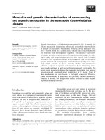
We evaluated survival curves for time to first transfusion
for four groups defined by the quartiles of the distribution
of the linear predictor score (i.e. the sum of the product
between covariate values and their corresponding parame-
ter estimates). Lower values (first quartile) corresponded to
different combinations of mostly positive predictors (82
cases - linear predictor score values below 1.45), while high-
er values (fourth quartile) corresponded to different combi-
nations of mostly negative predictors (76 cases - linear pre-
dictor score values above 2.70). Intermediate groups includ-
ed 78 cases with linear predictor score values between 1.45
and 2.05 (second quartile) and 80 cases with linear predictor
score values between 2.05 and 2.70 (third quartile).
Following this classification, 50% of patients in the fourth
quartile group underwent their first transfusion within 6
months of life, whereas only 3% of patients in the first
quartile group had started transfusions by the same age. In
this group it took more than 6 years for 50% of the patients
to start transfusions, whereas by the same age all patients in
the fourth quartile group had undergone their first transfu-
sion. In the first quartile group, 47% of patients never start-
ed red blood cell transfusion (Figure 1).
All survival curves were significantly different from each
other (P<0.01, Breslow’s test). In particular, the third quar-
tile group was significantly different from the fourth quar-
tile group (P<0.001) and the second quartile group was sig-
nificantly different from both the first and third quartile
risk-groups (P<0.001 and P=0.007, respectively).
Discussion
The purpose of this study was to measure the influence
of known genetic modifiers of β
0
-thalassemia on phenotype
severity, assessed as time to first transfusion. To this aim,
SNP in the BCL11A gene and HBS1L-MYB intergenic region
were selected based on previous studies and genotyped in a
group of 316 patients, as were α-globin gene defects and the
HBG2:g 158C>T polymorphism. All these variables,
together with gender, were included in a Cox proportional
F. Danjou et al.
992
haematologica | 2012; 97(7)
Table 3. Results of the Cox proportional hazards model.
Locus P Hazard ratio Harrell’s C-index Predictor for later
transfusion start
HBG2:g 58C>T <0.001 0.081 0.54 +/-
α gene defects <0.001 0.514 0.61 class 2 1
BCL11A rs1427407 <0.001 2.391 0.63 T allele
rs10189857 0.005 1.312 G allele
HBS1L/MYB rs4895441 <0.001 1.979 0.57 G allele
rs6904897 0.020 0.697 TT genotype
Gender 0.016 0.738 0.52 Male
1
Definitions of classes are reported in Table 1.
Figure 1. Kaplan-Meier survival curves for patients with different
combinations of predictors for later or earlier transfusion need.
First quartile (patients with combinations
of more positive predictors)
1
Second quartile
1
Third quartile
1
Fourth quartile (patients with combinations
of more negative predictors)
1
012345678910 11 12
Age(years)
1.0
0.9
0.8
0.7
0.6
0.5
0.4
0.3
0.2
0.1
0.0
Transfusion-free survival
1
Statistical tests of differences between the curves are reported in the text.
©Ferrata Storti Foundation
hazard model for time to first transfusion, and their respec-
tive effects were measured. The results showed that Hb F
production variants and α-globin gene defects had a sub-
stantial impact on the severity of β-thalassemia phenotype,
allowing prediction of the risk of patients to start transfu-
sion at different times of their life.
In this study we assumed that the time to first transfusion
accurately reflects variations in β-thalassemia phenotype
severity. This hypothesis seems to be supported by our
results, as all variables and the hierarchy of their effects
agree with previous studies on genetic modifiers of both Hb
F levels and the clinical severity of β-thalassemia, even
though other unknown genetic factors and clinical condi-
tions might be co-responsible for the need for transfu-
sions.
3,11,15,23,24
To the best of our knowledge, the present study is the
first to analyze the severity of β-thalassemia in a quantita-
tively defined manner and to include such a complete set of
known predictors. In a previous study, Galanello et al.
11
studied the effect of two SNP (rs11886868 in BCL11A and
rs9389268 in the HBS1L-MYB intergenic region) and α-glo-
bin gene defects on the phenotypic expression (defined as
major versus intermedia status) of Sardinian patients with
β
0
-thalassemia. A recent study by Badens et al.
24
further
extended this analysis accounting for the HBG2:g 158C>T
polymorphism and β
0
/β
+
status, in addition to the previous-
ly mentioned markers, in a heterogeneous cohort of 106
patients with 30 different β-globin gene mutations. The
present analysis expands these results by including the
effect of different independent predictors in each gene,
selected to be the strongest reported to date, in a homoge-
neous cohort of β
0
-thalassemia patients. This, we believe,
enables a better definition of the respective effects of each
predictor. Above all this work relates genetic modifiers to
time to first transfusion, a key event that characterizes dis-
ease severity regardless of patients’ major or intermedia
phenotype, thus notably increasing our knowledge on the
specific effects of genetic modifiers of the clinical severity of
β-thalassemia.
While it is likely that future whole genome sequencing
studies will better define the genetic polymorphisms that
modulate the effect of the BCL11A and HBS1L-MYB loci,
the results from the present study could already be of sup-
port in clinical settings, by providing clear probabilities for
the need to start transfusion at different ages as a function
of the personal genetic background of individual patients.
Authorship and Disclosures
The information provided by the authors about contributions from
persons listed as authors and in acknowledgments is available with
the full text of this paper at www.haematologica.org.
Financial and other disclosures provided by the authors using the
ICMJE (www.icmje.org) Uniform Format for Disclosure of
Competing Interests are also available at www.haematologica.org.
Genetic modifiers of β-thalassemia
haematologica | 2012; 97(7)
993
References
1. Cao A, Galanello R. Beta-Thalassemia. In:
Pagon RA, Bird TC, Dolan CR, Stephens K,
editors. GeneReviews [Internet]. Seattle
(WA): University of Washington, Seattle;
1993-2000 [updated 2010].
2. Cao A, Rosatelli C, Pirastu M, Galanello R.
Thalassemias in Sardinia: molecular patholo-
gy, phenotype-genotype correlation, and
prevention. Am J Pediatr Hematol Oncol.
1991;13(2):179-88.
3. Taher A, Musallam KM, Cappellini MD.
Thalassaemia intermedia: an update.
Mediterr J Hematol Infect Dis. 2009;1(1):
e2009004.
4. Cao A, Galanello R, Rosatelli MC.
Genotype-phenotype correlations in beta-
thalassemias. Blood Rev. 1994;8(1):1-12.
5. Lettre G, Sankaran VG, Bezerra MAC,
Araújo AS, Uda M, Sanna S, et al. DNA poly-
morphisms at the BCL11A, HBS1L-MYB,
and beta-globin loci associate with fetal
hemoglobin levels and pain crises in sickle
cell disease. Proc Natl Acad Sci USA. 2008;
105(33):11869-74.
6. Sedgewick AE, Timofeev N, Sebastiani P, So
JCC, Ma ESK, Chan LC, et al. BCL11A is a
major HbF quantitative trait locus in three
different populations with [beta]-hemoglo-
binopathies. Blood Cells Mol Dis. 2008;41
(3):255-8.
7. Garner C, Tatu T, Reittie JE, Littlewood T,
Darley J, Cervino S, et al. Genetic influences
on F cells and other hematologic variables: a
twin heritability study. Blood. 2000;95(1):
342-6.
8. Creary LE, Ulug P, Menzel S, McKenzie CA,
Hanchard NA, Taylor V, et al. Genetic varia-
tion on chromosome 6 influences F cell levels
in healthy individuals of African descent and
HbF levels in sickle cell patients. PLoS One.
2009;4(1):e4218.
9. So CC, Song YQ, Tsang ST, Tang LF, Chan
AY, Ma ES, et al. The HBS1L-MYB intergenic
region on chromosome 6q23 is a quantita-
tive trait locus controlling fetal haemoglobin
level in carriers of beta-thalassaemia. J Med
Genet. 2008;45(11):745-51.
10. Menzel S, Thein SL. Genetic architecture of
hemoglobin F control. Curr Opin Hematol.
2009;16(3):179-86.
11. Galanello R, Sanna S, Perseu L, Sollaino MC,
Satta S, Lai ME, et al. Amelioration of
Sardinian beta0 thalassemia by genetic mod-
ifiers. Blood. 2009;114(18):3935-7.
12. Menzel S, Garner C, Gut I, Matsuda F,
Yamaguchi M, Heath S, et al. A QTL influ-
encing F cell production maps to a gene
encoding a zinc-finger protein on chromo-
some 2p15. Nat. Genet. 2007;39(10):1197-
9.
13. Solovieff N, Milton JN, Hartley SW, Sherva
R, Sebastiani P, Dworkis DA, et al. Fetal
hemoglobin in sickle cell anemia: genome-
wide association studies suggest a regulatory
region in the 5’ olfactory receptor gene clus-
ter. Blood. 2010;115(9):1815-22.
14. Bhatnagar P, Purvis S, Barron-Casella E,
Debaun MR, Casella JF, Arking DE, et al.
Genome-wide association study identifies
genetic variants influencing F-cell levels in
sickle-cell patients. J. Hum. Genet. 2011;56
(4):316-23.
15. Galarneau G, Palmer CD, Sankaran VG,
Orkin SH, Hirschhorn JN, Lettre G. Fine-
mapping at three loci known to affect fetal
hemoglobin levels explains additional
genetic variation. Nat Genet. 2010;42(12):
1049-51.
16. Farrell JJ, Sherva RM, Chen Z-Y, Luo H-Y,
Chu BF, Ha SY, et al. A 3-bp deletion in the
HBS1L-MYB intergenic region on chromo-
some 6q23 is associated with HbF expres-
sion. Blood. 2011;117(18):4935-45.
17. Uda M, Galanello R, Sanna S, Lettre G,
Sankaran VG, Chen W, et al. Genome-wide
association study shows BCL11A associated
with persistent fetal hemoglobin and amelio-
ration of the phenotype of beta-thalassemia.
Proc Natl Acad Sci USA. 2008;105(5):1620-5.
18. Sutton M, Bouhassira EE, Nagel RL.
Polymerase chain reaction amplification
applied to the determination of beta-like glo-
bin gene cluster haplotypes. Am J Hematol.
1989;32(1):66-9.
19. Galanello R, Sollaino C, Paglietti E, Barella S,
Perra C, Doneddu I, et al. Alpha-thalassemia
carrier identification by DNA analysis in the
screening for thalassemia. Am J Hematol.
1998;59(4):273-8.
20. T. I. H. Consortium, The International
HapMap Project. Nature. 2003;426(6968):
789-96.
21. Price AL, Patterson NJ, Plenge RM, Weinblatt
ME, Shadick NA, Reich D. Principal compo-
nents analysis corrects for stratification in
genome-wide association studies. Nat
Genet. 2006;38:904-9.
22. Purcell S, Neale B, Todd-Brown K, Thomas
L, Ferreira MAR, Bender D, et al. PLINK: a
tool set for whole-genome association and
population-based linkage analyses. Am J
Hum Genet. 2007;81(3):559-75.
23. Weatherall DJ, Clegg JB. The Thalassemia
Syndromes. 4th 2001 Blackwell Science
Ltd.
24. Badens C, Joly P, Agouti I, Thuret I, Gonnet
K, Fattoum S, et al. Variants in genetic modi-
fiers of beta-thalassemia can help to predict
the major or intermedia type of the disease.
Haematologica. 2011;96(11):1712-4.
©Ferrata Storti Foundation