Facile synthesis of leaf like Cu(OH)2 and its conversion into cuo with nanopores
Bạn đang xem bản rút gọn của tài liệu. Xem và tải ngay bản đầy đủ của tài liệu tại đây (1.34 MB, 6 trang )
ACTA PHYSICO-CHIMICA SINICA
Volume 24, Issue 12, December 2008
Online English edition of the Chinese language journal
Cite this article as: Acta Phys. -Chim. Sin., 2008, 24(12): 2257−2262.
Received: June 25, 2008; Revised: September 8, 2008.
*Corresponding author. Email: ; Tel: +8621-66132663.
The project was supported by the National Natural Science Foundation of China (20503015) and the Science and Technology Commission of Shanghai, China
(0852nm00700).
Copyright © 2008, Chinese Chemical Society and College of Chemistry and Molecular Engineering, Peking University. Published by Elsevier BV. All rights reserved.
Chinese edition available online at www.whxb.pku.edu.cn
ARTICLE
Facile Synthesis of Leaf-like Cu(OH)
2
and Its Conversion
into CuO with Nanopores
Liangmiao Zhang, Wencong Lu*, Yongli Feng, Jipeng Ni, Yong Lü, Xingfu Shang
Department of Chemistry, Shanghai University, Shanghai 200444, P. R. China
Abstract: Leaf-like Cu(OH)
2
single crystals were synthesized via the controlled emulsion interface method using Span80 (sorbitan
monooleate) as the stabilizer of the emulsion system. CuO products with nanopores could be simply obtained by the dehydration of
Cu(OH)
2
, while maintaining the strip-shaped architecture. The phase structures and morphologies were measured by X-ray
diffraction (XRD), Fourier transform infrared (FTIR) spectra, scanning electron microscopy (SEM), and transmission electron
microscopy (TEM). Experimental results showed that Cu(OH)
2
microleaves were single crystals and the growth direction seemed to
be in [111] crystal plane of the orthorhombic Cu(OH)
2
. The formation of the nanopores should be attributed to the water loss of the
transformation from Cu(OH)
2
to CuO. The formation process of Cu(OH)
2
was investigated by taking TEM images at different stages
of the reaction. The formed nanoparticles began to rearrange to form nanorods and microleaves possibly via edge-by-edge and
side-by-side oriented-attachments because of the formation of larger crystals greatly reducing the interfacial energy. Besides, CuO
microarchitectures exhibit blue shifts in UV-Vis spectra and possess larger band gaps compared with those of bulk crystals.
Key Words:
Leaf-like Cu(OH)
2
; Emulsion; Soft template; CuO; Nanopores
In the recent years, since the dimensional and structural
characteristics of inorganic nanostructures endowed them with
potential applications in catalysis, medicine, electronics, cos-
metics, etc., synthesis of inorganic nanostructures with spe-
cific size and well-defined morphologies has attracted consid-
erable attention
[1]
. Considerable effort has been focused on the
assembly of lower dimensional building blocks into two- and
three-dimensional (2D and 3D) ordered superstructures, such
as snowflakes
[2]
, nanoribbons
[3]
, nanodendrites
[4]
, nanospin-
dles
[5]
, hollow structures
[6]
, and hierarchical structures
[7−10]
.
The methodology for controllable organization with structural
diversity from various nanobuilding blocks is a hot research
topic in the recent material research fields. Recently, we suc-
ceeded in synthesizing hierarchical cantaloupe-like and hol-
low microspherical AlOOH superstructures based on self-as-
sembly of one-dimensional (1D) nanorods
[11]
. To date, several
self-assembly processes driven by chemical or physical prin-
ciples (such as vapor-liquid-solid growth, laser-assisted cata-
lytic growth, template-based liquid-chemistry methods, etc.)
have been developed and employed for fabrication of the
complex nano- and micro-structures
[12−14]
. However, process
simplification, size, and shape control still remain tremendous
challenges in the fabrication of these architectures.
Recently, copper-based nanomaterials have received in-
creasing attention because of their potential application in op-
toelectronic devices, catalysis, and superconductors. The
magnetic properties of Cu(OH)
2
are remarkably sensitive to
the intercalation of molecular anions
[15−17]
, making the mate-
rial a candidate for sensor applications. More importantly,
CuO can be obtained through the dehydration of Cu(OH)
2
,
and the original size and morphology of Cu(OH)
2
nanostruc-
tures can be retained. As an important transition metal oxide
with a narrow band gap (E
g
=1.85 eV)
[18]
, CuO has been
widely exploited for diverse applications such as heterogene-
ous catalysts, gas sensors, superconductors, optical switches,
lithium ion electrode materials, and field-emission emitters
[19]
.
In addition, CuO was demonstrated to have a complex mag-
netic phase, which formed the basis for several high T
c
(criti-
Liangmiao Zhang et al. / Acta Physico-Chimica Sinica, 2008, 24(12): 2257
−
2262
cal temperature) superconductors and materials with giant
magnetoresistance
[20]
. On the basis of the practical applica-
tions of CuO nanomaterials, well-defined CuO nanostructures
with various morphologies, such as nanowires
[21−23]
, nanorib-
bons
[23]
, nanosheets
[24]
, nanoellipsoids
[24,25]
, pricky micro-
spheres
[26]
, flowerlike
[27,28]
, and dendritic assemblies
[29]
, have
been documented. To the best of our knowledge, self-assem-
bling preparation of CuO microleaves with highly porous
structure has not been reported until now.
Herein, the fabrication of Cu(OH)
2
microleaves through
wet-chemical method is introduced. This method requires
neither high temperature nor long synthesis time, wherein
CuO microstrips with porous structure are prepared from the
dehydration of Cu(OH)
2
.
1
1 Experimental
Analytic grade chemicals of CuSO
4
, ammonia, n-hexane,
and surfactant Span80 were purchased from Shanghai Chemi-
cal Reagents Company (China) and used as received.
A typical synthesis of Cu(OH)
2
microstructure was carried
out using the following three solutions: water phase 1 (WP-1):
a 1.0 mol·L
−1
aqueous solution (36 mL) from ion-exchange
water and CuSO
4
solution (18 mmol of Cu); oil phase (OP):
n-hexane solution (72 mL) of Span80 (1.50 g) for the stabili-
zation of the water-in-oil (W/O) emulsion; water phase 2
(WP-2): 0.2 mol·L
−1
ammonia solution (126 mL). The liquid
phase system was generated by adding WP-1 solution into the
OP, which was mixed using a homogenizer at 10000 r·min
−1
.
After being emulsified for at least 1 min at room temperature,
the mixture was poured into WP-2 in one portion, and the fi-
nal pH of the above liquid phase system was adjusted to 4 and
further stirred for 2 h for aging. Subsequently, the precipitates
were separated by centrifugation, washed with distilled water
and absolute alcohol, and finally dried at 60 °C for 12 h. For
the dehydration of Cu(OH)
2
microleaves to get CuO, the as-
prepared samples were heated at 400−900 °C for 1 h with a
heating rate of 5 °C·min
−1
.
Powder X-ray diffraction (XRD) measurements of the as-
prepared samples were carried on a Japan Rigaku D/Max-RB
X-ray diffractometer with Cu K
α
radiation (λ=0.1542 nm).
Transmission electron microscope (TEM) and high-resolution
transmission electron microscope (HRTEM) images were
captured on JEOL JEM-1200EX II and JEOL JEM-2010F at
an acceleration voltage of 200 kV, respectively. The mor-
phology of the as-prepared samples was characterized by field
emission scanning electron microscopy (FE-SEM, JEOL JSM-
6700F). The UV-Vis absorption spectra of the as-prepared
products were recorded by a Shimadzu UV-2051PC photo-
spectrometer. Fourier transform infrared (FTIR) spectra were
obtained on an AVATAR370 spectrometer.
2 Results and discussion
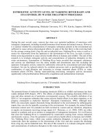
The typical morphologies of the final Cu(OH)
2
architectures
were examined by SEM. Fig.1 shows the SEM photographs of
the samples. The low magnification image (Fig.1a) shows the
panoramic of the product indicating that the Cu(OH)
2
crystal-
lites self-organize into assemblies. The assemblies tend to ag-
gregate with each other to form large agglomerates. To further
examine the surface morphologies of the microarchitecture, a
high magnification SEM of a single assembly was recorded,
as shown in Fig.1b. The entangled architecture is actually
comprised of leaf-like particles with the average thickness of
100 nm, width of 200 nm, and various lengths up to several
micrometers.
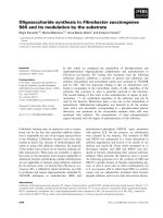
To probe the 3D hierarchical nanoarchitectures in more de-
tail, we analyzed the agglomerates by means of TEM. Fig.2a
is a TEM image of the microstructures. From Fig.2a, it is evi-
dent that these architectures consist of individual leaf- like
microstructures that are bundled. These leaves are about
100−300 nm wide in the middle section and connect to each
other to form 3D architectures. Furthermore, we found that a
prolonged ultrasonication for up to 20 min could absolutely
disrupt these assemblies (Fig.2b), implying that the interaction
among the constituent 3D microstructures was particularly
weak. Fig.2c is a TEM image of an individual leaf; it is esti-
mated to be ca 150 nm in width and ca 1.2 μm in length. The
leaf-like structure of the products was further examined by
high-resolution TEM (HRTEM). Fig.2d shows the magnifica-
tion of selected area of the leaf shown in Fig.2c. The fringe
spacing measures 0.25 nm, which concurs well with the d
value of the orthorhombic Cu(OH)
2
[111] crystal plane
[30]
.
Thus, the growth direction of the microleaves seems to be in
the [111] direction. Fig.2e shows the selected-area electron
diffraction (SAED), which reveals that Cu(OH)
2
microleaves
are single crystals. When the Cu(OH)
2
sample was calcined in
air by applying a heating rate of 5 °C·min
−1
and holding the
calcination temperature at 800 °C for 1 h, porous CuO was
obtained, as confirmed by Fig.2f. Compared to the samples
without calcination, some newly created pores are observed,
while the strip-shaped architecture is still maintained. It
should be emphasized here that this leaf-shaped architecture
has a high thermal stability and is stable even after calcination
at 800 °C. Potentially, this highly thermally stable and porous
nanostructure has applications in catalysis.
Fig.1 Low magnification (a) and high magnification (b)
FE-SEM images of Cu(OH)
2
particles
Liangmiao Zhang et al. / Acta Physico-Chimica Sinica, 2008, 24(12): 2257
−
2262
The samples were further examined by IR analysis (Fig.3).
As seen in Fig.3a, the IR spectrum indicated the existence of
surface hydroxyls and coordinated Span80 molecules on
Cu(OH)
2
microleaves. The broad band at 3000−3700 cm
−1
is
deconvolved to make clear the existence of Span80 in the
products and two peaks centered at 3394.6 and 3571.4 cm
−1
appeared (Fig.3a), which can be assigned to the stretching
mode of hydroxyl of pure Span80 (Fig.3b). The bands at
3488.2 and 1631.1 cm
−1
correspond to the stretching and
bending modes of the hydroxyls of adsorbed water
[31]
. The
band at 1077.6 cm
−1
corresponds to the C−O stretching vibra-
tion coordinating to metal cations
[32]
, which shifts about 10
cm
−1
to lower wavenumbers compared to the IR spectrum of
pure Span80, suggesting the formation of hydrogen bonds
between Span80 and the inorganic components. The band at
424.2 cm
−1
can be assigned to Cu−O stretching mode and may
prove that Cu(OH)
2
is formed
[33]
. Generally speaking, anneal-
ing can decompose impurity groups in the sample and im-
prove the crystal quality. Fig.3c shows the room-temperature
infrared absorption spectrum of the annealed CuO products.
Except for the absorption peak at around 580 cm
−1
owing to
Cu−O stretching along [
0
1
1 ] direction and the mode at 535
cm
−1
owing to Cu−O stretching along [101] direction
[34,35]
, all
absorption bands corresponding to the Span80 impurities dis-
appear, clearly demonstrating that the impurities have been
removed.
XRD analysis was used to determine the structure and
phase of the samples. Fig.4a shows the XRD pattern of the
as-prepared blue products. The prepared material was identi-
fied as the orthorhombic Cu(OH)
2
(JCPDS No. 13-0420), con-
firming that the inorganic component was Cu(OH)
2
. A con-
spicuous feature of the Cu(OH)
2
crystals is their broadness,
which indicates the small size of the Cu(OH)
2
crystals.
Fig.4(b−f) presents the XRD patterns of the products prepared
under the same reaction conditions except different calcination
temperatures. At 400 °C, a slow transformation to CuO has
already started, but it is amorphous because no peaks can be
observed. When the temperature is increased to 600 °C, the
Fig.2 (a) Panoramic of Cu(OH)
2
assemblies, (b) with 20 min
sonication, (c) an individual leaf-like particle, (d) HRTEM
image of the single particle in (c), (e) its corresponding
SAED pattern, and (f) CuO microleaves with nanopores
Fig.3 Infrared spectra of Cu(OH)
2
(a), Span80 (b), and CuO (c)
Fig.4 XRD patterns of the as-prepared Cu(OH)
2
leaf-like
particles (a) and particles obtained after calcination at
different temperatures for 1 h (b−f)
Liangmiao Zhang et al. / Acta Physico-Chimica Sinica, 2008, 24(12): 2257
−
2262
transformation rate increases significantly. Finally, the trans-
formation at 800 °C is complete with no Cu(OH)
2
signal re-
maining. In a word, slow conversion of Cu(OH)
2
microleaves
to CuO microleaves can occur above 400 °C. We observed the
surface morphology of the product, which was calcined at 400
°C when the phase transformation occurred. It indicated that
there were nanopores on the leaves′ surface (Fig.5), and no
pores appeared below this temperature. Combined with the
compact morphology and single crystal structure of Cu(OH)
2
by TEM observation, the formation of the nanopores should
be attributed to the water loss of the transformation from
Cu(OH)
2
to CuO.
Other conditions, such as growth temperature and pH val-
ues, are also important factors affecting the morphologies of
the structures. By control of these aspects, different Cu(OH)
2
nanoarchitectures can be realized. If the same reaction is car-
ried out at 60 °C, only randomly packed rod-like structure,
rather than a leaf-like pattern, is the dominant morphological
configuration (Fig.6a). It is therefore apparent that a relatively
higher temperature does not favor the formation of well-
defined Cu(OH)
2
crystal leaves. For the systems with increas-
ing pH value (pH=10), it is found that several particles sized
about 100 nm are obtained (Fig.6b).
To investigate the formation process and the growth
mechanism of the hierarchical microarchitecture, time-de-
pendent experiments were carried out. Fig.7 shows the TEM
images of the samples obtained after the reaction has pro-
ceeded for 1, 5, 30, and 120 min, respectively. These images
clearly exhibit the evolution of Cu(OH)
2
nanostructures from
nanoparticles to nanorods and finally to microleaves over the
time at 25 °C. Therefore, the formation of Cu(OH)
2
leaf-like
microstructures is proposed to be a process composed of the
following stages: (i) generation of W/O micelle templates with
small, nanometer-sized drops of liquid Cu
2+
, (ii) release of
OH
–
ions from ammonia, which react with the Cu
2+
ions near
the surface of the micelles to form Cu(OH)
2
nuclei surround-
ing the spherical water pool. In short, the micelles act as soft
templates for the formation of Cu(OH)
2
nanoparticles. There-
fore, the initial formation of a nanometer-sized micelle tem-
plate is of great importance for the initial formation of
Cu(OH)
2
nanonuclei. (iii) The formed nanoparticles began to
rearrange to form nanorods and microleaves possibly via a
side-by-side oriented-attachment, which were energetically
favored, because of the formation of larger crystals greatly
reducing the interfacial energy.
A comparative experiment without Span80 or n-hexane, but
with other conditions kept constant, only irregular particles
were obtained. Fig.8 shows the morphologies of the products
synthesized with the assistance of different amounts of
Span80. It seems that leaf-like products can only be synthe-
sized under the assistance of enough Span80. We believe that
Span80 plays a key role in the formation of the leaf-like shape.
In the present study, Span80 was not only used for the prepa-
ration of W/O type emulsion, but also served as the structure-
directing agent for the formation of superstructures. The first
nucleation seeds plus Span80 as an initial nucleus will absorb
little particles along the growth direction. In this process,
Span80 absorbs the small particles by the hydrophilic sorbitan
group acting as a soft template for the formation of nanorods.
Self-assembly of nanocrystals is driven by van der Waals
forces and hydrogen bonding among the certain organic
molecules on the surface of particles
[36]
. Therefore, the initial
Fig.5 TEM image of the products calcined at 400 °C for 1 h
Fig.6 TEM images of the products obtained at (a) 60 °C and
(b) pH=10
Fig.7 TEM images of the products obtained with the reaction proceeding for different times
t/min: (a) 1, (b) 5, (c) 30, (d) 120
Liangmiao Zhang et al. / Acta Physico-Chimica Sinica, 2008, 24(12): 2257
−
2262
formation of a nanometer-sized micelle template and the role
of Span80 as structure-directing agent are both important for
the formation of Cu(OH)
2
microleaves. Further theoretical and
experimental investigations must be done to determine the
exact nature of the growth mechanism.
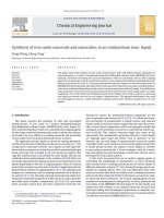
UV-Vis absorption measurement is one of the most widely
used techniques to reveal the energy structures and optical
properties of semiconductor nanocrystals. The optical absorp-
tion properties of well-aligned CuO leaf-like particles dis-
persed in ethanol solution are investigated at room tempera-
ture by UV-Vis spectroscopy. The spectrum is presented in
Fig.9a. There is a broad absorption peak centered at 256 nm.
Moreover, a classical Tauc approach is further employed to
estimate the band gap value of CuO crystals according to the
equation αE
p
=A(E
p
−E
g
)
1/2
(where, α is the absorption coeffi-
cient, E
p
is the discrete photon energy, E
g
is the band gap en-
ergy, and A is a constant)
[37]
. The plot of (αE
p
)
2
−E
p
for CuO is
shown in Fig.9b, exhibiting a linear relationship between 3.28
and 4.00 eV. The extrapolated value (the dot straight line to
the x axis) corresponding to the band gap of as-prepared CuO
is estimated to be 2.20 eV, which is apparently larger than the
reported value for bulk CuO (1.85 eV)
[18]
. The increase in the
band gap of CuO architectures is an indication of quantum
confinement effects
[38]
.
3
3 Conclusions
In summary, a sophisticated production of Cu(OH)
2
micro-
leaves has been successfully synthesized with micelles acting
as soft templates. The addition of Span80 molecules is be-
lieved to facilitate the formation of the oriented attachment
structures. Furthermore, we also demonstrated that leaf-like
CuO products with nanopores can be simply obtained by the
dehydration of Cu(OH)
2
. It is expected that the novel CuO ar-
chitectures may offer exciting opportunities for potential ap-
plications in catalysis, electrochemistry, superconductivity,
and superhydrophobic coating. Although the detailed mecha-
nism is not very clear and still needs more investigation, it is
no doubt a pretty simple and easily controlled route for pro-
ducing other metal hydroxide nanoarchitectures.
References
1 Pan, Z. W.; Zu, R. D.; Wang, Z. L. Science, 2001, 291: 1947
2 Chen, X. Y.; Lee, S. W. Chem. Phys. Lett., 2007, 438: 279
3 Kar, S.; Satpati, B.; Satyam, P. V.; Chaudhuri, S. J. Phys. Chem.
B, 2005, 109: 19134
4 Lu, L.; Kobayashi, A.; Kikkawa, Y.; Tawa, K.; Ozaki, Y. J. Phys.
Chem. B, 2006, 110: 23234
5 Xu, X.; Cortie, M. B. J. Phys. Chem. C, 2007, 111: 18135
6 Li, L. Y.; Wang, J. G.; Sun, P. C.; Liu, X. H.; Ding, D. T.; Chen, T.
H. Acta Phys. -Chim. Sin., 2008, 24: 359
7 Cheng, Y.; Wang, Y.; Jia, C.; Bao, F. J. Phys. Chem. B, 2006, 110:
24399
8 Chen, X. Y.; Huh, H. S.; Lee, S. W. Nanotechnology, 2007, 18:
285608
9 Bouizi, Y.; Majano, G.; Mintova, S.; Valtchev, V. J. Phys. Chem.
C, 2007, 111: 4535
10 Tang, J.; Alivisatos, A. P. Nano Lett., 2006, 6: 2701
11 Feng, Y. L.; Lu, W. C.; Zhang, L. M.; Bao, X. H.; Yue, B. H.; Lv,
Y.; Shang, X. F. Cryst. Growth Des., 2008, 8: 1426
12 Trentler, T. J.; Hickman, K. M.; Goel, S. C.; Viano, A. M.;
Gibbons, P. C.; Buhro, W. E. Science, 1995, 270: 1791
13 Gudiksen, M. S.; Lieber, C. M. J. Am. Chem. Soc., 2000, 122:
8801
14 Sa
p
p, S. A.; Lakshmi, B. B.; Martin, C. R. Adv. Mater., 1999, 11:
Fig.8 TEM images of the products obtained with
different amounts of Span80
m(Span80)/g: (a) 0.25, (b) 0.50, (c) 0.75, (d) 1.00
Fig.9 (a) UV-Vis absorption spectrum of CuO microleaves
and (b) the corresponding (αE
p
)
2
−E
p
curve
Liangmiao Zhang et al. / Acta Physico-Chimica Sinica, 2008, 24(12): 2257
−
2262
402
15 Fujita, W.; Awaga, K. Synth. Met., 2001, 122: 569
16 Fujita, W.; Awaga, K. Inorg. Chem., 1996, 35: 1915
17 Fujita, W.; Awaga, K. J. Am. Chem. Soc., 1997, 119: 4563
18 Wang, H.; Xu, J. Z.; Zhu, J. J.; Chen, H. Y. J. Cryst. Growth,
2002, 244: 88
19 (a) Musa, A. O.; Akomolafe, T.; Carter, M. J. Sol. Energy Mater.
Sol. Cells, 1998, 51: 305
(b) Zheng, X. G.; Xu, C. N.; Tomokiyo, Y.; Tanaka, E.; Yamada,
H.; Soejima, Y. Phys. Rev. Lett., 2000, 85: 5170
20 Prabhakaran, D.; Subramanian, C.; Balakumar, S.; Ramasamy, P.
Physica C, 1999, 319: 99
21 Zhong, Z. Y.; Vivien, N.; Luo, J. Z.; Teh, S. P.; Teo, J.; Gedanken,
A. Langmuir, 2007, 23: 5971
22 Song, X. Y.; Sun, S. X.; Zhang, W. M.; Yu, H. Y.; Fan, W. L.
J. Phys. Chem. B, 2004, 108: 5200
23 Lu, C. H.; Qi, L. M.; Yang, J. H.; Zhang, D. Y.; Wu, N. Z.; Ma, J.
M. J. Phys. Chem. B, 2004, 108: 17825
24 Liu, J. P.; Huang, X. T.; Li, Y. Y.; Sulieman, K. M.; He, X.; Sun,
F. L. Cryst. Growth Des., 2006, 6: 1690
25 Zhang, Z. P.; Sun, H. P.; Shao, X. Q.; Li, D. F.; Yu, H. D.; Han,
M. Y. Adv. Mater., 2005, 17: 42
26 Xu, Y. Y.; Chen, D. R.; Jiao, X. L. J. Phys. Chem. B, 2005, 109:
13561
27 Liu, J. P.; Huang, X. T.; Li, Y. Y.; Sulieman, K. M.; He, X.; Sun,
F. L.
J. Mater. Chem., 2
006
, 16: 4427
28 Liu, Y.; Chu, Y.; Zhuo, Y. J.; Li, M. Y.; Li, L. L.; Dong, L. H.
Cryst. Growth Des., 2007, 7: 467
29 Zhang, Z. P.; Shao, X. Q.; Yu, H. D.; Wang, Y. B.; Han, M. Y.
Chem. Mater., 2005, 17: 332
30 Luo, Y. H.; Huang, J. G.; Jin, J.; Peng, X. S.; Schmitt, W.;
Ichinose, I. Chem. Mater., 2006, 18: 1795
31 Nakamoto, K. Infrared spectra of inorganic and coordination
compound. 4th ed. Trans. Huang, D. R.; Wang, R. Q. Beijing:
Chemical Industry Press, 1991: 251
32 Bernson, A.; Lindgren, J.; Huang, W.; Frech, R. Polymer, 1995,
36: 4471
33 Nyquist, R. A.; Kagel, R. O. Infrared spectra of inorganic com-
pounds. 1st ed. New York and London: Academic Press, 1971:
220
34 Zou, G. F.; Li, H.; Zhang, D. W.; Xiong, K.; Dong, C.; Qian, Y. T.
J. Phys. Chem. B, 2006, 110: 1632
35 Kliche, K.; Popovic, Z. V. Phys. Rev. B, 1990, 42: 10060
36 Banfield, J. F.; Welch, S. A.; Zhang, H. Z.; Ebert, T. T.; Penn, R.
L. Science, 2000, 289: 751
37 Tsunekawa, S.; Fukuda, T.; Kasuya, A. J. Appl. Phys., 2000, 87:
1318
38 Yang, J. P.; Meldrum, F. C.; Fendler, J. H. J. Phys. Chem., 1995,
99: 5500