Modulation of nuclear factor b signaling attenuates allergic airway inflammation 2
Bạn đang xem bản rút gọn của tài liệu. Xem và tải ngay bản đầy đủ của tài liệu tại đây (1.93 MB, 74 trang )
1. Introduction
1
1.1. Asthma
1.1.1.
Epidemiology of Asthma and impetus to develop novel anti- inflammatory
agents
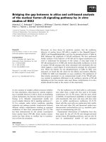
Globally, 300 million people suffer from asthma and the prevalence of asthma still continues
to increase. With this rising trend, it is predicted that there will be at least 400 million
asthmatic patients by 2025 (Masoli et al., 2004). The prevalence is highest in developed
countries — UK (> 15%), USA (~11%) and, Australia (~15%) (Figure 1.1) (Pawankar et al.,
2012). The increase in prevalence of asthma could be attributable to urbanization and shift
away from “naturalistic” diet and lifestyle as explained by hygiene hypothesis (Fishbein and
Fuleihan, 2012). According to the hygiene hypothesis, excessive Th2 response is mediated by
the absence of recurrent microbial infection, while Th1 response is mediated by microbial
infection. Allergies are less common in children growing up in rural environment, particularly
in the farms, as compared to those growing up in urban environment (Ege et al., 2011).
Protection against allergy correlates positively with the level of exposure to bacterial and
fungal microbes. In addition, allergy in children is inversely related to the habit of drinking
unpasteurized milk and the exposure to high level of endotoxin in house dust mite (HDM)
during infancy (Waser et al., 2007). However, hygiene hypothesis theory has oversimplified
the nature of allergic asthma (Fishbein and Fuleihan, 2012; Haschke and Klassen, 2009).
Although exposure to microbes may offer children protection against allergy, viral infection
predisposes children to wheezing and asthma. Rhinovirus-induced wheezing in the first three
years of life is the greatest risk factor for developing asthma by six years of age (Holgate,
2012). Several postulations have been made to explain for this controversy. These
postulations shall be elaborated in later sections. Briefly, it is postulated that viral infection
increases the sensitivity of airway epithelial cells to allergens (Monick et al., 2003). A
disturbed immune regulation involving T-regulatory (Treg) cells, rather than a mere shift
towards Th2 immunity, was the key behind allergic asthma (Haschke and Klassen, 2009).
2
Although asthma is not considered to be a life-threatening disease, it is the key factor behind
one out of every 250 deaths worldwide. Complexity and severity of asthma continue to
increase in children and young adults. Severe or uncontrolled asthma presents high socioeconomic burden on the countries. The healthcare costs correlate positively with severity of
asthma. The financial burden of asthma ranges from US$300 to US$1, 300 per patient per
year in the developed countries. In the United States, 23 million people including seven
million children suffer from asthma. As a result of asthma attacks, these seven million
children miss 14 million days of school each year. Caregivers of these children would have to
take time off work to attend to them, which would result in lost wages for the caregivers
(Pawankar et al., 2011). In developing countries like Vietnam, with gross domestic product
per capita of US$1, 411, the financial burden of asthma estimates to be US$184 per patient
per year. In India, the medication for an asthmatic child can cost a third of the family’s
income (Table 1.1) (Pawankar et al., 2012).
In summary, the rising prevalence, mortality, and high economic burden of asthma are having
a huge impact on the health-care systems worldwide. Although current therapies for asthma
are relatively safe and effective at controlling symptoms, these therapies do not change the
chronic course of disease. Currently, there is no established method to prevent asthma. The
major unmet needs of this area include better management of the severe forms of the disease
and the developments of curative therapies (Akdis, 2012). Therefore, much research has been
done to better understand the pathophysiology of asthma and to explore novel therapies for
this asthma. One attractive target for therapeutic intervention would be the nuclear factor
(NF)-κB signaling pathway, which plays an important role in Th2-mediated inflammation
(Edwards et al., 2009).
3
USA
UK
Australia
Figure 1.1: World map of the prevalence of clinical asthma (Adapted from Matthew Masoli
2004) (Matthew Masoli, 2004)
4
Country
Year cost
calculated
Population (2010)
(million)
Cost estimate
South Korea
Israel
Mexico
USA
2005
2007
2007
50
8
103
310
US$2 billion
US$250 million
US$0.35 billion
US$20 billion
Table 1.1 The economic burden of asthma (Adapted from Pawankar et. al., 2011)
5
1.1.2 Pathophysiology/ Development of asthma
Asthma is considered to be a heterogeneous disease with numerous distinct clinical
phenotypes. The most common form of asthma — allergic asthma — affects 60 percent of the
asthmatics (Kim et al., 2010).
Allergic asthma is a chronic airway inflammatory disease (Fishbein and Fuleihan, 2012;
Lambrecht and Hammad, 2012; Pawankar et al., 2012). The inflamed airway, similar to a
chronic wound, is susceptible to a wide range of environmental insults (for example,
biologically active allergens, viruses, air pollutants, certain drugs, and chemicals) and has an
altered repair response that involves growth factors secretion, which induces goblet cell
metaplasia (GCM), smooth muscle proliferation, angiogenesis, fibrosis, and nerve
proliferation (Barnes, 2011; Holgate, 2012; Lambrecht and Hammad, 2012).
Allergic asthma is often initiated when one is sensitized to inhaled allergens from the
environment such as HDM, cockroaches, animal danders, fungi, and pollen (Barnes, 2011;
Holgate, 2012; Lambrecht and Hammad, 2012).
During initial sensitization, the inhaled allergens interact with epithelial cells and result in the
release of endogenous danger signal, including chemokine ligand (CCL)-2 and CCL-20 to
recruit more dendritic cells progenitors and dendritic cells from the bone marrow. The role of
epithelial cells in the pathogenesis of asthma shall be discussed in details in section 1.1.2.1 on
“Airway Epithelial Cells”. Besides interacting with the airway epithelium, the inhaled
allergens also interact with the dendritic cells. Degradation of airway epithelium by
proteolytic activity of allergens breaches the epithelium barrier function and allows the
allergen to gain access to the dendritic network (Wan et al., 1999).
These antigens presenting dendritic cells express pattern recognition receptor (PRR) and takes
up the allergens. The interaction between PRR and allergens initiates the migration of
6
dendritic cells to the T cell area of the draining regional lymph nodes. During migration,
dendritic cells undergo further maturation and process the allergens into small peptides
(Lambrecht and Hammad, 2010). In the T cell area of the lymph node, mature dendritic cells
arrest and select the rare naïve antigen-specific T cells and present the processed peptides in
the context of major histocompatibility complex (MHC) class II to T cell receptor (TCR)
(Stoll et al., 2002). The antigen presentation leads to differentiation of naïve T-cells to
antigen-specific Th-2 cells (Figure 1.2) (Holgate, 2012). The mechanisms responsible for Th2 polarisation during initial allergen sensitization remain poorly understood. The possible
mechanisms shall be discussed in section 1.1.2.2 on Th2 cells (Holgate, 2012).
The differentiated antigen specific Th2 cells that mediate pathophysiology of subsequent
allergen exposure consist of two subsets: effector memory T-cells or resident memory T cells
and central memory T cells.
The effector memory T cells have reduced expression of lymph node homing receptor cluster
of differentiation (CD)-62L and migrate to the site of inflammation and serves as surveillance
for future re-exposure to allergen (Figure 1.2). Upon re-encounter with allergen, or during an
asthma attack, the effector memory T cells interact with antigen presenting dendritic cells and
can rapidly release pro-inflammatory cytokines into the airway (Robinson et al., 1992).
Unlike the effector Th2 cells, the central memory T cells express CD62L but lack immediate
effector function (Iezzi et al., 2001). These central memory T cells are localized to the lymph
node. Upon allergen re-exposure, dendritic cells release signals to the central memory cells in
the lymph nodes. Consequently, the memory cells proliferate and differentiate into effect T
cell and migrate to the site of allergen challenge to mediate airway inflammatory response
(Sallusto and Lanzavecchia, 2001).
7
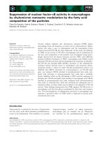
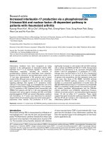
Figure 1.2 Inflammatory and immune cells involved in allergic airway inflammation.
(Adapted from Lambrecht and Hammad, 2012)
Lung epithelium expresses pattern recognition receptors (PRRs) such as toll-like receptors
(TLRs), nod-like receptors (NLR), C-type lectins, and protease-activated receptors (PARs).
PRRs when bound to allergens result in activation of signaling pathways, which would cause
the release of endogenous danger signals (uric acid, ATP, LPA, TSLP, IL-25, IL-33, GMCSF, IL-1 members). Epithelial cells also release CCL-2 and CCL-20 to attract dendritic cell
progenitors — monocytes — to the lung. These signals from epithelial cells activate dendritic
cells. Activated dendritic cells migrate to T cell region in the draining lymph nodes. In the
lymph nodes, dendritic cells interact with naïve T cells and induce T cell differentiation.
Depending on the signals released by dendritic cells, T cells can differentiate into Th1, Th2,
or Th17 cells. For example, interaction between OX40L of dendritic cells and OX40 of T cell
would enhance differentiation of naïve T cells to Th2 cells. Differentiated and activated Th2
cells would activate B cells and result in IgE production by B cells. Notably, basophil is
considered to be an antigen presenting cell and has been reported to be an important source of
IL-4, which supports differentiation of naïve T cell to Th2 cell.
Abbreviations: CCL, chemokine (C-C motif) ligand; GM-CSF, granulocyte-macrophage
colony-stimulating factor; IL, interleukin; LPA, lysophosphatidic acid; NOD 1/2, nucleotidebinding oligomerization domain; ROS, reactive oxygen species; TGF-β, transforming growth
factor-β; and TSLP, thymic stromal lymphopoietic protein.
8
1.1.2.1 Airway Epithelial Cells
The airway epithelial cells are located at the interface between the host and the environment;
therefore, they are the first line of defense against foreign antigens (Xiao et al., 2011).
Epithelial cells express many PRRs (Toll-like receptor [TLR], NOD-like receptor [NLR], Ctype lectins, and protease-activated receptor [PAR]) that interact with pathogen associated
molecular pattern (PAMP)s and danger-associated molecular pattern (DAMP)s. Activation of
PRR by PAMPs or DAMPs would result in the activation of NF-κB signaling (Lambrecht and
Hammad, 2012). Studies showed that tonic activation of NF-κB in airway epithelial cells is
sufficient to activate dendritic cells, breach inhalation tolerance and enhance sensitization to
innocuous inhaled allergen, such as OVA (Ather et al., 2011; Poynter et al., 2004). On the
other hand, the inhibition of NF-κB in airway epithelial cells attenuates Th2 cell infiltration
and airway remodeling (Das et al., 2001). Activated NF-κB signaling pathway results in the
release of an array of cytokines and chemokines, such as TSLP and IL-8 (Edwards et al., 2009;
Lambrecht and Hammad, 2012). These cytokines contribute to pathogenesis of allergic
airway inflammation. TSLP is hought to mediate polarization of Th-2 immune response
(Holgate 2012). Its role shall be discussed in details in section 1.1.2.2. on Th2 cells. On the
other hand, IL-8 contributes to airway inflammatory cell infiltration (Kunkel et al., 1991;
Lampinen et al., 1999). The roles of these cytokines in the pathophysiology of asthma shall be
discussed in the following paragraphs.
IL-8 is a pivotal chemoattractant for neutrophils as well as eosinophils (Kunkel et al., 1991;
Lampinen et al., 1999). This chemoattractant is a marker for asthma because it can be
detected in the serum of asthmatic patients but not in control subjects (Shute et al., 1997). It
was also reported that asthmatic patients with severe neutrophilic asthma phenotype have
elevated IL-8 levels in the supernatant of their sputum as compared to mild asthmatic patients
or control subjects (Bonnans et al., 2002). In addition, IL-8 also contributes to mucus
hypersecretion (section 1.1.2.5) (Bautista et al., 2009).
9
Activation of NF-κB signaling pathway in epithelial cells triggers the release of endogenous
danger signals, which in turn activate dendritic cells (Lambrecht and Hammad, 2009). The
threshold for activation of epithelial cells is dependent on the expression of PRRs and the
expression of signaling proteins downstream of PRRs. The greater the number of PRRs, the
greater the sensitivity of the epithelial cells is to the allergen. For example, viral infection of
the airway epithelial with respiratory syncytial virus (RSV) increases the expression of TLR-4.
This increased sensitivity of airway epithelial cells may in part explain how RSV infection
predisposes children to wheezing and asthma (Monick et al., 2003).
Besides functioning as a receptor for foreign particles, the airway epithelium is also a physical
barrier that prevents the access of allergens to lung dendritic cells (Lambrecht and Hammad,
2012). Based on bronchial biopsy studies, the airways of subjects with asthma have fragile
epithelial (Lackie, 1997; Lambrecht and Hammad, 2012). The integrity of airway epithelial is
maintained by apical tight junctions and adherent junctions, which keep the cells together and
maintain their apicobasal polarity (Xiao et al., 2011). The main component of adherent
junction is E-cadherin, which constantly releases inhibitory signals to dendritic cells and
thereby suppresses dendritic cell-mediated allergic sensitization. As compared to normal
individuals, asthmatic patients have lower expression of E-cadherin; this is possibly due to
epithelial-to-mesenchymal transitions (Jiang et al., 2007; Nawijn et al., 2011). The loss of Ecadherin may be caused by exposure to inhaled allergens — HDM, cockroaches, pollen, fungi,
respiratory viruses, or environmental pollutants (cigarette smoke, ozone) (Lambrecht and
Hammad, 2012). Therefore, exposure to these allergens induces disruption of epithelial
junctional proteins and the barrier function of the airway epithelium. Once the integrity of the
epithelial cells has been destroyed, inhaled allergens can gain access to the dendritic cell
network and activate immune responses (Lambrecht and Hammad, 2012). In particular, HDM
results in epidermal growth factor receptor (EGFR)-induced tyrosine phosphorylation and
delocalization of junctional protein. Degradation of E-cadherin and destruction of the intact
epithelial barrier function subsequently allows for the EGF on the basolateral side of the
10
epithelium to bind with of EGF on EGFR on the epithelial of the apical side of the epithelium
(Heijink et al., 2007; Heijink et al., 2010; Lambrecht and Hammad, 2012). Activation of EGF
signaling pathway results in goblet cell metaplasia, where goblet cells increase in number, and
thus causes mucus hypersecretion. The role of epithelial cells in mucus hypersecretion shall
be elaborated in section 1.1.2.5.
Fifteen years ago, based on the observations of asthma biopsies and cultured bronchial
epithelial cells, Stephen Holgate was the first to suggest epithelial cell as a major culprit in
asthma. Years of intense research that follows confirm that airway epithelium controls many
aspects of allergic sensitization and is a central player in allergic airway inflammation,
remodeling and bronchial hyperreactivity.
1.1.2.2 T cells
T cells constitute the majority of lung lymphocytes in normal individuals and could be found
within the airway, alveolar epithelium, and interstitium (Baraldo et al., 2007; Barnes, 2011).
Naïve T cells require three sources of signals for activation: (1) signals from antigen-MHC
class II; (2) signals from costimulatory molecules; and (3) signals from autologous IL-2.
TCR interacts with peptide antigen MHC class II on the surface of antigen presenting cells.
The T cell also interacts with inducible co-stimulatory molecules (ICOS) such as B7 and
CD40 expressed on antigen presenting cell. At the same time, these activated T cells
synthesizes IL-2 along with α chain of the IL-2 receptor (also known as CD25). Unlike
activated T cell IL-2 receptors, resting T cells IL-2 receptors only composed of β and γ chains.
This form of receptor binds IL-2 with moderate affinity only. However, in activated T cells, α
chain synthesized associates with the existing β and γ heterodimer to create a receptor with
much higher affinity for IL-2. Binding of IL-2 to the receptor triggers the activation,
differentiation and proliferation of T cells (Janeway et al., 2005). Once activated, T cells can
differentiate and proliferate into mature Th1, Th2, Th17, Th9, Treg, natural killer (NK)T cells,
11
or γδ T cells depending on the cytokines released by antigen presenting cells (Kaiko et al.,
2008). These different subtypes of T cells contribute to development of allergic asthma in
various ways (Figure 1.3) (Lloyd and Hessel, 2010).
Th2 cells
Twenty years ago, studies showed that there is an increased presence of Th2 cells in the
airways of patients with asthma. Since then, asthma has been categorized as a Th2 cell
associated inflammatory disease (Robinson et al., 1992). Subsequent studies on human
samples confirmed that the number of Th2 cells present in the airway correlates positively
with the severity of asthma. These data suggest an essential role for Th2 cells in human
asthma (Larche et al., 2003). Although Th2 response plays an important role in development
of asthma, the mechanism responsible for initiating Th2 response remains to be elucidated.
Nonetheless, recent studies have shown that Th2 response is likely to be initiated by IL-25,
IL-33, and TSLP (Figure 1.2) (Kaiko and Foster, 2011; Lambrecht and Hammad, 2012; Lloyd
and Hessel, 2010). These Th2 priming cytokines are postulated to be released by epithelium
upon protease allergen or parasitic antigens stimulation (Kaiko et al., 2010).
IL-25 is also known as IL-17E. It is a member of the IL-17 family (Kim et al., 2010). IL-25
was originally thought to be released by Th2 cells only. However, recent studies confirm that
this cytokine is also released by epithelial cells upon allergen challenge. Upon exposure to
allergen, both human and animal cell lines have dysregulated IL-25 expression (Hammad et
al., 2009). Studies have shown that IL-25 amplifies Th2 cytokine production (Wang et al.,
2007). In addition, administration of soluble IL-25 receptor fusion protein or antibody to
mouse asthma model before allergen sensitization and challenge results in attenuation of
airway inflammation (Tamachi et al., 2006). More importantly, administration of recombinant
IL-25 results in the production of Th2 cytokines by innate cells — nuocytes — and leads to
expulsion of helminths in both wildtype and recombination-activating-gene-deficient mice
(Fallon et al., 2006). These innate helper cells can be found in the lungs (Moro et al., 2010).
12
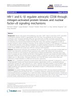
Figure 1.3 T cells involved in the induction of allergic phenotype (Lloyd and Hessel, 2010).
Asthma is a heterogeneous disease that is associated with AHR, inflammatory cell infiltration,
goblet cell metaplasia, and tissue remodeling. An array of T cell subsets are proposed to
influence the nature and severity of allergic airway inflammation.
Abbreviations: AHR, airway hyperresponsiveness; IFN-γ, interferon-γ; and TH, Th or T
helper
13
IL-33 is another cytokine released by epithelial cells (Figure 1.2). IL-33 is a member of the
IL-1 cytokines. Its expression is increased in airway epithelium of asthmatic patients and
correlates positively with the severity of asthma (Prefontaine et al., 2010). Studies have
shown that IL-33 activates cell through its receptor ST2, which is expressed on cell surface of
dendritic cells, mast cells, and epithelial cells (Besnard et al., 2011; Kurowska-Stolarska et al,
2009; Smithgall et al., 2008). Activation of ST2 results in activation of NF-κB and mitogenactivated protein kinase (MAPK) signaling pathway (Milovanovic et al., 2012). In a study by
Besnard et al., using OVA-induced mouse asthma model, IL-33 was shown to plays a critical
role on dendritic cell activation, maturation, and initiation of Th-2 mediated allergic airway
inflammation (Besnard et al., 2011c). This suggests the potential role that IL-33 may play in
the initiation of Th-2 immune response. Antibodies that block IL-33 or ST2 are currently in
clinical development (Barnes, 2011).
Compelling evidence implicates TSLP as a potential initiator of Th-2 bias allergic airway
inflammation. Blockade of TSLP has been shown to reduce airway dendritic cells migration
and thus diminishes CD4+ T cell priming (Fernandez et al., 2011). In line with this
observation, another study demonstrated that TSLP activates dendritic cells by inducing the
expression of OX40L, enhancing Th2 inflammatory response (Ito et al., 2005). Furthermore,
in the bone marrow, TSLP promotes the growth and differentiation of basophils, which
produce IL-4 that enhances Th2 inflammation (Figure 1.2) (Siracusa et al., 2010; Lambrecht
and Hammad, 2012). Corresponding well with the data obtained, it has been demonstrated
that over-expression of TSLP and GM-CSF in mice lungs results in spontaneous Th2
sensitization to harmless inhaled allergen OVA (Stampfli et al., 1998; Zhou et al., 2005).
Genetic polymorphisms in the promoter region of TSLP are linked to increased risk of asthma
(Harada et al., 2011). However, there is no strong evidence that dendritic cells release IL-4 in
response to TSLP activation, such discrepancy suggests the existence of an alternate
14
hematopoietic antigen presenting cell source for IL-4, which is needed to initiate the
differentiation of naïve T cells to Th2 cells (Kaiko and Foster, 2011).
The alternative source of IL-4 has been postulated to be basophils. Basophils have been
reported to rapidly release IL-4, IL-9, IL-25, and TSLP upon activation (Lambrecht and
Hammad, 2012; Siracusa et al., 2010). Recent reports demonstrate that basophil-derived IL-4
is essential for the induction of Th2 response to papain. In addition, basophil, rather than
dendritic cell, is a crucial antigen presenting cells for the Th2 response. Furthermore, an
observational study shows that basophil number is higher in peripheral blood of asthmatic
patients as compared to healthy individual (Gilmartin et al., 2007). However, these
observations contradict the conventional idea that dendritic cells play an important role as an
antigen presenting cell. Therefore, the mechanism behind the initiation of Th2 response
remains to be verified. Nonetheless, 20 years after the discovery of Th2 subsets, the
mechanism behind the synthesis of Th2 cytokines by T cells is clearer now.
Upon activation by IL-4 released by antigen presenting cells, Janus Kinase (JAK) molecules
in T cells are activated, leading to dimerization and nuclear translocation of transcription
factor signal transduction and activator of transcription (STAT)-6. In the nucleus, STAT-6
results in the expression of GATA-3, a transcription factor of Th2 cytokines (Nelms et al.,
1999). In addition to mediating the expression of Th2 cytokines, GATA-3 also inhibits the
activation of T-Bet, a transcription factor of Th1 cytokines (Ouyang et al., 1998). Notably, the
expression of GATA-3 activation is dependent on NF-κB signaling pathway (Das et al., 2001).
The role of NF-κB in the pathophysiology of asthma shall be discussed in section 1.2.2.
Activation of IL-4 receptor signaling, in turn, induces T cell to release the Th2 cytokines IL-4,
IL-5, and IL-13 (Figure 1.4). These Th2 cytokines initiate and maintain key
pathophysiological features of the disease (Lloyd and Hessel, 2010).
15
IL-4 is a key cytokine that influences naïve T cells to differentiate into Th2 cells (Figure 1.4)
(Long, 2009). Other sources of IL-4 include certain population of invariant natural killer
(NKT) cells (Lai et al., 2011), eosinophils (Piehler et al., 2011), basophils (van Panhuys et al.,
2011), and mast cells (Gessner et al., 2005). The IL-4 receptor consists of two chains: (1) high
affinity IL-4 binding chain (p140, α chain) which binds to IL-4 and (2) IL-2Rγ chain which
amplifies the signaling of IL-4 receptor (IL-4R). Notably, expression of high affinity IL-4Rα
binding chain is localized to the airway epithelial cells, lymphocytes, and mast cells in the
airway mucosa. As compared to normal individuals, asthmatic patients are reported to have
higher expression of high affinity IL-4Rα binding chain (Slager et al., 2012). Also, IL-4 is
essential signal for isotype switching of B-cells for IgE production. In addition, IL-4 has been
reported to promote the expression of low-affinity IgE receptors (FcεRII) and FcεRI on mast
cells and basophils. Finally, IL-4 contributes eosinophilia (Figure 1.4) (Long, 2009; PalmerCrocker and Pober, 1995). Results from mouse asthma model suggest that removal or
blockade
of
IL-4
attenuates
eosinophilia,
mucus
hypersecretion,
and
airway
hyperresponsiveness (AHR) (Brusselle et al., 1994; Long., 2009).
IL-4 and IL-13 share the same receptor subunit IL-4Rα. Therefore, these two cytokines share
many functional properties. For example, IL-4 and IL-13 mediate the migration of eosinophil
(Hogan et al., 2008; Stone et al., 2010; Wegmann, 2011). These Th2 cytokines up-regulates
the expression of: (a) eotaxin (CCL11 and CCL26), which are chemoattractant for eosinophils;
and (b) endothelial cell vascular cell adhesion molecule (VCAM) - 1, adhesion molecule that
interacts with integrin (very late antigen [VLA]-4) expressed on eosinophils, resulting in
eosinophilia (Kotowicz et al., 1996; Long, 2009 Matsukura et al., 2001; Palmer-Crocker and
Pober, 1995) (Figure 1.4). Also, both IL-4 and IL-13 play central role in IgE production;
16
PRR Agonist
PAR Agonist
Protea se
PRR Agonist
PAR Agonist
Dendritic cell
TSLP, GM-CSF,
IL-25, IL-33
1. MHC-Ag-TCR
2. Co-stimulatory
Molecules
Differentia ting
cytokines
Immunological synapse
IFN-γ, IL-12
IL-33
naïve T-cell
Th-1 cell
TGF-β, IL-2
IL-13
IL-4
TSLP
IL-5,TGF-β,
IL-23
Treg cell
IL-9
IgE
IL-4
IL-13
Th-17 cell
IL-17
IgE
IL-4
IL-5, IL-9,
GM-CSF, IL-3
Neutrophil
Basophil
Eosinophil
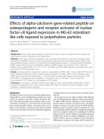
Figure 1.4 Components of airway allergic inflammation (Taken from Long, 2009)
Allergens stimulate and activate epithelial cells to mediate the release of these cytokines.
TSLP is a product of NF-κB activation and is known to activate dendritic cells and upregulate
the expression of OX40L, which plays an important role in mediating Th-2 inflammatory
response. Dendritic cells internalise and process the antigen into fragments, then present the
processed fragments to antigen specific T-cells in the context of MHC class II. Differentiation
of naïve T-helper cells to Th2 cells is favoured by the presence of IL-4. Th-2 cells release IL4, IL-13, and IL-5. IL-4 and IL-13 mediates the production of IgE. IgE plays an important
role in the activation and degranulation of mast cell and basophil. Degranulation of these cells
release biologically active mediators that act on airway smooth muscle and cause
17
bronchoconstriction. IL-13 mediates mucus hypersecretion; while IL-5 enhances the
proliferation and survival of eosinophils.
Abbreviations: PRR, pattern recognition receptor; PAR protease activated receptor; TSLP
thymic Stromal Lymophopoietin; GM-CSF, granulocyte-macrophage colony stimulating
factor. Arrows indicate the direction in which the identified signal is acting. Red lines indicate
the discharge of cellular mediators (including granular contents, lipid mediators and
cytokines).
18
goblet cell metaplasia, which results in mucus hypersecretion; and AHR development (Figure
1.4) (Long, 2009). IL-4 and IL-13, together with TGF-β and IL-6, induce airway remodeling
(Cosmi et al., 2011; Long, 2009). Nonetheless, IL-4 and IL-13 also have distinct functions.
For example, IL-13 is also involved in the recruitment of pro- inflammatory cells such as
monocytes, macrophages, and T cells (Long., 2009). In animal studies, blockade of IL-13 has
been shown to result in the attenuation of airway inflammation, airway responsiveness, and
mucus hypersecretion (Yang et al., 2004). Monoclonal anti-IL-13 antibodies are currently
under clinical trial phase I and II (Holgate, 2011)
IL-5 is an eosinophilopoietin; therefore, IL-5 is crucial for the differentiation of hematopoietic
progenitor cells to eosinophil. In addition, it also plays an important role in mediating the
maturation of eosinophil and the survival of eosinophil (Figure 1.4). Eosinophil express
transforming growth factor (TGF)-β, a pro-fibrotic growth factor; and therefore, IL-5 has also
been linked to airway remodeling and development of AHR (Long, 2009; Wegmann, 2011).
Although these Th2 cytokines are mainly released by Th2 cells, the sources of these Th2
cytokines are not limited to Th2 cells. Recent reports show that these Th2 cytokines can also
be produced by nuocytes. The function of nuocyte is inferred based on the observation that
Th2 response still develops in mice in the absence of T and B cells (Moro et al., 2010).
In order to suppress the unwanted Th2 response in asthma, studies have also focus on the
inhibitory effect that Th1 response might have on asthma. The deletion of Th1 cell master
transcription factor T-bet in mice resulted in spontaneous development of AHR and
eosinophilia (Finotto et al., 2002). However, the administration of Th1 associated cytokine
interferon (IFN)-γ resulted in no improvement of disease symptoms (Boguniewicz et al.,
1995). Recent cluster analysis of asthma clinical phenotypes demonstrates that eosinophilic
and non-eosinophilic subtypes of asthma exist (Holgate, 2012). Although atopic asthma has a
substantial Th2 cell component, this disease comprises of different subtypes, and recent
19
evidence suggests that other T cells are contributors to different stages and different
phenotypes of asthma (Lloyd and Hessel, 2010).
Th17 cells
Th17 is CD4+ T cell that is developed from a different T helper cell lineage. The
development of Th17 cell in mice is mediated by TGF-β and inflammatory cytokines IL-6,
IL-21, and IL-23. However, it is still unclear which cytokines prime the differentiation of
naïve T cells to Th17 cells. Th17 cells express key transcription factors orphan retinoic acid
nuclear receptor (ROR)γt and RORα, as well as pro-inflammatory cytokine IL-17. IL-17 also
mediates the recruitment of neutrophils (Figure 1.3) (Lloyd and Hessel, 2010). In line with
these functions, there are reports showing that there is marked Th17 cell infiltration in the
lungs of asthmatic patients with high neutrophil counts (Al-Ramli et al., 2009). The role that
Th17 plays in the pathogenesis of asthma is supported by other evidence. Although allergen
sensitization via the airway primes only modest Th2 responses, such route of sensitization
primes strong Th17 cell responses that promote airway neutrophilia and acute AHR (Wilson
et al., 2009). In addition, Th17 cells also exacerbate Th2 mediated eosinophilic airway
inflammation (Wakashin et al., 2008). Also, the transfer of Th17 cells promotes steroid
resistant airway inflammation and AHR in mice (Figure 1.3) (McKinley et al., 2008).
Moreover, a polymorphism in IL-17F that that leads to a loss-of-function mutation is
inversely related to asthma risk (Kawaguchi et al., 2006). Finally, abrogation of IL-17 during
allergen sensitization provides protection against allergic airway inflammation (Nakae et al.,
2002). However, administration of exogenous of IL-17A during allergen re-challenge
attenuates airway inflammation. This attenuation suggests that under certain conditions there
is a regulatory role for IL-17 in the allergic lung (Murdoch and Lloyd, 2010; SchnyderCandrian et al., 2006). Therefore, whether IL-17 mediates inflammatory or suppress
inflammation would depend on its cellular source, which includes γδ T cell (Murdoch and
Lloyd, 2010).
20
Th9 cells
IL-9 was originally thought to be released by Th2 cells. However, recent studies show IL-9 is
produced by a discrete population of CD4+ T cells — Th9 cells (Lloyd and Hessel, 2010). In
addition, studies have also shown that other sources of IL-9 include eosinophils and mast cells
(Angkasekwinai et al., 2010; Chang et al., 2010). Th9 cells depend on IL-4 and TGF-β for
their differentiation (Veldhoen et al., 2008). TGF-β re-programmes Th2 cells to Th9 cells,
which enforces the responsiveness of Th9 cells to IL-25. IL-25 is essential for IL-9
production (Angkasekwinai et al., 2010). Notably, Th9 cells produce IL-9 and IL-10, and not
Th2 cytokines (Wong et al., 2010). Generation of Th9 cells requires the expression of PU.1
transcription factor (Lloyd and Hessel, 2010). There is evidence indicating that IL-9 is
involved in allergic inflammation in lungs. IL-9 is detected in lung biopsies of asthmatic
patients and IL-9-transgenic mice have eosinophilia and develop AHR (Figure 1.3) (McLane
et al., 1998; Shimbara et al., 2000). Although PU.1 deficient T cells mice (mice with nonfunctional Th9 cells) were reported to develop normal Th2 responses after allergen exposure,
these PU.1 deficient T cells mice have reduced pulmonary inflammation. IL-9 is postulated to
be involved in the remodeling processes of asthma due to the role of TGF-β in Th9 cells
development. In addition, IL-9 is known to induce differentiation and proliferation of mast
cells (Barnes, 2008) (Figure 1.3). Recently, a humanized monoclonal antibody for blockade
of IL-9 is undergoing phase I clinical trial as a potential treatment for moderate to severe
asthma (Antoniu, 2010).
CD8+ T cells
CD8+ T cells are present in the airway and sputum of asthmatic patients and are reported to
release IL-4, IL-5 and IFN-γ (Ying et al., 1997). The number of CD8+ T cells correlates
positively with the decline in (forced expiratory volume in 1 second) FEV1 (van Rensen et al.,
2005).
Interestingly, CD8+ cells in the airways are different from CD8+ cells in the
peripheral blood. Briefly, airway CD8+ cells produce IL-5 and IFN-γ in the sputum. The
amount of cytokine released correlates positively to the severity of asthma. However, such
21
trend could not be observed in peripheral blood CD8+ cells (Cho et al., 2005). It is noted that
patients who died from asthma have higher proportion activated CD8+ T cell as compared to
asthmatic patients who died from other causes (O'sullivan et al., 2001). In addition, virus
specific CD8+ T cells, in the presence of Th2 cells, switch off the production of IFN-γ and
secrete IL-5, resulting in eosinophilia (Coyle et al., 1995).
Results from animal studies show that adoptive transfer of CD8+ αβ T cells results in
worsening of airway inflammation (Isogai et al., 2004). In addition, CD8 knockout mice are
less susceptible to allergic airway inflammation. The transfer of in vitro generated antigenprimed effector memory T cells into sensitized CD8 knockout mice increased AHR,
eosinophilic inflammation, and levels of IL-13 in bronchoalveolar lavage fluid (BALF)
(Miyahara et al., 2004a). However, the transfer of CD8+ T cells from IL-13 knockout mice or
non-challenged mice failed to result in airway inflammation, indicating that the CD8+ T cells
produced IL-13 locally and are activated within the airway (Miyahara et al., 2004b)
Treg
Treg cells play a crucial role in modulating and regulating immune responses by promoting
tolerance, suppressing inflammatory reactions, and maintaining homeostasis. It is well
established that the number and functions of Treg cells are impaired or altered in allergic
patients as compared with healthy individuals, providing possible explanation for the
inappropriate immune response to innocuous allergens observed in asthmatic patients (Lloyd
and Hessel, 2010; Thorburn and Hansbro, 2010). Evidence suggests the protective effect of
Treg cells in asthma. To date, two Treg cell subsets have been identified — (1) forkhead box
(FOX)P3+CD4+CD25+ cells and (2) inducible Treg cells (Lloyd and Hessel, 2010). In vivo
transfer of FOXP3+CD4+CD25+ Treg cells attenuates the development of airway
inflammation and AHR and prevents the allergen induced activation of dendritic cells in the
airways (Kearley et al., 2005). In addition, as compared to healthy children, Treg in the
airways of asthmatic children have reduced expression of FOXP3 (Lin et al., 2008).
22
Furthermore, the reduced chemotactic response of CD4+CD25+ Treg correlates positively
with FEV1 (Nguyen et al., 2009). Finally, glucocorticoid resistant asthmatic patients have
non-functional CD4+CD25+ Treg cells, i.e. their Treg cell fail to produce IL-10 upon
stimulation (Xystrakis et al., 2006).
Treg cells exert their regulatory effect through releasing IL-10 and TGF-β. IL-10 prevents the
synthesis of pro-inflammatory cytokines and down-regulates the expression of effector T cells
cytokines as well as antigen presentation and costimulatory properties of antigen presenting
cells (Figure 1.3) (Saraiva and O'Garra, 2010). On the other hand, TGF-β inhibits the
proliferation and differentiation of T cells, B cells, and macrophages; while promotes the
apoptosis of these cells. TGF-β helps to maintain the regulatory functions of Treg cells and
promotes the differentiation of adaptive Treg (Yoshimura et al., 2010).
The regulatory effect of Treg is also achieved by expressing CTLA-4. CTLA-4 closely
resembles T cell costimulatory molecules (CD28). However, as compared to CD28, CTLA-4
has higher ligand binding affinity to CD80/86 on dendritic cell. Consequently, CTLA-4
effectively competes with CD28 for CD80/86 binding site, inhibiting dendritic cells mediated
T cells activation and resulting in formation of anergic T cells (Thorburn and Hansbro, 2010).
The ligation of CTLA-4 to CD80/86 on dendritic cells induces the production of indoleamine
2, 3 dioxygenase, causing dendritic cells to be more immune-suppressive. Indoleamine 2, 3
dioxygenase is an enzyme that degrades tryptophan and induces cellular apoptosis, leading to
immunosuppressive activity (Fallarino et al., 2003; Thorburn and Hansbro, 2010).
Finally, Treg ameliorate effector responses by competing with effector cells for essential
growth factor — IL-2, which is essential for both Treg and effector cell function. Effector
cells deprived of IL-2 undergo apoptosis (Chen et al., 2011).
NKT cells
23
Majority of the NKT cells express an invariant TCR and recognize CD1d, while a small
population of NKT cells express do not express the invariant TCR but still recognize CD1dassociated antigens. Activation of the invariant TCR on NKT cells results in the production of
pro-inflammatory cytokines, including IL-4 and IFN-γ (Iwamura and Nakayama, 2010).
These pro-inflammatory cytokines help to activate dendritic cells, macrophages, T cells, and
B cells, driving the development of adaptive immunity (Bendelac et al., 2007; Iwamura and
Nakayama, 2010). NKT cells have been demonstrated to play an important role in the
pathogenesis of asthma. Although mice deficient in NKT cell had some degree of
eosinophilia, these mice failed to develop AHR (Umetsu and DeKruyff, 2010). Nonetheless,
it was reported that pulmonary instillation of NKT cell activating glycolipid (αgalactosylceramide) promoted production of IL-13 and TSLP and promoted AHR (Figure
1.3). In addition, exposing the mice to the same glycolipid together with another antigen
enhanced NKT-dependent allergen sensitization in the subsequent allergen exposure (Akbari
et al., 2003). Similar results were obtained in non-human primate asthma model
(Matangkasombut et al., 2008).
Although the studies of NKT cells in animal models have been compelling, the role of NKT
cells in the development of human asthma has been controversial. Up till now, there are at
least 14 studies that examine the presence of NKT cells in BALF, endobronchial biopsy
specimens, or both from patients with asthma (Matangkasombut et al., 2009). Although most
of these studies (10 reports) found that NKT cell numbers were increased in the lungs of
patients with asthma, four of these studies concluded that NKT cell numbers were not
increased (Umetsu and DeKruyff, 2010). These differences are possibly caused by variation
in experimental techniques and patient population (Lloyds and Hessel, 2010).
γδ T cells
γδ T cells are generally found near the airway epithelium, where they exert an
immunosurveillance function, responding to endogenous stress signals and migrating to site
24
of tissue damage (Wands et al., 2005). Interestingly, evidence from mouse asthma model
suggests that γδ T cells have dual contrasting roles in the pathogenesis of asthma. γδ T cells
can be divided into two subsets — Vγ1+γδ T cell and Vγ4+γδ T cells. The Vγ1+γδ T cells
release IL-5 and IL-13 into the airway and act together with NKT cells to induce AHR (Hahn
et al., 2004; Lloyd and Hessel, 2010). On the other hand, Vγ4+γδ T cells may release IL-17A
(Hahn et al., 2003). Interestingly, the IL-17A released by Vγ4+γδ T cells may exert
suppressive effect on established airway inflammation and AHR (Murdoch and Lloyd, 2010).
Therefore, γδ T cells derived IL-17 has regulatory role; whereas Th17 cells derived IL-17 has
pro-inflammatory role (Figure 1.3). Notably, AHR develops in non-allergic mice that are
depleted of γδ T cells, indicating a role for γδ T cells in the regulation of normal airway
function (Zuany-Amorim et al., 1998). Taken together, these results indicate that the tissue
localization of γδ T cells and their capability to detect stress or damage-associated antigen
allows them to exert protective immune responses. On the other hand, signals from local
inflammatory environment requires the γδ T cells to exert regulatory functions, controlling
inflammation and stimulating resolution (Lloyd and Hessel, 2010).
1.1.2.3 B cell IgE production and mast cell activation
B cell is an immune cell that contributes to the pathophysiology of asthma. B cells develop
and undergo maturation in the bone marrow. This process involves nuclear factors such as Ebox factors, early B cell factors, and NF-κB signaling (Schutte et al., 2012). In particular,
some components of NF-κB signaling influence several stages of late B cell differentiation
and maturation. For example, p50-deficient mice lack marginal zone V-cells; while c-Rel (vrel reticuloendotheliosis viral oncogene homolog) deficient mice show reduced numbers of
marginal zone B cells (Cariappa et al., 2000; Köntgen et al., 1995). In addition, p50/p52
double knockout mice fail to generate B cells and are unable to produce immunoglobulin,
including IgE (Franzoso et al., 1997).
25