Molecular mechanisms of mechanosensing at cell cell and cell matrix adhesion 2
Bạn đang xem bản rút gọn của tài liệu. Xem và tải ngay bản đầy đủ của tài liệu tại đây (14.21 MB, 115 trang )
MOLECULAR MECHANISMS OF
MECHANOSENSING AT CELL-CELL
AND CELL-MATRIX ADHESIONS
YAO MINGXI
NATIONAL UNIVERSITY OF
SINGAPORE
2014
MOLECULAR MECHANISMS OF
MECHANOSENSING AT CELL-CELL
AND CELL-MATRIX ADHESIONS
YAO MINGXI
B. Sci. (Hons.), NUS, 2009
A THESIS SUBMITTED
FOR THE DEGREE OF DOCTOR OF
PHILOSOPHY
MECHANOBIOLOGY INSTITUTE
NATIONAL UNIVERSITY OF
SINGAPORE
2014
DECLEARATION
I hereby declare that this thesis is my original work and it has been
written by me in its entirety.
I have duly acknowledged all the sources of information which have been
used in the thesis.
This thesis has not been submitted for any degree in any university
previously.
Yao Mingxi
August 18, 2014
Acknowledgment
It is truly a rewarding experience in the past five years as an PhD
student in mechanobiology institute. It is such a vibrant institute where
exciting research takes place. I am grateful for the opportunity to work in
this dynamic environment surrounded by excellent colleagues.
I would like to thank my supervisor Dr Yan Jie for his guidance and
support over the years. He has been instrumental for creating a warm
and stimulating atmosphere in the lab. I keep being amazed by his work
altitude and passion for science. It is a great pleasure working with him
and I learned a lot from him both academically and in life.
I also want to thank my collaborators - Dr Rene-Marc Mege, Dr Benoit
Ladoux, Dr Benjamin T Goult, Dr Mike Sheetz and Qiu Wu for their in-
sightful suggestions and great work. Two excellent undergraduate students,
Guo Yingjian and Kasper Graves Hvid, have helped me in many aspect of
experiments. I am grateful for their contributions.
I would like to express my gratitude to friends and colleagues in Yan
Jie’s lab - Chen Hu, Fu Hongxia, Peiwen Cong, Yuan Xin, Lim Ciji, Zhang
Xinghua, Le Shimin, Qu Yuanyuan, Chen Jin, Artem, Rickson, Lee Xinyi,
Wong Weijuan, Zhao Xiaodan, Li You, Li Yanan, Rangit, Dugarao. They
make the lab a warm and fun place to be. I am very grateful for their
friendship and support along the way.
December 15, 2014
iv
Contents
1 Introduction 1
1.1 Mechanosensitivity of cells . . . . . . . . . . . . . . . . . . . 1
1.2 Review of cell adhesions . . . . . . . . . . . . . . . . . . . . 3
1.2.1 Cadherin based adherens junctions . . . . . . . . . . 4
1.2.2 integrin based cell-matrix adhesions . . . . . . . . . . 7
1.3 Literature survey on mechanosensing related proteins at cell-
adhesions . . . . . . . . . . . . . . . . . . . . . . . . . . . . 9
1.3.1 vinculin . . . . . . . . . . . . . . . . . . . . . . . . . 10
1.3.2 talin . . . . . . . . . . . . . . . . . . . . . . . . . . . 12
1.3.3 α-catenin . . . . . . . . . . . . . . . . . . . . . . . . . 14
1.3.4 other mechanosensing proteins at cell adhesions . . . 16
1.4 Key question: Mechanosensing mechanisms of talin and α-
catenin . . . . . . . . . . . . . . . . . . . . . . . . . . . . . . 16
2 Strategies and Methods 19
2.1 Theory of force induced structural transitions of protein . . . 20
2.1.1 Structural states during two state protein unfoding
and refolding transitions . . . . . . . . . . . . . . . . 20
2.1.2 Force-extension curves of the structural states . . . . 22
2.1.3 Force dependent free energy differences between states 23
2.1.4 Free energy landscape along the transition coordinate 28
2.2 Magnetic tweezers . . . . . . . . . . . . . . . . . . . . . . . . 31
2.2.1 Magnetic tweezers setup . . . . . . . . . . . . . . . . 32
2.2.2 Force determination for magnetic tweezers . . . . . . 33
2.3 Other Methods . . . . . . . . . . . . . . . . . . . . . . . . . 37
2.3.1 Protein Expression . . . . . . . . . . . . . . . . . . . 37
2.3.2 Force calibration . . . . . . . . . . . . . . . . . . . . 38
2.3.3 Data-analysis . . . . . . . . . . . . . . . . . . . . . . 39
2.3.4 Hidden Markov models . . . . . . . . . . . . . . . . . 39
v
2.3.5 Bioconjugation and surface chemistries . . . . . . . . 40
3 Force response of talin rod and α-catenin 43
3.1 Introduction . . . . . . . . . . . . . . . . . . . . . . . . . . . 43
3.2 Results and Discussion . . . . . . . . . . . . . . . . . . . . . 44
3.2.1 The force response of talin rod domain . . . . . . . . 44
3.2.2 The force response of αE-catenin . . . . . . . . . . . 52
4 vinculin binding to talin and α-catenin fine-tuned by me-
chanical forces 61
4.1 Introduction . . . . . . . . . . . . . . . . . . . . . . . . . . . 61
4.2 Results and discussion . . . . . . . . . . . . . . . . . . . . . 62
4.2.1 The effects of V
D1
domain binding on the effect of
talin rod . . . . . . . . . . . . . . . . . . . . . . . . 62
4.2.2 Force dependent interaction of vinculin head to αC
M
70
4.2.3 The effect of full length vinculin on the samples of
talin and α-catenin . . . . . . . . . . . . . . . . . . . 77
5 Discussion and Conclusions 81
vi
Summary
Over the past decade, mechanical forces have been identified to take
part in many important biological processes ranging from embryo develop-
ment to tissue maintenance and cancer. A novel class of proteins, termed
mechanosensing proteins, is found to be able to convert mechanical forces
into biochemical signals that direct cellular responses. These proteins are
particularly enriched at cell adhesion sites where cells’ cytoskeleton con-
nects with their micro-environment and mechanical forces are transmitted
and sensed making cell adhesion sites signaling hubs for detecting mechani-
cal cues. Established mechanosensing mechanisms of these proteins include
force dependent channel opening, phosporylation and catch bond forma-
tion.
Talin and α-catenin, two mechanosensing proteins located at focal ad-
hesions and adherens junctions respectively, are critical for the force depen-
dent initialization and growth cell adhesions.It has been suggested by cell
and structural studies that mechanical force applied to the two proteins
will increase their binding affinity to vinculin, a protein that promotes
cytoskeleton linkage leading to growth and maturation of cell adhesions.
Unlike many mechanosensors, accumulating data suggests that talin and
α-catenin respond to applied force by expose their cryptic vinculin binding
sites. However, molecular level mechanisms of this process have not been
quantitatively understood with direct experimental evidence.
In this thesis work, I used state-of-art magnetic-tweezers technology to
study the mechanosensing mechanism of talin and α-catenin. The hypoth-
esis is that the two proteins change their conformations upon application
of force and modulate their binding affinity to vinculin. In Chapter 1, I
review the biological background on mechanosensing, focusing on the role
talin and α-catenin plays during initiation of cell adhesions. In Chapter 2,
I describe the methods used for my thesis, introducing magnetic tweezers
and theoretic background of force induced protein unfolding. In chapter 3
I study the mechanical stability of the rod domains of talin and central do-
main of α-catenin using both wild type and mutant constructs. Both talin
vii
rod domain and α-catenin central domain undergo well-defined conforma-
tion changes at forces greater than 5 pN, suggesting physiological relevant
forces could expose cryptic vinculin binding sites in the two proteins. In
Chapter 4, I compare the mechanical responses of talin rod domain and α-
catenin central domain to show that vinculin binding to talin and α-catenin
only upon application of force and vinculin binding inhibits the refolding of
these proteins. In addition, at forces larger than 30 pN, bound vinculin can
be displaced from these proteins, implying the binding of vinculin is bipha-
sic with force. Finally in Chapter 5 I discuss the biological implications of
the findings.
The work in this thesis establishes a molecular mechanism of mechanosens-
ing at early adhesion formations where the force dependent conformational
changes of talin and α-catenin play key role in the initiation of adhesion-
cytoskeleton linkage. Besides providing novel mechanistic insights into
the function mechano-sensitive proteins,the single molecule manipulation
methods developed in this work opens up possibility to study other force-
dependent protein-protein interactions such as ligand receptor interaction,
which has important implication in many biological and pathological pro-
cesses.
viii
List of Figures
1.1 Anchoring junctions . . . . . . . . . . . . . . . . . . . . . . . 4
1.2 Types of cell-matrix adhesions . . . . . . . . . . . . . . . . . 8
1.3 Vinculin domain map . . . . . . . . . . . . . . . . . . . . . . 11
1.4 The domain map of talin . . . . . . . . . . . . . . . . . . . . 12
1.5 α-catenin domain map . . . . . . . . . . . . . . . . . . . . . 14
2.1 The conformational states of protein under force . . . . . . . 21
2.2 Energy landscape of two state model . . . . . . . . . . . . . 21
2.3 Calculated force-extension curve of folded and unfolded i27 . 23
2.4 Calculated force-dependent Gibbs free energy folded and un-
folded I27 . . . . . . . . . . . . . . . . . . . . . . . . . . . . 25
2.5 Calculated force-dependent unfolding and refolding rate of I27 27
2.6 Calculated force-dependent free energy landscape of I27 as
a function of extension . . . . . . . . . . . . . . . . . . . . . 30
2.7 Force geometry affect the free energy of transition states . . 31
2.8 Photo of the vertical magnetic tweezers used in this study . 32
2.9 Illustration of magnetic tweezers setup . . . . . . . . . . . . 34
2.10 Calibration of permanent magnets using λ-DNA . . . . . . . 36
2.11 Calibration of the strength of individual magnetic beads . . 38
3.1 Sketch of the conformation changes of talin R1-R3 domain
under mechanical force . . . . . . . . . . . . . . . . . . . . . 44
3.2 The schematic figure of experimental setup for talin stretch-
ing experiment . . . . . . . . . . . . . . . . . . . . . . . . . 45
3.3 Force cycle experiments of the talin R1-R3 domain. . . . . . 46
3.4 2-D histogram of the unfolding force and unfolding size of
WT talin R1-R3 domain . . . . . . . . . . . . . . . . . . . . 47
3.5 Unfolding force histograms of two R1-R3 talin domains . . . 47
3.6 Two state fluctuations of talin R3 domain . . . . . . . . . . 49
ix
3.7 Unfolding force histogram of wildtype and IVVI mutant talin
R1-R3 domains of talin at 5 pN/s constant loading rate. . . 50
3.8 Unfolding force responses of the R9-R12 region of talin rod . 51
3.9 Unfolding force responses of the R7-R9 region of talin rod . 52
3.10 Experimental setup of αC
M
stretching. . . . . . . . . . . . . 53
3.11 Force responses of wild type αC
M
. . . . . . . . . . . . . . . 55
3.12 Repeated unfolding-refolding force cycle experiments on a
single αC
M
tether . . . . . . . . . . . . . . . . . . . . . . . . 56
3.13 Unfurling of αC
M
and ∼ 5 pN forces . . . . . . . . . . . . . 57
3.14 The force responses of L344P mutant of αC
M
. . . . . . . . 59
4.1 Mechanosensitivity of talin R1-R3 . . . . . . . . . . . . . . 64
4.2 Concentration dependence of V
D1
binding to talin R1-R3
domain . . . . . . . . . . . . . . . . . . . . . . . . . . . . . . 65
4.3 V
D1
dissociate from talin rod at high forces . . . . . . . . . . 66
4.4 Observation of five V
D1
dissociation steps . . . . . . . . . . . 68
4.5 Detecting the binding of V
D1
to the peptide chain of unfolded
vinculin binding α-helices at high force in 100 nM V
D1
. . . 69
4.6 Effect of V
D1
on the unfolding/refolding of αC
M
. . . . . . . 71
4.7 Correlation between vinculin dissociation and αC
M
folding . 73
4.8 The mechanosensitivity of αC
M
folding on vinculin binding . 75
4.9 High force displaces the bound V
D1
from the vinculin binding
site in αC
M
. . . . . . . . . . . . . . . . . . . . . . . . . . . . 76
4.10 Detecting the binding of full length vinculin to talin R1-R3
and αC
M
. . . . . . . . . . . . . . . . . . . . . . . . . . . . . 78
4.11 Dissociation of full length vinculin from the αC
M
at high
force . . . . . . . . . . . . . . . . . . . . . . . . . . . . . . . 79
5.1 Model of fore-dependent talin-vinculin interaction . . . . . . 87
5.2 Model of fore-dependent α-catenin-vinculin interaction . . . 88
x
List of Abbreviations
AFM Atomic Force Microscopy
CAM Cell adhesion molecules
ECM Extra-cellular matrix
magnet distance distance between the permanent magnet and the mag-
netic bead
V
D1
The D1 domain of vinculin head
VBS vinculin binding sites
xi
xii
Chapter 1
Introduction
1.1 Mechanosensitivity of cells
Mechanobiology is an emerging field in biomedical research, driven by the
realization that mechanical forces play a major role in a wide range of
biological and pathogenic processes [1,2].
A century ago, biologists started to conceptualize that physical forces
can play in determining the morphology of life [3]. Later on, the study
of biomechanics has revealed the role of forces in tissue development such
as the bone strengthening and muscle growth [4–6]. In the 80s, Harris et
al. demonstrated that non-muscle cells can apply mechanical deformation
to their environment and deform elastic film they residing on started the
era of mechanobiology at cellular level [7].Subsequent studies revealed that
cells actively sense and respond to their mechanical environment - a process
with profound biological and pathological consequences.
For example, mechanical cues were sensed collectively at tissue level
that could direct the migration behavior of cells [8]. When pluropotent
embryonic stems cells were grown on substrates varying in rigidity, they
tend to differentiate into tissue cells with comparable rigidity - neuron
cells on soft substrate, bone cells on hard substrates and adipocyte cells
on substrate with intermediate rigidity [9]. In addition, how cells interact
1
with their mechanical environment is important factor in many diseases
as well [1]. A defining feature of metastatic cancer cells is their ability to
escape apoptosis and proliferate in foreign environments [10]. Metastatic
breast cancer cells that invade lung or liver tissues were shown to grow most
efficiently on substrate with same rigidity to the invading tissue, suggesting
the ability to adapt to different substrate rigidities is critical for metastasis
process [11].
It has been proposed that cells sense the rigidity by actively deform-
ing their substrate and measure the stress-strain relationship [2, 12, 13].
In order to translate these mechanical cues into cellular signals directing
cells’ responses, there must exist mechanisms at molecular level that de-
tect the amount of forces that are applied on them. Molecules that ex-
hibit this properties are termed mechanosensing proteins. Over the years
many mechanosensing protein have been identified in the cells with diverse
mechanisms. Cells could directly translate applied force to a biochemical
modification such as phosphorylation [14]. The expression level and local-
ization of many individual proteins were shown to be directly regulated
by mechanical related signals [15, 16]. However, the mechanotransduction
and mechanosensing functions in the cell are not necessarily performed at
individual proteins level. Instead, various active protein assemblies with
defined constitutions and underlying molecular mechanisms, often referred
to as functional modules, work together to govern these complex and robust
processes [2,17].
This thesis work devotes to understand the molecular mechanisms of
a key mechanosensing functional modules centered around the vinculin
protein at cell-matrix and cell-cell junctions. Part of the work is already
published [18, 19] and some materials from the papers are reused in the
thesis.
2
1.2 Review of cell adhesions
In advanced organisms such as animal or plants, one or more types of
specialized cells organize into tissues and carry out biological functions
collectively. The ability of cells to physically adhere with each other and to
their environment is essential for the formation and functioning of tissues.
Cell-cell adhesion is one of the corner stones in the arising of multicel-
lular organisms. Depending on the context, cell-cell adhesion could serve
different roles such as supporting mechanical integrity of tissues, signal
transduction across cells, cellular recognition and triggering of immune re-
sponses. Tissue organizations do not only depend on cell-cell adhesions. In
epithelium and muscles, cells are surrounded by a fibrous protein network
called extra-cellular matrix (ECM) . Main components of ECM include col-
lagen, proteoglycans and multiadhesive matrix proteins such as fibronectin.
ECM acts as an organization scaffold for tissues and is responsible for the
signaling and regulation of variety of cellular processes such as cell growth,
migration and gene-expression [20].
To fulfill such a set of functions, eukaryotic cells have evolved delicate
and robust molecular apparatus for the fine control of cell adhesions cen-
tered around several families of trans-membrane cell adhesion molecules
(CAMs) such as cadherins,Immunoglobins, integrins and selectins. Build-
ing upon these CAMs, cells develop several types of highly specialized cell
junctions for different purposes such as attachment (Anchoring Junctions),
barrier formation (Occluding Junctions), inter-cellular channel formation
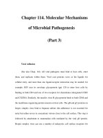
and signal transduction (Gap Junctions) [20]. Among them, anchoring
junctions, particularly the junctions linked to cytoplasmic actin cytoskele-
ton, play key roles in maintaining the mechanical integrity of cells(Fig. 1.1).
They are also signaling hubs where the chemical properties and rigidity of
cells’ micro-environment are sensed [21].
3
Figure 1.1: Anchoring junctions play key roles in maintaining mechanical
integrity of cells. Used by permission from MBInfo: www.mechanobio.
info; Mechanobiology Institute, National University of Singapore.
1.2.1 Cadherin based adherens junctions
There are three main types of cell-cell adhesions present in cells - tight
junctions, desmosomes and adherens junctions. Tight junctions (zonula
occludens) are found in epithelial and endothelial cells that act as diffusion
barrier forming a tight seal. Desmonsomes are responsible for anchoring
intermediate filaments to the cell-cell contact sites and are present in cells
that experiencing high shear stress [20, 22]. The most well-studied cell-
cell adhesions that play critical role in maintaining mechanical integrity of
tissues and regulate cell fates are cadherin based adherens junctions.
The cadherin family is the most diverse class of CAMs for cell-cell ad-
hesions. It contains over 100 members with distinct functions that can be
classified into 6 groups (Summarized in Table 1.1). Among them, type I
4
classical cadherins is well-recognized for their roles in cell-cell adhesions.
They are mostly transmembrane proteins featuring a conserved extracel-
lular cadherin repeats domains. Despite the similar domain features, indi-
vidual members of cadherin family are highly specialized and only present
in specific tissue types or sub-cellular structures.
Subfamilies Functions Examples
Type I classical
cadherins
Formation of adherens
junctions
E-cadherin, N-cadherin,
P-cadherin
Type II atypical
cadherins
Maintenance of
specialized tissues
VE-cadherin
Desmosomal
cadherins
Formation of
desmosomes
desmoglein, desmocollin
Flamingo
cadherins
Planar cell polarity
regulation
flamingo
Proto-cadherins Neuron
development [23]
Pcdhα,Pcdhβ
Ungrouped
cadherins
Mostly unknown CDH9 CDH10
Table 1.1: Subfamily of Cadherins
Cadherins interact by extracellular homophilic interactions - they only
interact with proteins of the same type with rare but important exceptions
most notably in the immune responses of epithelial cells where E-cadherin
interacts with integrins of immune cells [24]. The interactions between
cadherin molecules are carried out by calcium sensitive associations of the
extracellular cadherin repeats. This interaction can happen between two
adjacent cells (trans interactions) or two cadherin molecules from the same
cell (cis interactions). Both trans and cis interactions are shown to play
important roles in the formation of stable cell-cell contacts [25].
The formation of adherens junction is a complex and tightly regulated
process. At onset, type I cadherins are recruited to the membrane by direct
exocytosis along with the β-catenin that can be assisted by nectins . At
the membrane, cadherins are diffusive initially and they become less immo-
bile upon engagement with cadherins of neighboring cells through calcium
5
dependent trans interactions of their extracellular cadherin repeats [26].
After the establishment of trans interactions, cadherins from the same cells
can also form lateral cis interactions that are thought to be important in
the formation of stable junctions [27]. Following the establishment of cad-
herin interactions, conserved set of key cytoplasmic components such as
α-catenin, β-catenin, p120-catenin and Eplin are recruited to the nascent
adherens junctions. These proteins in turn form the basis of the dense
complex molecular assembly at adherens junctions that is also referred to
as adherens junction plaques [28]. These proteins mediate the dynamics
of adhesions including assembly/disassembly as well as interaction and re-
modeling of the actin cytoskeleton. Building upon these core proteins, cell-
cell adhesion can adopt different morphology and molecular compositions
depending on the circumstances and cell types [29].
The mechanical linkage between cadherins and actin cytoskeleton is
critical for the formation of stable adherens junctions [30]. Inhibition of
actomyosin contraction by blebbistatin inhibit the recruitment of adherens
junctions protein that is essential for their growth and maturation such as
vinculin [31]. The homophilic trans interactions between individual cad-
herin molecules can withstand forces up to 100 pN before rupture [32, 33].
E-cadherins in vivo were shown directly experiencing stretching forces in
the pN range and actomyosin contractility greatly influences the formation,
maturation and remodeling of adherens junctions [34]. The composition
and topology of this mechanical link is still not fully resolved [35]. How-
ever, it is well-established that cytoplasmic proteins such as α-catenin was
shown to be at center stage for the establishment of this mechanical link-
age [36]. In addition, cytoskeletal proteins such as vinculin are recruited to
adherens junctions in a force dependent manner in by α-catenin, demon-
strating the mechanosensitivity of adherens junctions [37].
Adherens junction mechanosensing has been shown to be involved in im-
6
portant biological processes such as development, tissue repair and diseases.
Great efforts have been made to elucidate the key players and underlying
mechanosensing mechanisms. (Reviewed in [38]).
1.2.2 integrin based cell-matrix adhesions
Integrin based cell-matrix adhesions are the most-studied cell adhesions so
far. Integrins are a class of conserved transmembrane proteins that links
cytoskeleton to the ECM. Integrins typically consist of a large extracellular
domain and a relatively small cytoplasmic domain [39]. In the activated
functional form, two subunits of integrins α and β will form heterodimers
that bind to specific amino acid sequences of ECM proteins such as RGD
peptide of fibronectins, LDV peptide of VCAM-1 or GFOGER motif of
collagen. There are 18 α and 8 β subunit genes in mammalian cells and
among them 24 α-β pairs are identified so far that recognize wide variety
of ECM ligands [40]. The short cytoplasmic domains of integrins act as
a molecular interaction hub that can interact, directly or indirectly, with
hundreds of adhesion and cytoskeleton proteins that form the optically
dense matrix adhesion plaques [41].
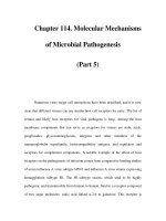
Integrin based cell-matrix adhesions can generate a highly diverse set of
adhesive structures that have distinct morphology and behaviors depend-
ing on the micro-environment and phases of adhesion maturation. In 2D
culture system, in the initialization phase of cell spreading, when a cell is
just in contact with its substrate, cells form nascent dynamic focal com-
plexes. When the focal complex evolved, it can mature and form contractile
adhesions such as focal adhesions. In some specific cell types and circum-
stances, other types of adhesions can arise such as ring like, highly dynamic
podosomes (See Fig. 1.2). Cells in their native environment are believed
to form adhesion structures reminiscent to these structures formed in 2D
culture systems in terms of molecular architecture and functions [42].
7
Figure 1.2: Different cell-matrix adhesions have distinct morphologies and
functions depending on cell type and spatial-temporal phases of the cell.
Used by permission from MBInfo: www.mechanobio.info; Mechanobiology
Institute, National University of Singapore.
Cell-matrix adhesions start to form upon stimulation, either external
when cells gets in contact with ECM, or internal, by the activation of sig-
naling cascades such as Ras. The autoinhibited integrin on te tensile mem-
brane gets activated. During activation, integrin will undergo significant
structure changes that allow the binding of adhesive proteins such as talin
and paxillin, initiating the formation of focal complexes. Overtime, focal
complex mature and more proteins are recruited such as vinculin, FAK and
α-actinin. At this stage, focal complexes start to engage with contractile
actin cytoskeleton and detect their substrate rigidity through a actomyosin
contraction dependent mechanism that has not been fully resolved to date.
If the rigidity of the substrate is high enough, the focal complex mature
into focal adhesions [43]. In this process, more components such as zyxin
and tensin are recruited while the linkages between focal adhesion and actin
cytoskeleton are enhanced. Mature focal adhesion complex is a highly or-
8
ganized structure that has well defined molecular architecture facilitated
by specific interactions between adhesion proteins [44].
Mechanical force is a critical factor that couples tightly with the forma-
tion and dynamics of cell-matrix adhesions. Most cell-matrix adhesions,
such as focal complex and focal adhesions, are contractile that actively
exert forces to their substrate [45]. Stable focal adhesions are lost when
actomyosin contraction of cells is inhibited by blebbistatin. In addition,
the size of focal adhesion is proportional to the forces they exerted to
the substrate [46] and many focal adhesion proteins show force dependent
localization to focal adhesions [47]. Cell-matrix adhesions are also respon-
sible for sensing the rigidity of the substrate, directing the behaviors of cell
spreading (Reviewed in [12]).
As cell-matrix adhesions are force bearing structures, members of cell-
matrix adhesion proteins must be under tension and transmit mechanical
forces generated from cytoskeleton. Many studies have devoted to measure
the strengths of interactions between members of adhesion proteins as well
as their mechanical integrities. The tension exerted on single proteins can
exceed 100 pN before breaking (Reviewed in [48]).
1.3 Literature survey on mechanosensing re-
lated proteins at cell-adhesions
As cell adhesions play a pivotal role in the proper function of cells, mis-
regulation of cell adhesions often leads to severe pathological consequences.
Altered cell-cell and cell-matrix adhesion function is one of the hall marks
of cancer [49, 50]. The proper functions of cell adhesions, both in terms
of adhesion strength and underlying signaling pathways are critical for al-
most every aspect of development (reviewed in [51]). Mechanosensitivity
has been increasingly recognized for their importance in cell adhesions reg-
9
ulation and related diseases [1]. In recent years, great efforts have been
focused on identifying the mechanosensors within cells. Number of known
force sensitive molecular responses are fast growing in dept to the rapid
growth of available tools that can probe and alter cells’ mechanical path-
ways such as traction force microscopy, deformable substrates, micro/nano-
fabrication and myosin inhibitors. However, the molecular mechanisms of
mechanosensing for these proteins are largely unknown. In this section, key
protein players identified in the mechanosensing pathways are reviewed.
1.3.1 vinculin
Vinculin is a 120kD cytoplasmic actin binding protein enriched in both
focal adhesions and adherens junctions. It is essential for development as
vinculin deletion is embryonic lethal due to heart and brain defects [52].
Vinculin does not possess enzymatic activity - all its biological functions
are carried out through highly regulated molecular interactions that can
be fine-tuned by conformation changes. At subcellular level, vinculin plays
important roles in regulating cell-matrix and cell-cell adhesions. Cells lack-
ing vinculin develop less stable focal adhesions and migrate faster in wound
healing assays [53]. vinculin deletion also led to impaired adherens junction
reinforcement upon mechanical stimulation [54].
Vinculin is a compact globular protein composed of successive 4 α-helix
bundles. Five of these α-helix bundles constitute the vinculin head bind-
ing to various partners such as talin, while the C-terminal constitutes the
vinculin tail binding to F-actin [55] (See Fig. 1.3).
The localizations of vinculin to both cell-matrix adhesions and adherens
junctions are mediated by mechanical force [37,46,54,56]. Vinculin recruit-
ment is generally related to the enhancement of cytoskeleton linkage and
leads to the formation of stable adhesions [57]. FRET force-sensor has
shown that vinculin is under mechanical tension inside cells [58].
10
Figure 1.3: The domain map of vinculin. The N-terminal head domain of
vinculin contain binding cite for proteins in cell adhesions such as α-catenin
and talin and a C-terminal tail domain that binds to F-actin. In cytosol,
vinculin exist in an auto-inhibited conformation where its head and tail
domains bind with nanomolar affinity. Used by permission from MBInfo:
www.mechanobio.info; Mechanobiology Institute, National University of
Singapore.
In the cytosol, vinculin is under an inactive head-to-tail conformation
presenting only weak affinity for actin. In contrast, vinculin captured at
focal adhesions by force-dependent activated talin is stabilized under an
open conformation characterized by relaxation of head to tail dissociation
that is stabilized by binding of the head to talin, and high affinity binding
of the tail domain to F-actin. in vivo study suggested that vinculin is
recruited to focal adhesions in the auto-inhibited conformation and gets
activated in situ that orchestrate downstream signaling events [59].
However currently there are conflicting data regarding the mechanism
of vinculin activation. One line of literature suggests that due to the tight
head-tail association, full-length vinculin can only be activated in the pres-
ence of multiple ligands such as talin and F-actin simultaneously. Support-
ing this idea, using a FRET vinculin probe, Chen et. al. show that talin
11
can not activate vinculin alone without the presence of F-actin [60]. In-
consistent with these findings, some other studies suggest that interaction
of the N-terminal domain can trigger vinculin activation. The N-terminal
binding sites for talin and α-catenin are not blocked by the head-to-tail
interactions [61,62]. Moreover, the affinity of talin and α-actinin’s vinculin
binding sites to vinculin has comparable affinity measured by Surface Plas-
mon Resonance (SPR) methods [63]. The mechanism of vinculin activation
is important for the understanding of the establishment of mechanical links
between actin cytoskeleton and cell adhesions.
1.3.2 talin
Talin is a focal adhesion protein that plays an important role in the ini-
tialization of focal complexes. It is one of the earliest proteins that gets
recruited to the nascent adhesion sites and is related to the activation of
integrins [64]. Depletion studies identified that talin is essential for the me-
chanical connection between focal adhesions to the actin cytoskeleton [65].
Figure 1.4: The domain map of talin. (A) The structural illustration of
the full length talin. Zoom in shows the detailed structure of the R1-R3
region of talin rod domain. High lighted in yellow is the Trp residues at
the center of talin domain. (B) The compact R1-R3 part of talin rod is
hypothesized to be the core mechanosensing region of talin
12
Talin comprises an N-terminal FERM domain (50 kDa) that binds
integrin cytoplasmic tails and acidic membrane phospholipids coopera-
tively [66,67]. The C-terminal of talin contains a long rod domain (220kDa)
consisting 13 helical bundles (R1-R13) and a terminal helix involved in
the formation of talin homodimer (Fig. 1.4(A)) [68]. In the talin rod do-
main, there are eleven putative vinculin binding sites (VBS) , each defined
by hydrophobic residues on a single helix, but these are normally buried
within the helical bundles [69]. The talin-vinculin interaction occurs pri-
marily through the association of the vinculin head (V
D1
) domain with
these VBS [58, 70]. Talin rod domains can directly interact with F-actin
through its three F-actin binding sites [71]. The engagements with actin
cytoskeleton put talin under the mechanical influences of actomyosin con-
traction [72].
As talin is stretched inside of cells and many of its vinculin binding sites
are hidden in the folded talin rods, It has been long proposed that talin
rod domain is mechanosensitive. Talin is critical for the force-dependent
recruitment of vinculin to focal adhesions [56]. Steered full-atom molecular
dynamics (MD) simulations indicate that the cryptic VBS may be exposed
by mechanical force resulting from actomyosin contractions in vivo [73,74].
This hypothesis is supported by experiments that revealed substantial in-
creases in the vinculin-talin interaction when talin was subjected to forces of
12 pN with magnetic tweezers [75]. in vivo single molecule measurements on
the end-to-end distances of talin also suggested that it is constantly under
unfolding-refolding fluctuations inside the cells [70]. However, open ques-
tions remain on the force-sensing mechanisms of talin such as the amount
of forces that required to trigger vinculin binding and how the different
talin rod domains work in synergy to regulate vinculin binding.
The recent structural characterization of full-length talin (Fig. 1.4(A))
shows that the talin rod is comprised of 13 helical bundles (R1-R13) orga-
13