The mechanistic studies of the anticancer potential of artesunate in human cancer cells
Bạn đang xem bản rút gọn của tài liệu. Xem và tải ngay bản đầy đủ của tài liệu tại đây (7.11 MB, 171 trang )
THE MECHANISTIC STUDIES OF THE
ANTICANCER POTENTIAL OF ARTESUNATE IN
HUMAN CANCER CELLS
YANG NAIDI
(M.Sc. Zhejiang University, P.R. China)
A THESIS SUBMITTED
FOR THE DEGREE OF DOCTOR OF PHILOSOPHY
DEPARTMENT OF PHYSIOLOGY
NATIONAL UNIVERSITY OF SINGAPORE
2014
iii
ACKNOWLEDGEMENTS
I would like to express my most sincere and deepest gratitude to my
supervisor, A/Prof. Shen Han-Ming for his professional and enthusiastic
guidance throughout the past four years. This study would not have been
possible without his excellent guidance, great supports and continuous
encouragements. His enthusiasm and dedication to science have impressed and
inspired me deeply. I have indeed gained fruitful experience for the ropes of
biological research. What I have learned from him will not only benefit my
future career but also my life.
I would like to take this opportunity to delicate my sincere thanks to my
thesis advisory committee members: A/Prof. Kevin, Tan Shyong Wei, and
A/Prof. Reshma Taneja for their instructive suggestions and continuous
supports on my study.
I would also like to extend my gratefulness to the following people for
providing materials for this study:
Dr. N Mizushima (Tokyo Medical and Dental University, Japan) for
providing the HeLa cells with stable expression of GFP-LC3;
Dr. A Ballabio (Telethon Institute of Genetics and Medicine, Italy) for
providing the TFEB-luciferase construct;
Dr. DJ Kwiatkowski (Harvard University, USA) for providing the pair of
Tsc2 WT and KO MEFs;
Dr. Huang Jingxiang (National University Hospital, Singapore) for
providing the pair of TSC2 WT and shTSC2 HeLa cells;
Dr. TW Soong (National University of Singapore, Singapore) for
providing Flag-FTH plasmid.
iv
It has been a great honor and fortune for me to work in such a warm and
harmonious laboratory. I would like to specially thank Dr. Ng Shukie for her
immense help in my study as well as daily life and also Dr. Tan Shi Hao for
his helpful suggestions to my research. Special thanks also go to Mr. Ong
Yeong Bing and Ms Su Jin for their logistical help. I would also like to
express my deep appreciation to other lab members: Dr. Zhou Jing, Dr. Cui
Jianzhou, Dr. Chen Bo, Ms Zhang Yin, Ms Shi Yin, Mr. Zhang Jianbin and
Ms Mo Xiaofan for their supports and the friendship. Also, thank all other
staffs in Saw Swee Hock School of Public Health and Department of
Physiology, Yong Loo Lin School of Medicine. Especially, I would like to
thank Dr. Tai Yee Kit (Department of Physiology, NUS) for the insightful
discussions on my research project.
Finally, I would like to extend my deep appreciation to my parents,
younger sister for their endless love. Also numerous thanks to my husband Dr.
Jiang Bo for his continuous love, support and understanding.
v
PUBLICATIONS AND PRESENTATIONS AT CONFERENCES
PUBLICATIONS
1. Yang ND, Tan SH, Ng S, Shi Y, Zhou J, Tan K SW, Wong WS F, Shen
HM. (2014). Artesunate induces cell death in human cancer cells via
enhancing lysosomal function and lysosomal degradation of ferritin. J Biol
Chem 289, 33425-33441.
2. Zhang J, Ng S, Wang J, Tan SH, Zhou J, Yang ND, Lin Q, Xia D, Shen
HM. (In press). Histone Deacetylase Inhibitors Induce Autophagy through
FoxO1-Dependent Pathways. Autophagy.
3. Shi Y, Tan SH, Ng S, Yang ND, Zhou J, McMahon KA, Del Pozo MA,
Hill MM, Parton RG, Kim YS, Shen HM. (In press). Caveolin-1 and lipid
rafts in modulation of lysosomal function and autophagy in breast cancer
cells. Autophagy.
4. Zhou J, Tan SH, Nicolas V, Bauvy C, Yang ND, Zhang J, Xue Y,
Codogno P, and Shen HM. (2013). Activation of lysosomal function in the
course of autophagy via mTORC1 suppression and autophagosome-
lysosome fusion. Cell Res 23, 508-523.
5. Zhang Y, Yang ND, Zhou F, Shen T, Duan T, Zhou J, Shi Y, Zhu XQ, and
Shen HM. (2012). (-)-Epigallocatechin-3-gallate induces non-apoptotic
cell death in human cancer cells via ROS-mediated lysosomal membrane
permeabilization. PLoS One 7, e46749.
vi
PRESENTATIONS AT CONFERENCES
Yang ND, Tan SH, Ng S, Shi Y, Zhou J, Shen HM. Artesunate induces cancer
cell death via enhancing the lysosomal degradation of ferritin. Gordon
Research Conference, Autophagy in Stress, Development & Disease 16 –
21 Mar 2014, Renaissance Tuscany Il Ciocco Resort in Lucca (Barga) Italy.
Yang ND, Tan SH, Ng S, Shi Y, Zhou J, Shen HM. Artesunate induces cancer
cell death via enhancing the lysosomal degradation of ferritin. 7th Asia
Pacific Organization of Cell Biology (APOCB) Congress & American
Society for Cell Biology (ASCB) Workshops. 24 -27 Feb 2014, Biopolis,
Singapore.
Yang ND, Tan SH, Ng S, Shi Y, Zhou J, Shen HM. Artesunate induces cancer
cell death via enhancing the lysosomal degradation of ferritin. International
Conference on Natural Products and Health. 5-7 Sep 2013, Nanyang
Technological University, Singapore. (Silver Best Poster Award)
vii
THE MECHANISTIC STUDIES OF THE
ANTICANCER POTENTIAL OF ARTESUNATE IN
HUMAN CANCER CELLS
Table of Contents
DECLARATION ii
ACKNOWLEDGEMENTS iii
PUBLICATION AND PRESENTATIONS AT CONFERENCES v
PUBLICATIONS v
PRESENTATIONS AT CONFERENCES vi
SUMMARY xi
LIST OF TABLES xiii
LIST OF FIGURES xiii
LIST OF ABBREVIATIONS xvi
CHAPTER 1. INTRODUCTION 1
1.1. ARTESUNATE 2
1.1.1 Overview of artemisinin and artesunate 2
1.1.2 Pharmacological effects of artesunate 3
1.1.3 Molecular mechanisms underlying ART-mediated cell death
in cancer cells 14
1.2. AUTOPHAGY 19
1.2.1 Overview of autophagy 19
1.2.2 Stages of autophagy 20
1.2.3 Biological functions of autophagy 25
1.2.4 Implications of autophagy in human diseases 29
1.3. REGULATION OF AUTOPHAGY BY MTORC1 AND LYSOSOMES 34
viii
1.3.1 Regulation of autophagy by mTOR1 34
1.3.2 Regulation of autophagy by lysosomes 37
1.3.3 Regulation of lysosomal function 38
1.4. IRON 42
1.4.1 Overview the role of iron in human body and cells 42
1.4.2 Iron uptake regulated by TfR1 43
1.4.3 Iron storage protein ferritin 44
1.4.4 Iron responsive protein (IRP)/Iron responsive element (IRE)
system 46
1.5. GAP OF KNOWLEDGE AND OBJECTIVES 48
CHAPTER 2. MATERIAL AND METHODS 50
2.1. CELL CULTURE 51
2.2. CHEMICALS, REAGENTS, AND ANTIBODIES 51
2.3. WESTERN BLOTTING 52
2.4. CONFOCAL IMAGING 53
2.5. CELL COLLECTION FOR FLOW CYTOMETRY 54
2.6. DETECTION OF CELL DEATH 54
2.7. DETECTION OF THE INTRACELLULAR LOCALIZATION OF ART 54
2.8. LYSOTRACKER RED (LTR), LYSOTRACKER GREEN (LTG) AND
MITOTRACKER RED (MTR) STAINING 55
2.9. MAGIC RED CATHEPSIN B AND L ACTIVITY ASSAY 55
2.10. DETERMINATION OF PROTEIN PROTEOLYSIS USING DQ RED BSA
STAINING
56
2.11. IMMUNOFLUORESCENCE STAINING 56
ix
2.12. USE OF IN SITU PROXIMITY LIGATION ASSAY (PLA) ASSAY TO
CHECK THE INTERACTION BETWEEN
V
1
AND V
0
IN SITU 57
2.13. SMALL INTERFERING RNA (SIRNA) AND TRANSIENT
TRANSFECTION
57
2.14. MEASUREMENT OF ROS PRODUCTION 58
2.15. LUCIFERASE ASSAYS 59
2.16. REVERSE TRANSCRIPTION AND QUANTITATIVE REAL-TIME PCR 60
2.17. STATISTICAL ANALYSIS 60
CHAPTER 3. ARTESUNATE INDUCES AUTOPHAGY AND
ACTIVATES LYSOSOMAL FUNCTION 61
3.1. INTRODUCTION 62
3.2. RESULTS 63
3.2.1 ART induces autophagy 63
3.2.2 ART inhibits mTORC1 activity via the PI3K-Akt-TSC
pathway 65
3.2.3 Accumulation of ART in the lysosomes is independent of
lysosomal pH 69
3.2.4 Artesunate activates lysosomal function 73
3.2.5 ART treatment does not increase lysosomal number 79
3.2.6 Mechanisms of lysosomal activation by ART 81
CHAPTER 4. FERRITIN DEGRADATION IS REQUIRED FOR
ART-INDUCED CANCER CELL DEATH 89
4.1. INTRODUCTION 90
4.2. RESULTS 92
x
4.2.1 ART inhibits cell proliferation and induces cell death in
human cancer cells 92
4.2.2 ART induces apoptotic cell death in human cancer cells 95
4.2.3 ART induces oxidative stress 98
4.2.4 Lysosomes functions as the upstream of mitochondrial ROS
production 100
4.2.5 Lysosomal activation, ROS production and cell death
induced by ART is dependent on lysosomal iron 104
4.2.6 ART promotes ferritin degradation in the lysosomes 107
4.2.7 Overexpression of FTH reduces ART-induced cell death . 112
4.2.8 Autophagy plays a marginal role in ART-induced cell death
114
4.2.9 Lysosomal delivery and degradation of ferritin is required for
ART-induced cell death 116
CHAPTER 5. GENERAL DISCUSSION AND CONCLUSIONS 118
5.1. THE EFFECT OF ART ON AUTOPHAGY 119
5.2. THE EFFECT OF ART ON LYSOSOMES 121
5.3. THE IMPORTANCE OF ROS IN ART-INDUCED CELL DEATH 122
5.4. THE ROLE OF IRON IN ART-INDUCED LYSOSOMAL ACTIVATION 123
5.5. THE ROLE OF LYSOSOME, FERRITIN AND AUTOPHAGY IN ART-
INDUCED LYSOSOMAL ACTIVATION AND CELL DEATH 125
5.6. CONCLUSIONS 127
Reference. 129
xi
SUMMARY
Artesunate (ART) is an anti-malaria drug that has been shown to
exhibit anti-tumor activity. Autophagy is an evolutionarily conserved process
that degrades cytoplasmic proteins and organelles via the lysosomes. The
effect of ART on autophagy is controversial. Moreover, whether autophagy is
involved in ART-induced cell death has not been investigated. In addition,
lysosomes have been shown to be required for ART-induced cell death while
the mechanisms remain largely elusive. Therefore, we hypothesize that ART
promotes cell death via regulating lysosomal functions. To test this hypothesis,
we aimed to (i) examine the effects of ART on autophagy and lysosomes; (ii)
investigate the implication of lysosomes and the underlying molecular
mechanism in ART-mediated cell death.
In the first part of our study, we showed that ART induced autophagy
in human cervical cancer HeLa cells evidenced by the increase of autophagic
flux. In the search of the mechanisms for ART-induced autophagy, we found
that ART inhibits mechanistic/mammalian target of rapamycin complex 1
(mTORC1) activity. To further explore the effect of ART on mTOR, we
utilized tuberous sclerosis complex 2 (Tsc2)+/+ mouse embryonic fibroblasts
(MEFs) and Tsc2-/- MEFs and found that mTORC1 activity was not impaired
in Tsc2-/- MEFs, indicating that ART may inhibit mTORC1 via the class I
PI3K-Akt-Tsc pathway. Interestingly, ART was found to be dominantly
accumulated in the lysosomes and able to activate lysosomal function.
Moreover, the activation of lysosomal function by ART was found to be
independent of mTORC1 and transcription factor EB (TFEB) but involves V-
ATPase.
xii
In the second part of our study, we first confirmed the earlier findings
that ART induces apoptotic cell death in cancer cells. We also found that
lysosomes function upstream of mitochondria in reactive oxygen species
(ROS) production, as the lysosomal inhibitor, bafilomycin A1 (BAF), was
able to inhibit ART-induced mitochondrial ROS production. Importantly, we
provided evidence that lysosomal iron is required for the lysosomal activation
and mitochondrial ROS production induced by ART. Consistently, ART-
induced cell death is fully protected by lysosomal iron chelator deferoxamine
mesylate (DFO) and BAF. Finally, we showed that ART-induced cell death is
mediated by the release of iron in the lysosomes, which is resulted from the
lysosomal degradation of ferritin, an iron storage protein. Meanwhile, over-
expression of ferritin heavy chain (FTH) significantly protects cells from
ART-induced cell death. In addition, knockdown of nuclear receptor
coactivator 4 (NCOA4), the adaptor protein for ferritin degradation, is able to
rescue the ART-induced lysosome activation as well as cell death.
In summary, our results demonstrate that ART induces autophagy via
inhibition of mTORC1 activity and activation of lysosomal function.
Degradation of ferritin in the lysosomes is required for ART-induced
lysosomal activation as well as cell death, via a sequence of events including
release of iron and enhanced ROS production from mitochondria. Thus, our
study clarifies the effect of ART on autophagy and reveals a new mechanistic
action underlying ART-induced cancer cell death.
xiii
LIST OF TABLES
Table 1.1 Anti-cancer potential of ART tested in vitro 11
Table 1.2 Anti-cancer potential of ART tested in vivo 13
Table 1.3 Clinical trials of hydroxychloroquine (HCQ) in human patients 33
LIST OF FIGURES
Figure 1.1 Molecular structures of artemisinin and its main derivatives. 3
Figure 1.2 Anticancer effects of ART. 8
Figure 1.3 The sources and cellular responses to ROS. 15
Figure 1.4 Different stages of autophagy in mammals. 20
Figure 1.5 Regulation of mTORC1 signaling. 36
Figure 1.6 Regulation of lysosomal function 41
Figure 1.7 Regulation of intracellular iron. 43
Figure 1.8 The expression of ferritin and TfR regulated by IPR/IRE system.
48
Figure 3.1 ART induces autophagy. 64
Figure 3.2 ART inhibits mTORC1 in a time- and dose- dependent manner.
66
Figure 3.3 ART inhibits mTORC1 via inhibition PI3K-Akt-TSC pathway. 68
Figure 3.4 ART accumulates in the lysosomes. 70
Figure 3.5 Accumulation of ART in the lysosomes is independent of
lysosomal pH. 72
Figure 3.6 ART decreases lysosomal pH. 74
Figure 3.7 ART increases lysosomal cathepsin B and L activity. 76
xiv
Figure 3.8 ART promotes lysosomal protein proteolysis. 78
Figure 3.9 ART treatment does not increase lysosomal number. 80
Figure 3.10 ART increases lysosomal function independent of mTORC1.
82
Figure 3.11 ART increases lysosomal function independent of
Autophagy. 84
Figure 3.12 ART increases lysosomal function indepedent of TFEB. 86
Figure 3.13 ART increases lysosomal function via increasing V-ATPase
assembly. 88
Figure 4.1 ART inhibits cell proliferation and induces cell death in cancer
cells. 94
Figure 4.2 ART induces apoptosis in HeLa cells. 96
Figure 4.3 ART induces apoptosis in HepG2 cells. 97
Figure 4.4 ART increase intracellular ROS production. 99
Figure 4.5 Mitochondrial ROS production induced by ART can be
inhibited by lysosomal inhibitor. 102
Figure 4.6 Cell death induced by ART can be inhibited by lysosomal
inhibitors. 103
Figure 4.7 ROS production as well as cell death induced by ART can be
inhibited by lysosomal iron chelator. 105
Figure 4.8 Chelating of lysosome iron is able to inhibit the lysosome
activation induced by ART. 106
Figure 4.9 ART promotes ferritin degradation in the lysosomes. 110
Figure 4.10 DFO promote ferritin degradation in the lysosomes. 111
xv
Figure 4.11 Overexpression FTH rescues the cell death induced by ART.
113
Figure 4.12 Autophagy does not play an major role in ART-induced cell
death. 115
Figure 4.13 Cell death induced by ART can be inhibited by NCOA4
knockdown. 117
Figure 5.1 Illustration showing the mode of action by ART on autophagy
and cancer cell death. 128
xvi
LIST OF ABBREVIATIONS
4E-BP1 eIF4E binding protein 1
AMBRA1 beclin-1-regulated autophagy
AMPK AMP activated protein kinase
ART artesunate
BAF bafilomycin A1
Bif1 Bax-interacting factor 1
BMK big mitogen-activated protein kinase
BSA bovine serum albumin
CHX cycloheximide
CMA chaperone-mediated autophagy
DFO deferoxamine mesylate
DHA dihydroartmisinin
DMEM Dulbecco's Modified Eagle Medium
DMT1 divalent metal transporter 1
ERK extracellular regulated kinases
FAC ferric ammonium citrate
FBS fetal bovine serum
FoxO3 Forkhead Box O3
FTH ferritin heavy chain
FTL ferritin light chain
GAP GTPase activating protein
GAPDH Glyceraldehyde-3-phosphate dehydrogenase
GTPase guanosine triphosphatases
H
2
O
2
hydrogen peroxide
HBsAg HBV surface antigen
HBV hepatitis B virus
HCMV human cytomegalovirus
HCQ hydroxychloroquine
hTNFα Human TNFα
IRE Iron responsive element
IRP the iron responsive protein
JNK Jun N-terminal kinases
KHS Krebs-Henseleit solution
LAMP-1 lysosome-associated membrane protein 1
LAMP-2A lysosome-associated membrane protein type 2A
LAMTOR1 late endosomal/lysosomal adaptor, MAPK and mTOR activator 1
LTG LysoTracker Green
LTR LysoTracker Red
lysoNaATP endolysosomal ATP-sensitive Na+ channel
MAPK mitogen-activated protein kinase
MEF mouse embryonic fibroblasts
MSR MitoSOXTM Red
xvii
MtF
mitochondria ferritin
Mtorc mammalian target of rapamycin complex
MTR MitoTrakcer Red
NAC N-acetylcysteine
NCOA4
nuclear receptor coactivator 4
NF- κB nuclear factor κB
Nrf2 nuclear factor erythroid 2–related factor 2
O
2
.− superoxide radicals
OVA
ovalbumin
PAS phagophore assembly site
PBS phosphate buffer saline
PDK phosphoinositide-dependent kinase
PE
phosphatidylethanolamine
PFA paraformaldehyde
PI3K phosphoinositide 3- kinase
PKB protein kinase B
PKC
protein kinase C
PLA proximity ligation assay
qRT-PCR quantitative real-time PCR
RAPTOR regulatory-associated protein of mTOR
RICTOR
rapamycin-insensitive companion of mTOR
ROS reactive oxygen species
Rubicon Beclin-1 interacting and cystein-rich containing
S6K S6 kinase
siRNA
Small interfering RNA
SNARE
soluble N-ethylmaleimide-sensitive factor attachment protein
receptor
STEAP3 six-transmembrane epithelial antigen of prostate-3
TBST Tris Buffered Saline with Tween 20
TCA tricarboxylic acid
TFEB transcription factor EB
TfR transferrin receptor
TRAIL tumor necrosis factor-related apoptosis-inducing ligand
TSC tuberous sclerosis complex
ULK Unc-51-like kinases
UTR untranslated region
UVRAG UV radiation resistance-associated gene
V-ATPase Vacuolar (H+)-ATPase
VEGF vascular endothelial growth factor
1
CHAPTER 1.
INTRODUCTION
2
1.1. ARTESUNATE
1.1.1 Overview of artemisinin and artesunate
Artemisinin, an active ingredient of a traditional Chinese medicinal
plant Artemisia annua L. (qinhao), has been widely used for treatment of fever
and chills caused by malaria infections (Klayman, 1985). Artesunate (ART), a
water soluble derivate of artemisinin, was found to be one of the most
effective and safe drugs for treatment of malaria (Sinclair et al., 2011). There
are also several other derivatives of artemisinin including artemether, arteether
and dihydroartemisinin (DHA) are also wildly used as anti-malaria drugs. The
endoperoxide bridges of artemisinins are believed to be responsible for the
mechanism of action. The successful identification of artemisinin and
development of ART as the first-line drug for treatment of malaria has made a
huge contribution to the control of this deadly disease, especially in some of
the developing countries in Africa and Asia (Rosenthal, 2008; Woodrow et al.,
2005).
3
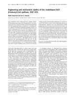
Figure 1.1 Molecular structures of artemisinin and its main
derivatives.
1.1.2 Pharmacological effects of ART
In addition to the anti-malaria function, ART has been found to possess
a wide spectrum of pharmacological activities, including anti-cancer
(Chaturvedi et al., 2010; Efferth, 2006), anti-viral (Efferth et al., 2008), anti-
inflammatory (Wang et al., 2007; Xu et al., 2007), anti-allergic and asthmatic
activities (Cheng et al., 2011). There are continuous efforts and increasing
interests in uncovering the underlying mechanisms of the above functions.
1.1.2.1 Anti-malaria
As shown in Figure 1.1, the basic structure of artemisinin and its
monomers including ART is a sesquiterpene lactone. All of them contain an
4
endoperoxide bridge which is believed to be essential for its anti-malarial
activity (Krishna et al., 2004). It is also widely accepted that cleavage of the
endoperoxide bridge of artemisinins by ferrous iron results in carbon-centered
free radicals production, which is essential for their anti-malarial activity
(Eckstein-Ludwig et al., 2003; Klonis et al., 2013). The underlying
mechanisms of the anti-malaria function of artemisinins have been extensively
studied, including: (i) inhibition the PfATP6 of Plasmodium falciparum in
Xenopus oocytes, the orthologue of mammalian SERCA (sarco/endoplasmic
reticulum Ca
2+
-ATPase) (Eckstein-Ludwig et al., 2003); (ii) alkylation of
cytosolic proteins such as translationally controlled tumor protein (PfTCTP)
(Meshnick, 2002); (iii) interference with the heme and alkylation of heme
(Cazelles et al., 2002), (iv) disruption of digestive vacuole membrane (del
Pilar Crespo et al., 2008). Currently, chemical modifications and the
development of multimeric artemisinin conjugates have led to the improved
efficacies as well as the decreased adverse effects of the drugs (Ho et al.,
2014).
1.1.2.2 Anti-inflammatory
In the early 1980s, a water soluble derivative of artemisinin,
hemisuccinate NA, was shown to possess immunosuppressive action against
mitogen-stimulated mouse spleen cells and human peripheral lymphocytes
(Shen et al., 1984). At present, there is increasing evidence to support the anti-
inflammation function of artemisinin and its derivatives, mainly in the
following models: rheumatoid arthritis, allergic anaphylaxis; systemic lupus
erythematosus and sepsis (Ho et al., 2014). Besides, ART has been shown to
5
possess anti-inflammatory effects in experimental colitis and Alzheimer's
disease model (Shi et al., 2013; Yang et al., 2012).
One of the most well studied mechanisms of the anti-inflammation
function of artemisinins is via inhibition of the nuclear factor kappa B (NF-κB)
signaling pathway (Li et al., 2006; Li et al., 2013). NF-κB has been shown to
play a key role in inflammatory and immune responses via the regulation of
genes encoding pro-inflammatory cytokines, chemokines, adhesion molecules
and etc (Tak and Firestein, 2001; Xu et al., 2007; Yamamoto and Gaynor,
2001). Therefore, inactivation of NF-κB leads to the repression of production
of key proinflammatory cytokines of such as TNF-α, IL-1, IL-6, IL-12,
reduction of the expression of enzymes such as nitric oxide synthase and
inhibition of the activation of immunocompetent cells (Lawrence et al., 2001).
It has been suggested that ART is capable of suppressing TNF-α induced
production of IL-1, IL-6 and IL-8 via inhibiting NF-κB signal pathway in
human rheumatoid arthritis fibroblast-like synoviocytes without affecting the
phosphorylation of mitogen-activated protein kinase (MAPK), extracellular
regulated kinases (ERK) and Jun N-terminal kinases (JNK) (Xu et al., 2007).
Moreover, a recent study showed that ART has the inhibitory effect on
collagen-induced arthritis through NF-κB and MAPK signaling pathway in
rats (Li et al., 2013).
The anti-inflammation effects of ART have also been shown to be
involved in the inhibition of the phosphoinositide 3- kinase (PI3K)-Akt
signaling pathway and activation of the NF-E2-related factor 2
(Nrf2)/antioxidant responsive element (ARE) pathway (Cheng et al., 2011; Ho
et al., 2012; Lee et al., 2012; Xu et al., 2007). Because of the key role of PI3K-
6
Akt signaling pathway in the activation and immune responses of eosinophils,
T and B lymphocytes, and mast cells, Cheng et al. investigated the anti-
inflammatory effect of ART in ovalbumin (OVA)-induced inflammatory mice
as well as in house dust mite induced mouse asthma model (Cheng et al.,
2011). They found that ART inhibited the OVA-induced phosphorylation of
Akt. They further made use of primary human bronchial epithelial cells and
found that EGF-induced PI3K-Akt activation is also inhibited by ART
treatment. Later on, the same group found that ART significantly enhanced
nuclear Nrf2 protein level in lung tissues from OVA-challenged mice and in
TNF-α-stimulated human bronchial epithelial cells (Ho et al., 2012). In
addition, the activation of Nrf2 pathway by ART treatment has been shown in
an ERK-dependent manner in microglial BV2 cells (Lee et al., 2012).
1.1.2.3 Anti-viral
The anti-viral functions of ART were mainly demonstrated in Herpes
viruses as well as Hepatitis B and C viruses (Efferth et al., 2008; Ho et al.,
2014). ART with a concentration of 15 μM inhibits more than 80% of the
DNA replication in human cytomegalovirus (HCMV) and herpes simplex
virus type 1 (HSV-1) in vitro (Efferth et al., 2002; Efferth et al., 2008).
Shapira et al. first reported that oral treatment with ART (100 mg/day)
reduced the viral load (1.7–2.1 log reduction) and improved hematopoiesis in
a 12-year-old patient within 10 days who suffered from late drug-resistant
HCMV infection after receiving haploidentical T cell–depleted hematopoietic
stem cells from his father (Shapira et al., 2008). It has been shown that co-
treatment with ferrous iron on HCMV-infected fibroblasts enhanced the
antiviral effect of ART, which is similar to the findings that ferrous iron is
7
required for the anti-malaria function (Kaptein et al., 2006). One of the
possible mechanisms underlying its anti-HCMV function is that ART inhibits
the PI3K pathway and thus diminishes the activation of NF-κB and Sp1,
which is linked to the replication of HCMV (Efferth et al., 2002).
The anti-hepatitis B virus (HBV) activity was first reported by Romero et
al. They found that ART was able to suppress HBV surface antigen (HBsAg)
secretion and reduce HBV-DNA levels in HepG2 2.2.15 cells (Romero et al.,
2005). The dose of ART was similar to its activity against HCMV, which is
less than 10 μM (Efferth et al., 2002). Although the effect is weaker than that
of lamivudine, a widely used inhibitor for chronic hepatitis B, a synergic
inhibitory effect in HBsAg release has been shown by the combined treatment
of ART and lamivudine (20 nM each) without inducing toxicity in host cells
(Romero et al., 2005). However, at present the underlying mechanisms of its
anti-HBV activity are not clear and need to be further investigated.
Artemisinin has been reported to inhibit HCV replication, with an EC50
(50% effective concentration) about 78 μM without affecting the proliferation
of Huh5-2 cells (Paeshuyse et al., 2006). Moreover, the anti-HCV activity of
artemisinin was enhanced 5-fold by combination with hemin, an iron donor,
without observing cytotoxic effect (Paeshuyse et al., 2006).
1.1.2.4 Anti-cancer
The anti-cancer effect of artemisinin was first described in 1993, in which
the researchers showed that the artemisinin as well as its derivatives such as
ART and dihydroartemisinin exhibits toxicity to Ehrlich ascites tumor (EAT)
cells (Woerdenbag et al., 1993). At present, there is extensive evidence
8
suggesting the anti-cancer function of artemisinins, especially ART and DHA
(Ho et al., 2014). Here we focus on the anti-cancer function of ART. Up to
date, the anti-cancer function of ART is mainly based on the following
observations: (i) induction of cell cycle arrest (Longxi et al., 2011; Zhao et al.,
2011), (ii) induction of cell death and sensitization to tumor necrosis factor-
related apoptosis-inducing ligand (TRAIL)-induced apoptosis (He et al., 2010;
Thanaketpaisarn et al., 2011), (iii) inhibition of angiogenesis (Chen et al.,
2004; Dell'Eva et al., 2004), (iv) reduction of cell invasion and metastasis
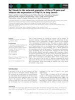
(Rasheed et al., 2010). These anti-cancer effects of ART are summarized in
Figure 1.2 as below. Moreover, the anti-cancer potential of ART has also been
tested in animal models. Currently there is one clinical trial ongoing using
ART in metastatic breast cancer
( and the results are still
pending.
Figure 1.2 Anti-cancer effects of ART.