Effects of andrographolide and 14 deoxy 11, 12 didehydroandrographolide in obstructive respiratory disease mouse models
Bạn đang xem bản rút gọn của tài liệu. Xem và tải ngay bản đầy đủ của tài liệu tại đây (2.78 MB, 217 trang )
i
EFFECTS OF ANDROGRAPHOLIDE AND 14-DEOXY-11,12-
DIDEHYDROANDROGRAPHOLIDE IN OBSTRUCTIVE
RESPIRATORY DISEASE MOUSE MODELS
GUAN SHOU PING
BSc (Hons)
A THESIS SUBMITTED FOR THE DEGREE OF DOCTOR OF
PHILOSOPHY
DEPARTMENT OF PHARMACOLOGY
NATIONAL UNIVERSITY OF SINGAPORE
2012
ii
DECLARATION
I hereby declare that this thesis is my original work and it has been written by me in its
entirety. I have duly acknowledged all the sources of information which have been used in the
thesis.
This thesis has also not been submitted for any degree in any university previously.
__________________________
GUAN SHOU PING
26 September 2012
iii
ACKNOWLEDGEMENTS
First and foremost, I would like to extend my most sincere appreciation to my supervisor
Professor Wong Wai-Shiu Fred for his guidance that is so instrumental in my Ph.D studies.
Without his encouragement and assistance, I would never have completed my studies and
overcome all the obstacles in this project.
I also like to thank Professor Shen Han Ming, Dr. Leung Pui Lam, Bernard and Dr Gautam
Sethi for their kind willingness to be my thesis advisory committee. Their invaluable advice
assisted on my research works.
I especially want to thank Mr. Koh Hock Meng, Alan who always being very resourceful all
this years.
I would also like to extend my gratitude for my senior Dr Bao Zhang and Dr Liao Wu Peng
Winston for their guidance during my early PhD studies.
I would also like to thanks Dr Cheng Chang, Ryan Wu Song Lian, Kong Li Ren, Chan Tze
Khee, Wilson Tee, Samantha Li, David Ng Shen Wen and all other colleague in the lab for
their industrious effort in the various studies that we have gone through together, shared
experience and supported me.
I would like to thank NUS for offering me a Scholarship that makes my study on this project
possible.
Finally, I would like to thank my parent and my wife for their endless love, support, and
patience all along.
Guan Shou Ping
iv
TABLE OF CONTENTS
DECLARATION ii
ACKNOWLEDGEMENTS iii
TABLE OF CONTENTS iv
SUMMARY viii
LIST OF TABLES x
LIST OF FIGURES xi
LIST OF ABBREVIATIONS xiv
LIST OF PUBLICATIONS AND CONFERENCE ABSTRACTS xviii
1. INTRODUCTION 1
1.1 Obstructive Lung Disease 2
1.2 Asthma 3
1.2.1 Epidemiology and Burden of Asthma 4
1.2.2 Pathophysiology of Asthma 5
1.2.2.1 Inflammatory and Structural Cells 8
1.2.2.2 Mediators of Asthma 22
1.2.2.3 Airway Hyperresponsiveness (AHR) 32
1.2.3 The mouse model of asthma 33
1.2.4 Current Treatment 34
1.3 Chronic obstructive pulmonary disease (COPD) 36
1.3.1 Epidemiology and Burden of COPD 38
1.3.2 Etiology 38
1.3.3 Inflammatory cells 39
1.3.4 Mediators of COPD 43
v
1.3.4.1 Chemokines 43
1.3.4.2 Cytokine 46
1.3.4.3 Proteases and antiproteases 49
1.3.4.4 Oxidant and antioxidant 51
1.3.5 Mouse Models of COPD 55
1.3.6 Current Drug 58
1.4 Andrographolide and DDAG 59
1.4.1 Andrographis paniculata 59
1.4.2 Andrographolide 60
1.4.3 14-deoxy-11,12-didehydroandrographolide 64
2. RATIONALE AND OBJECTIVES 67
3. MATERIAL AND METHODS 70
3.1. Materials and reagents 71
3.2. Mouse Model 74
3.2.1. Asthma mouse model and DDAG treatment protocol 75
3.2.2. Cigarette smoke-induced lung injury and andrographolide treatment
protocol 78
3.3. Collection of bronchoalveolar lavage (BAL) fluid from mice 78
3.4. Total and differential BAL fluid cell counts 79
3.5. Lung total protein extraction 82
3.6. ELISA 82
3.6.1. Cytokines and chemokine levels in BAL fluid 82
3.6.2. Oxidative damage marker level in BAL fluid 83
3.6.3. Immunoglobulin levels in serum 85
3.6.4. Cotinine Measurement 85
vi
3.7. Measurements of airway hyperresponsiveness (AHR) 86
3.8. Histology 87
3.9. Cell cultures 89
3.9.1. Cell viability assay 89
3.9.2. In Vitro Inflammation model 90
3.9.3. In vitro cigarette smoke exposure model 90
3.10. Nuclear Protein extraction 91
3.10.1. NF-κB and Nrf2 DNA-transactivation Assay 91
3.11. Immunoblotting 92
3.12. RNA extraction and Reverse Transcription 93
3.13. Polymerase Chain Reaction (PCR) 94
3.14. Biochemical Assay Antioxidant Activities 97
3.14.1. Antioxidant Activities in Lung Tissue 97
3.14.2. Glutathione Assay 98
3.15. Statistical analysis 98
4. ANTI-INFLAMMATORY EFFECTS OF 14-DEOXY-11,12-
DIDEHYDROANDROGRAPHOLIDE IN ALLERGIC ASTHMA MOUSE MODEL 99
4.1. Results 100
4.1.1. DDAG is less cytotoxic than andrographolide 100
4.1.2. DDAG reduces bronchoalveolar lavage fluid Th2 cytokines 104
4.1.3. DDAG reduces serum immunoglobulins 104
4.1.4. DDAG reduces lung inflammatory biomarkers 107
4.1.5. DDAG suppresses allergic airway inflammation 109
4.1.6. DDAG prevents lung mast cell degranulation 109
4.1.7. DDAG reduces AHR 115
vii
4.1.8. DDAG inhibits NF-κB activation 115
4.2. Discussion 120
5. ANTI-OXIDATIVE STRESS EFFECTS OF ANDROGRAPHOLIDE IN CIGARETTE
SMOKE INDUCE LUNG INJURY MOUSE MODEL 128
5.1. Results 129
5.1.1. Andrographolide attenuates cigarette smoke-induced lung
inflammation 129
5.1.2. Andrographolide attenuates cigarette smoke-induced inflammatory
cytokine and chemokine Level 133
5.1.3. Andrographolide attenuates cigarette smoke-induced inflammatory and
proteolytic mediators’ gene expression 133
5.1.4. Andrographolide protects against cigarette smoke-induced oxidative
lung damage 136
5.1.5. Andrographolide augments the GPx and GR activities 136
5.1.6. Andrographolide promotes nuclear Nrf2 accumulation 139
5.1.7. Andrographolide promotes GSH level 139
5.1.8. Andrographolide augments Nrf2-regulated gene targets 143
5.2. Discussion 146
6. CONCLUSIONS 155
7. REFERENCES 159
viii
SUMMARY
Chronic obstructive pulmonary disease (COPD) and asthma account for most
obstructive lung diseases that place a huge burden on health services and society. There are
currently limited therapeutic options for severe asthmatic and COPD patients.
Andrographolide and 14-deoxy-11,12-didehydroandrographolide (DDAG) are the main
biologically active constituents isolated from Andrographis paniculata. Andrographolide has
been shown to activate nuclear factor erythroid-2-related factor 2 (Nrf2). As Nrf2 activity is
reduced in COPD, we hypothesize that andrographolide may have therapeutic value for
COPD. Our group has also recently reported novel anti-inflammatory effects of
andrographolide in a mouse asthma model as well. However, andrographolide has been
shown to possess cytotoxic activity towards tumour cell lines. As DDAG is an analogue of
andrographolide, we hypothesized that DDAG retains the anti-inflammatory effects for
asthma but is devoid of cytotoxicity.
Contrary to andrographolide, DDAG did not elicit any cytotoxic activity in A549 and
BEAS-2B human lung epithelial cells and rat basophilic leukemia (RBL)-2H3 cells using a
MTS assay. BALB/c mice sensitized and challenged with ovalbumin (OVA)-developed
allergic airway inflammation. DDAG dose-dependently inhibited OVA-induced increases in
total cell counts and eosinophil counts, IL-4, IL-5, and IL-13 levels in lavage fluid and serum
OVA-specific IgE level in a mouse asthma model. In addition, DDAG attenuated OVA-
induced airway eosinophilia, mucus production, mast cell degranulation, pro-inflammatory
biomarker expression in lung tissues, and airway hyperresponsiveness (AHR) to methacholine
in mice. DDAG also blocked p65 nuclear translocation and DNA-binding activity in the
OVA-challenged lung and in TNF-α -stimulate d human lung epithelial cells.
Andrographolide suppressed cigarette smoke-induced increases in BAL fluid cell
counts, levels of IL-1β, MCP-1, IP-10 and KC, and levels of oxidative biomarkers 8-
ix
isoprostane, 8-OHdG and 3-nitrotyrosine in a dose-dependent manner. Andrographolide also
promoted inductions of glutathione peroxidase (GPx) and glutathione reductase (GR)
activities in lungs from cigarette smoke-exposed mice. In BEAS-2B cells, andrographolide
markedly increased nuclear Nrf2 accumulation, promoted binding to antioxidant response
element (ARE), and total cellular glutathione level in response to CSE. Andrographolide up-
regulated ARE-regulated gene targets including glutamate-cysteine ligase catalytic (GCLC)
subunit, GCL modifier (GCLM) subunit, GPx, GR and heme oxygenase-1 (HO-1) in BEAS-
2B cells in response to CSE.
Taken together, current study demonstrated that andrographolide possesses anti-
oxidative properties against cigarette smoke-induced lung injury probably via augmentation
of Nrf2 activity. DDAG, on the other hand, retains the anti-inflammatory activities of
andrographolide for asthma probably through the inhibition of NF-κB, and thus, DDAG may
be considered as a safer analogue of andrographolide for the potential treatment of asthma.
x
LIST OF TABLES
Table Title Page
Table 3.1. Primer sets for RT-PCR analysis 95
Table 3.2. Primer sets for real time-PCR analysis 96
xi
LIST OF FIGURES
Figure Title Page
Figure 1.1. Tissue sections from the airway of a non-asthmatic person 6
Figure 1.2. Tissue sections from the airway of a patient with severe asthma 7
Figure 1.3. Cytokines involved in asthma. 21
Figure 1.4. Cytokines involved in COPD 44
Figure 1.5. Andrographis paniculata. 61
Figure 1.6. Andrographolide 61
Figure 1.7. 14-deoxy-11,12-didehydroandrographolide (DDAG) 65
Figure 3.1. Allergen aerosol delivery system. 76
Figure 3.2. Cigarette smoke and Sham Air delivery system 77
Figure 3.3. Type of cells found in BAL fluid of mice 80
Figure 3.4. Type of cells found in BAL fluid of mice 81
Figure 3.5. The FinePointe™ system 86
Figure 4.1. Effects of andrographolide and DDAG on cell viability
of A549 cells at 24 and 48 h time intervals 101
Figure 4.2. Effects of andrographolide and DDAG on cell viability
of BEAS-2B cells at 24 and 48 h time intervals. 102
xii
Figure 4.3. Effects of andrographolide and DDAG on cell viability
of RBL-2H3 cells at 24 and 48 h time intervals. 103
Figure 4.4. Effects of DDAG on BAL fluid Th2 cytokines. 105
Figure 4.5. Effects of DDAG on serum immunoglobulins. 106
Figure 4.6. Effects of DDAG on lung inflammatory biomarkers. 108
Figure 4.7. Inflammatory cell counts in BAL fluid obtained from
sensitized mice 110
Figure 4.8. Effects of DDAG on OVA-induced inflammatory cell
recruitment. 111
Figure 4.9. Effects of DDAG on OVA-induced inflammatory cell
recruitment. 112
Figure 4.10. Effects of DDAG on OVA-induced mucus hypersecretion 113
Figure 4.11. Effects of DDAG on OVA-induced lung mast cell
degranulation. 114
Figure 4.12. Effects DDAG on OVA-induced AHR. 116
Figure 4.13. Effects of DDAG on OVA-induced AHR. 117
Figure 4.14. Effects of DDAG on NF-κB activity in OVA-challenged lungs. 118
Figure 4.15. Effects of DDAG on NF-κB activity in in TNF-α-
stimulated A549 human lung epithelial cells. 119
Figure 5.1. Effects of cigarette smoke in mice. 130
xiii
Figure 5.2. Effects of cigarette smoke-induced inflammatory cell recruitment. 131
Figure 5.3. Effects of andrographolide on cigarette smoke-induced
inflammatory cell recruitment. 132
Figure 5.4. Effects of andrographolide on cigarette smoke BAL fluid
cytokine and chemokine levels. 134
Figure 5.5. Effects of andrographolide on cigarette smoke-induced lung
tissue pro-inflammatory and proteolytic mediator gene expression in mice. 135
Figure 5.6. Effects of andrographolide on cigarette smoke-induced BAL
fluid oxidative damage marker levels. 137
Figure 5.7. Effects of andrographolide on cigarette smoke-induced lung
antioxidant enzymatic activities. 138
Figure 5.8. Effects of andrographolide on nuclear Nrf2 level. 140
Figure 5.9. Effects of andrographolide on nuclear Nrf2 level. 141
Figure 5.10. Effects of andrographolide on cellular GSH levels. 142
Figure 5.11. Effects of andrographolide on antioxidant gene expression. 144
Figure 5.12. Effects of andrographolide on antioxidant gene expression. 145
xiv
LIST OF ABBREVIATIONS
3-NT 3-nitrotyrosine
8-OHdG
8-hydroxy-2-deoxyguanosine
α
1
-AT
α1-Antitrypsin
AERD
Aspirin-exacerbated respiratory disease
AHR Airway hyperresponsiveness
AM
Alveolar macrophages
AMCase Acidic mamalian chitinase
AMV
Avian myeloblastosis virus
ANOVA
Analysis of variance
AP Alkaline phosphatase
APC
Antigen presenting cell
ARE Antioxidant response elements
ASM
Airway smooth muscle
BAL
Bronchoalveolar lavage
BALF Bronchoalveolar lavage fluid
BCA
Bicinchonic acid
bFGF Basic fibroblast growth factor
BHC
Hexachlorocyclohexane
BSA
Bovine serum albumin
c/EBP CCAAT-enhancer-binding proteins
C5a
Complement component 5a
CAT Catalase
CCL3
Macrophage inflammatory protein-1α
CCR
C-C chemokine receptor
CD 4 Cluster of differentiation 4
CD 13
Cluster of differentiation 13 or Alanine aminopeptidase
CD 117 Cluster of differentiation 117 or Proto-oncogene c-Kit
CDDO
2-cyano-3, 12-dioxooleana-1, 9-dien-28-oic acid
Cdyn
Dynamic compliance
COPD Chronic obstructive pulmonary disease
COX-2
Cyclooxygenase-2/Prostaglandin-endoperoxide synthase 2
CRG-2 Cytokine Responsive Gene-2
CSE
Cigarette smoke extract
CSF
Colony-stimulating factor
CXCL8 Interleukin 8
CXCL10
Interferon-γ-inducible protein 10
CysLTs Cysteinyl leukotrienes
DAMP
Damage-associated molecular patterns
DC
Dendritic Cell
DDAG 14-deoxy-11,12-didehydroandrographolide
DEPC
Diethylpyrocarbonate
DMSO Dimethyl sulfoxide
xv
DTNB
5,5'-dithio-bis-2-nitrobenzoic acid
ECL Enhanced chemiluminescent
ECM
Extracellular matrix
ECP
Eosinophil cationic protein
ecSOD Extracellular superoxide dismutase
EDTA
Ethylenediaminetetraacetic acid
EPO Eosinophil peroxidase
FBS
Fetal bovine serum
FcεRI
high-affinity IgE receptor
FEV
1
Forced expiratory volume in 1 second
FIZZ1 Found in inflammatory zone-1
FOXP3
Forkhead box P3
FVC Forced vital capacity
GATA
GATA-binding protein (globin transcription factor)
GCLC
Glutamate-cysteine ligase, catalytic subunit
GCLM Glutamate-cysteine ligase, modifier subunit
GCP-2
Granulocyte chemotactic protein 2
G-CSF Granulocyte colony-stimulating factor
GM-CSF
Granulocyte macrophage colony-stimulating factor
GPE1
GST-P enhancer-1
GPx Glutathione peroxidise
GR
Glutathione reductase
GRO-α Growth-related oncogene-α
GSH
Glutathione
GST
Glutathione S transferase
H&E Haematoxylin and Eosin
HO-1
Heme oxygenase-1
HRP Horseradish peroxidase
i.p.
Intraperitoneal injection
ICAM-1
Intercellular adhesion molecule 1
IFN-γ Interferon-γ
IgE
Immunoglobulin E
IgG1 Immunoglobulin G1
IgG2a
Immunoglobulin G2a
IKK β
Inhibitory κB kinase-β
IL
Interleukin
IL1RL1
Interleukin 1 receptor-like 1
IM Interstitial macrophages
iNOS
Inducible nitric oxide synthase
IP-10
Interferon-γ-inducible protein 10
iTreg Inducible T regulatroy lymphocyte
KC
Keratinocyte-derived chemokine
KEAP1 Kelch-like ECH-associated protein 1
LPS
Lipopolysaccharide
xvi
LTB
4
Leukotriene B
4
LTC
4
Leukotriene C
4
Lyn
Tyrosine-protein kinase Lyn
M1 Classical activated macrophages
M2
Alternatively activated macrophages
mAb
Monoclonal antibody
MBP Major basic protein
MCP-1
Monocyte chemotactic protein-1
MIP-1α Macrophage inflammatory protein 1α
MMP
Matrix metalloproteinase
MPO
Myeloperoxidase
MTS
(3-(4,5-dimethylthiazol-2-yl)-5-(3-carboxymethoxyphenyl)-2-(4-sulfophenyl)-2H-
tetrazolium)
MyD88
Myeloid differentiation primary response gene (88)
NAC n-acetylcysteine
NADP
Nicotinamide adenine dinucleotide phosphate
NADPH
Nicotinamide adenine dinucleotide phosphate
NE Neutrophil elastase
NFAT
Nuclear factor of activated T-cells
NF-HEV Nuclear factor from high endothelial venules
NF-
κ
B
Nuclear factor kappa B
NGF
Nerve growth factor
NHBE Normal human bronchial epithelial cells
NLR
NOD-like receptor
NO Nitric oxide
Nrf2
Nuclear erythroid-2-related factor 2
nTreg
Natural T regulatory lymphocyte
OVA Ovalbumin
PAF
Platelet-activating factor
PAMP Pathogen-associated molecular patterns
PAR
Protease-activated receptors
PAS
Peroidic acid Schiff
PBMC Peripheral blood mononuclear cells
PBS
Phosphate buffered saline
PCR Polymerase chain reaction
PE
Pinacyanol Erythrosinate
PGD
2
Prostaglandin D
2
PGE
2
Prostaglandin E
2
PGP
N-acetyl proline-glycine-proline
PMA Phorbol 12-myristate 13-acetate
PRR
Pattern recognition receptors
PVDF Polyvinylidene difluoride
RANTES
Regulated upon Activation, Normal T-cell Expressed, and Secreted
RBL
Rat basophilic leukemia
xvii
rcf
Relative Centrifugal Force
Rl lung resistance
RNS
Reactive nitrogen species
RORγt
Receptor-related orphan receptor-γt
ROS Reactive oxygen species
rpm
Revolutions per minute
SCF Stem-cell factor
SDS
Sodium dodecyl sulfate
SEM
Standard error of the mean
SNP Single-nucleotide polymorphisms
SOD
Superoxide dismutase
STAT6 Signal transducer and activator of transcription 6
Syk
Spleen tyrosine kinase
TAE
Tris-acetate-EDTA
TARC Thymus and activation regulated chemokine
T-BET
T cell–specific T-box transcription factor
TBP TATA binding protein
Tc
Cytotoxic T lymphocyte
TCR
T Cell Receptor
TEMED Tetramethylethylenediamine
TGF-β
Tansforming growth factor-β
Th T helper lymphocyte
TIMP
Tissue inhibitor of metalloproteinases
TLR
Toll-like receptor
TMB 3,3´,5,5´-tetramethylbenzidine
TNB
5-thios-2-nitrobenzoic acid
TNF-α Tumour necrosis factor-α
Treg
Regulatory T lymphocytes
VCAM-1
Vascular cell adhesion molecule 1
VEGF Vascular endothelial growth factor
VLA-4
Very late antigen 4
xviii
LIST OF PUBLICATIONS AND CONFERENCE ABSTRACTS
Publication
Guan S, Tee W, Ng D, Chan T, Peh H, Ho W, et al. (2013). Andrographolide protects against
cigarette smoke-induced oxidative lung injury via augmentation of Nrf2 activity. Br J
Pharmacol 168(7): 1707-1718.
Guan SP, Mok YK, Koo KN, Chu KL, Wong WS (2009). Chitinases: biomarkers for human
diseases. Protein and peptide letters 16(5): 490-498.
Bao Z, Guan SP, Cheng C, Wu S, Wong SH, Kemeny DM, et al. (2009). A novel
antiinflammatory role for andrographolide in asthma via inhibition of the nuclear factor-
kappaB pathway. Am J Respir Crit Care Med 179(8): 657-665.
Guan SP, Kong LR, Cheng C, Lim JC, Wong WS (2011). Protective role of 14-deoxy-11,12-
didehydroandrographolide, a noncytotoxic analogue of andrographolide, in allergic airway
inflammation. Journal of natural products 74(6): 1484-1490.
Cheng C, Ho WE, Goh FY, Guan SP, Kong LR, Lai WQ, et al. (2011). Anti-malarial drug
artesunate attenuates experimental allergic asthma via inhibition of the phosphoinositide 3-
kinase/Akt pathway. PloS one 6(6): e20932.
Ng FS, Wong KY, Guan SP, Mustafa FB, Kajiji TS, Bist P, et al. (2011). Annexin-1-deficient
mice exhibit spontaneous airway hyperresponsiveness and exacerbated allergen-specific
antibody responses in a mouse model of asthma. Clin Exp Allergy 41(12): 1793-1803.
xix
Goh FY, Upton N, Guan SP, Cheng C, Shanmugam MK, Sethi G, et al. (2012). Fisetin, a
bioactive flavonol, attenuates allergic airway inflammation through negative regulation of
NF-kappaB. Eur J Pharmacol 679(1-3): 109-116.
Tang Y, Guan SP, Chua BY, Zhou Q, Ho AW, Wong KH, et al. (2012). Antigen-specific
effector CD8 T cells regulate allergic responses via IFN-gamma and dendritic cell function. J
Allergy Clin Immunol 129(6): 1611-1620 e1614.
Conference Abstract
Guan SP, Kong LR, Cheng C, Wong WS. (2010). An Anti-Inflammatory Role For 14-Deoxy-
11,12-Didehydroandrographolide In Asthma. Am. J. Respir. Crit. Care Med; 181: A5673.
(“2010 ATS International Conference”, May 14-19, 2010, New Orleans, Louisiana, USA)
Guan SP, Tee W, DSW Ng, TK Chan, HY Peh, EWX Ho, C Cheng, JC Mak and WSF
Wong. (2012) Andrographolide Attenuates Cigarette Smoke Induce Lung Inflammation and
Oxidative Stress By Augmenting Nrf2 and Attenuating NF-κB Response. (“Yong Loo Lin
School of Medicine Graduate Scientific Congress 2nd Annual Graduate Scientific Congress”,
15 Feb 2012, NUHS Tower Block, Main Auditorium, Singapore)
1
1. INTRODUCTION
2
1.1 Obstructive Lung Disease
Respiratory disease is a common and important cause of illness and death around the
world. Respiratory disease can be segregated into two major categories, which include
obstructive lung disease and restrictive lung disorders. Obstructive lung diseases which are
characterized by airway obstruction while restrictive lung disease is characterized by reduced
lung volume due to restricted lung expansion. Obstructive lung diseases are far more common
than restrictive diseases in a general population (Culver, 2011). To assess the pathophysiology
of obstructive lung disease, spirometrical measurement which provides the initial lung
function is commonly used. The main diagnostic criteria for a lung diseases lie in a
diminished forced expiratory volume in 1 second (FEV
1
)/ the total volume of air exhaled
during a forced manoeuvre, the forced vital capacity (FVC) (Macintyre, 2009).
A person is said to have airway obstruction when the ratio of FEV
1
/FVC is less than 70%
(Seemungal et al., 2008). Diseases resulting in obstructive pathophysiology primarily include
asthma, chronic obstructive pulmonary disease (COPD), bronchiectasis, and bronchiolitis
(Ryu et al., 2001). Although a broad spectrum of disorders is associated with airflow
obstruction, COPD and asthma account for most obstructive lung disease (Ryu et al., 2001).
Both diseases are characterized by airway obstruction, which is variable and reversible in
asthma but is progressive and largely irreversible in COPD (Barnes, 2008c). As the incidence
of asthma and COPD is increasing globally, both diseases are placing an increasing burden on
health services in industrialized and developing countries (Mannino et al., 2007; Pearce et al.,
2007). In asthma and COPD, there is chronic inflammation of the respiratory tract, and when
the intensity of the inflammation increases, there are acute exacerbations in both diseases as
well (Barnes, 2008c).
Nonetheless, there are marked differences in inflammation pattern in asthma and COPD
that occur within the respiratory tract. The differences are linked to the recruitment of
3
inflammatory cell types and production of mediators that underlie both of the diseases. Hence,
asthma and COPD also have different consequence to inflammation and respond differently to
therapy (Ichinose, 2009). There have been several recent important advances in our
understanding of the immunopathology of asthma and COPD (Murphy et al., 2010; Yao et al.,
2009). The understanding of the similar and different immune mechanisms that are involved
in both asthma and COPD has important implication for the development of new therapies for
these troublesome diseases.
1.2 Asthma
Asthma is a chronic respiratory disease characterized by episodic attacks of impaired
breathing (Akinbami et al., 2011). According to Global Initiative for Asthma’s (GINA)
guidelines, asthma is defined as a chronic inflammatory disorder of the respiratory airways in
which various cells and cellular elements play a role. The chronic inflammation is associated
with AHR that leads to recurrent episodes of shortness of breath, coughing, wheezing, and
chest tightness, particularly at night or in the early morning. These episodes are usually
associated with widespread, but variable, airflow obstruction within the lung that is often
reversible either spontaneously or with treatment (GINA, 2011). Asthma comprises a range of
heterogeneous phenotypes that differ in presentation, aetiology and pathophysiology. Allergic
(extrinsic) and non-allergic (intrinsic) asthma are 2 well characterized asthma phenotypes.
People with allergic asthma develop the disease early in life. They are atopic (producing IgE
specific to identifiable allergens) and have identifiable allergic triggers, and other allergic
diseases such as rhinitis or eczema or a family history of allergic diseases. Non-allergic
asthma develops later in life (after 40 years of age) and it is associated with aspirin-
exacerbated respiratory disease (AERD) but not with allergic sensitization. Hence, non-
allergic asthma is generally not as well understood. Other types of asthma phenotype include
4
exercise-induced asthma, obesity-related asthma and neutrophilic asthma. Nonetheless, in
patients with asthma of any severity, up to 50% of them have a T-helper Type 2 (Th2)-
predominant phenotype. Asthma that presents without evidence of Th2 immunity remains
poorly understood (Wenzel, 2012). Although several types of asthma have been classified
clinically, allergic asthma is the most common form of the disease (Mukherjee et al., 2011).
Allergic asthma is a chronic respiratory condition characterized by airway inflammation,
mucus hypersecretion, and AHR (Galli et al., 2008b). Increased mucus secretion and airway
hyper-responsiveness leads to recurrent wheezing and shortness of breath (Moreira et al.,
2011).
1.2.1 Epidemiology and Burden of Asthma
Asthma is a widespread disease. There are 300 million people affected by asthma
worldwide (Masoli et al., 2004). Nearly 30 million people in the United State alone are
affected by asthma, accounting for about 12% of children and 9% of adults in the US (Juhn,
2012). In Singapore, the prevalence of asthma has risen over the last 40 years but now appears
to have stabilized, with approximately 5.5% of children in 1969 to 20% of children in 1994
affected by the disease (Wang et al., 2004). Another recent survey estimated that almost 9%
of school attending children have asthma and about quarter of them is inadequately controlled
(Yang et al., 2007). In the United States, 10.5 million school days were reported to be missed
by asthmatic children aged 5-17 years in the year 2008 alone. Each day, an average of 9
persons in the United States dies from asthma. In addition, 2 out of 100 person with asthma
are hospitalize and 8.4 out of 100 person with asthma have emergency department visit in
2009 (Akinbami et al., 2012). The morbidity due to asthma, direct health care costs, indirect
costs such as lost productivity, and mortality due to asthma continue to pose a high burden to
the United States economy (Akinbami et al., 2011).
5
1.2.2 Pathophysiology of Asthma
Asthma is a chronic inflammatory disease accompanied by remodelling of the airways
and AHR that result in the clinical expression of airway obstruction that usually is reversible.
The symptoms and physiological changes in asthma often involved airway narrowing. Several
factors contribute to the development of airway narrowing in asthma. Airway smooth muscle
(ASM) contraction responding to multiple bronchoconstrictor mediators and neurotransmitter
is the predominant mechanism of airway narrowing and is largely reserved by brochodilators.
During acute exacerbations, airway oedema, which is due to increased microvascular leakage
in response to inflammatory mediators, also play a role in airway narrowing. Structural
changes, often termed "remodelling", lead to airway thickening that may play an important
role in more severe disease and is not fully reversible by current therapy. Mucus
hypersecretion, which is a product of increased mucus secretion and inflammatory exudates,
may lead to luminal occlusion that is also known as mucus plugging (GINA, 2011).
The structural changes are manifested by changes that involve all the layer of the
respiratory airway wall. Changes in the epithelium include an increased number of goblet
cells and epithelial detachment. Subepithelial fibrosis results from the development of
increased deposition of extracellular-matrix molecules in the lamina reticularis which lies
beneath the epithelial basement membrane and changes in fibroblasts with increased
development of myofibroblasts and increased vascularity. Blood vessels in the airway walls
proliferate under the influence of growth factors such as vascular endothelial growth factor
(VEGF) and may contribute to increased airway wall thickness. The increased size
(hypertrophy), number (hyperplasia) and function of ASM cells contribute to the increased
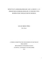
thickness of the muscular layer of the airways wall (Figs 1.1 and 1.2) (Galli et al., 2008b).
6
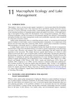
Figure 1.1. Tissue sections from the airway of a non-asthmatic person (a–c). A normal
small bronchus stained with haematoxylin and eosin (H&E) in a. There are few goblet cells
(black arrows in insets) in the epithelium. The basement membrane and underlying lamina
reticularis (at asterisk in a) are normal. Sections in b were stained with periodic acid–Schiff
(PAS) and diastase to stain mucus red. The submucosa (the length of the double-headed
arrows in a) contains few leukocytes and the occasional mast cell (blue arrows in c), and the
bronchial smooth muscle (SM) has few adjacent mast cells (red arrow in c) visualize by
staining with pinacyanol erythrosinate (PE) which stain mast cells purple. Adapted from Galli
(Galli et al., 2008b)