In vitro hair follicle engineering
Bạn đang xem bản rút gọn của tài liệu. Xem và tải ngay bản đầy đủ của tài liệu tại đây (5.38 MB, 137 trang )
IN VITRO HAIR FOLLICLE
ENGINEERING
PAN JING
NATIONAL UNIVERSITY OF SINGAPORE
2014
IN VITRO HAIR FOLLICLE
ENGINEERING
PAN JING
(B. Sc., China Pharmaceutical University)
A THESIS SUBMITTED FOR
THE DEGREE OF DOCTOR OF PHILOSOPHY
DEPARTMENT OF PHARMACY
NATIONAL UNIVERSITY OF SINGAPORE
2014
DECLARATION
I hereby declare that this thesis is my original
work and it has been written by me in its entirety.
I have duly acknowledges all the sources of
information which have been used in the thesis.
This thesis has also not been submitted for any
degree in any university previously.
Pan Jing
24 January 2014
I
ACKNOWLEDGEMENTS
I would like to thank and acknowledge many people for their contributions to
this thesis.
First of all, I am very grateful to my supervisor Dr Kang Lifeng. Thank you
for your encouragement, enthusiasm, positive attitude, staunch support and
guidance for my project which otherwise would not have accomplished.
I would also like to express my thanks to my co-supervisor A/P Chan Sui Yung
for her valuable suggestions and being always there for me. She has impressed
me with her ability to communicate optimism which has helped me grow both
personally and professionally.
I am grateful to the Department of Pharmacy at NUS, for providing me
scholarship and this wonderful opportunity. I thank other faculty members
who advised me and gave their insights at some point or the other.
I thank present and past members of the lab, Li Hairui, Dr Jaspreet Singh
Kochhar, Dr Li Fang, Yan Jun and Sara Dana, with whom I spent numerous
fun-filled hours at the lab.
I would like to thank the Final Year Project students Wong Xin Yi Cheryl,
Liew Xin Yi Cindy, Kuek Qi Min and Undergraduate Research Opportunities
Program student Hiew Tze Ning for the time spent together in research.
II
I also want to thank the lab-support and administrative staff of our Pharmily,
my research wouldn’t progress if not for your timely assistance.
Lastly, and most importantly, I am deeply thankful to my wonderful parents
for their love, support, and sacrifices. Without them, this thesis would never
have been written.
III
CONTENTS
ACKNOWLEDGEMENTS I
CONTENTS III
SUMMARY VI
LIST OF PUBLICATIONS IX
LIST OF TABLES X
LIST OF FIGURES XI
LIST OF ABBREVIATIONS XIX
Chapter 1 Background 1
1.1. Introduction 1
1.1.1. Hair follicle morphogenesis 2
1.1.2. Hair follicle generation from dissociated cells 3
1.1.3. Optimizing positional relationship and cell compartmentalization to
enhance EMIs 5
1.2. Literature Review 7
1.2.1. Human hair biology, pathophysiology and treatment 7
1.2.1.1. Hair size, shape and pigment 7
1.2.1.2 Human hair cycle 11
1.2.2. Androgenetic alopecia 15
1.2.2.1 Pathophysiology 15
1.2.2.2. Treatment 16
1.2.3. 3D microstructure fabrication in tissue engineering 18
1.3. Objectives and scope 21
Chapter 2 Poly (ethylene glycol) diacrylate (PEGDA) based 3D microstructural
hydrogel as potential substrate for hair follicle cells 25
2.1. Materials and Methods 26
2.1.1 Master fabrication 26
2.1.2. Polydimethylsiloxane (PDMS)-stamp fabrication 27
2.1.3. Microwell fabrication 28
2.1.4. Microwell stability 28
2.1.6. Mechanical testing 29
2.1.7. Cell culture 29
2.1.8. Toxicity exclusion tests 29
2.1.9. HaCaT cell seeding into microwells 31
2.1.10. Field emission scanning electron microscope (FE-SEM) study 31
2.1.11. Encapsulation of HDF cells in PEGDA hydrogel 32
2.1.12. Statistics 32
2.2. Results 33
2.2.1. Microwell Fabrication 33
2.2.2 Microwell stability 36
2.2.4. Mechanical testing 38
2.2.5. Toxicity exclusion tests 40
2.2.5.1. Investigating effects of PEGDA solution to HDF viability 40
IV
2.2.5.2. Effects of UV light exposure to HDF viability 41
2.2.5.3. Effects of photoinitiator (HHEMP) to HDF viability 42
2.2.5.4. Effects of combination of UV exposure and photoinitiator
(HHEMP) on HDF viability 43
2.2.6. Cell compatibility 45
2.2.7. HaCaT cell seeding into microwells 48
2.2.8. Cell growth (HaCaT) in the microwells 49
2.2.9. Cell growth (HDF) in the microstructured hydrogels 52
2.3. Discussion 54
2.3.1. Microwell fabrication 54
2.3.2. Microwell stability 56
2.3.4. HaCaT cell seeding into microwells 56
2.3.3. HDF cell encapsulation 57
2.4. Summary 58
Chapter 3 Hyaluronic acid based 3D microstructural hydrogel as potential substrate
for hair follicle cells 59
3.1. Materials and Methods 60
3.1.1. MeHA synthesis 60
3.1.2. Proton nuclear magnetic resonance (
1
H-NMR) spectroscopy 60
3.1.3. Hydrogel preparation 60
3.1.4. Hydrogel morphology by scanning electron microscopy 61
3.1.5. Contact angle measurement 61
3.1.6. Rheological study 61
3.1.7. Cell culture 62
3.1.8. HaCaT cell seeding 62
3.1.9. HDF cell encapsulation 63
3.1.10. Statistics 63
3.2. Results 64
3.2.1.
1
H-NMR characterization of MeHA 64
3.2.2. FE-SEM study 65
3.2.3. Contact angle measurement 65
3.2.4. Rheological properties 66
3.2.5. HaCaT cell seeding 67
3.2.6. HDF cell encapsulation 69
3.3. Discussion 71
3.3.1. Degree of methacrylation (DM) 71
3.3.2. Contact angle measurement 71
3.3.3. Rheology properties of MeHA hydrogels 72
3.3.4. HaCaT cell seeding 72
3.3.5. HDF cell encapsulation 73
3.4. Summary 74
Chapter 4 Tissue culture 76
4.1. Materials and Methods 76
4.1.1. HDF-HaCaT co-culture 76
V
4.1.2. Cell monitoring in 3D microenvironment 77
4.1.3. Immunofluorescence 77
4.1.4. Histology study 78
4.1.5. Real-time polymerase chain reaction (PCR) 79
4.1.6. Statistics 80
4.2. Results 80
4.2.1. HDF-HaCaT co-culture 80
4.2.2. Cell distributions in the microstructures 84
4.2.3. Cell proliferation and differentiation in the microenvironment 86
4.2.4. Multiple hair follicle specific genes expressing in microwell system . 89
4.3. Discussion 93
4.3.1. HDF-HaCaT co-culture 93
4.3.2. Histology study 95
4.3.3. Gene expression 95
4.4. Summary 98
Chapter 5 Conclusion 99
Chapter 6 Future study 101
REFERENCES 103
VI
SUMMARY
Hair is a complex mini-organ that is important for the integrity of skin. While
hair loss is usually not life threatening, it has a substantial psychosocial impact
on the sufferers and can severely undermine the confidence of affected
individuals and degrade their quality of life. As such, regenerating hair is of
great clinical interest.
Clinicians have resorted to transplanting hair follicles either from the patients’
own peripheral hair-bearing regions or from donor skin to bald regions.
Because of the inability to generate hair follicles de novo, there is a shortage
of human hair follicles for surgical transplantation. One potential solution is to
use tissue engineering approaches to generate large quantities of human hair
follicles in vitro to meet the clinical needs. From previously reported studies,
hair follicle-like structures can be reconstituted by combining and
transplanting mouse or rate epidermal and dermal papilla (DP) cells in
non-hair bearing skin of animal models. However, hair follicle-like structures
cannot be regenerated by using dissociated human cells, which may be due to
the difficulties in re-establishing the cellular interactions during hair follicle
development in vivo.
In this thesis, we aim to design and explore a 3D microstructure resembling
the architecture of the human hair follicles. Microwells with center islets were
fabricated by using a patterned polydimethylsiloxane (PDMS) stamp on a
glass substrate. Within the hydrogel microstructure, hair follicle inductive
dermal cells can be immobilized to grow close to, but separated from
epidermal cells. Poly (ethylene glycol) diacrylate (PEGDA) and hyaluronic
acid (HA) were both considered as the candidate materials of the
microstructure.
VII
PEGDA is a synthetic polymer, which has been commonly used in tissue
engineering due to its high hydrophilicity, photocrosslinkability and low
toxicity. Prior to encapsulating cells in PEGDA hydrogels, cytotoxicity of
various factors contributing to the photocrosslinking process, including
monomer solution, photoinitiator solution and ultraviolet (UV) intensity, were
tested. PEGDA with higher molecular weight was shown to be less toxic to
cells. Hydrogel stability was found to be inversely correlated with PEGDA
concentrations, i.e., microstructures made of higher PEGDA concentrations
were easier to detach from the underlying glass slide when immersed in PBS
over time. It was also shown that epithelial and dermal cells were accurately
compartmentalized within microstructures. Moreover, polymeric
microstructures were shown to support the cell growth over 14 days.
The natural polymer, hyaluronic acid (HA), was also studied as an alternative
material of the microstructural hydrogel because of its biocompatible and
biodegradable nature. HA was grafted with methacrylate groups to be
photocrosslinkable. It was found that the hydrophilicity of methacrylated HA
(MeHA) hydrogels decreased with increasing macromer concentration while
the stiffness of MeHA hydrogels increased with increasing the macromer
concentrations. These results are consistent with other reports. Also, the results
of field emission scan electron microscopy (FE-SEM) study showed that high
macromer concentration hydrogels possess smaller and more compact porous
structure. Similar to PEGDA hydrogels, MeHA hydrogels were also shown to
sustain cell survival and growth over time. However, gel swelling and weak
stability hindered the use of MeHA hydrogels in long-term study. Thus,
PEGDA was used as the material of microstructures for the study of cell-cell
co-culture, cell proliferation and differentiation, and gene expression.
Epithelial and dermal cells were co-cultured within PEGDA based
VIII
microstructures. Confocal images showed that dermal cells were distributed
homogeneously in the PEGDA hydrogel, while epithelial cells were
concentrated inside the microwells. Over time, the epithelial cells formed
cap-shaped aggregates enclosing center islets. The 3D microstructures were
also shown to maintain cell proliferation and may help to organize cell
differentiation. Furthermore, gene expression studies showed up-regulation of
genes relevant to epithelial-mesenchymal interactions in the native hair follicle.
Thus, the 3D hydrogel microstructure may serve as a suitable model for cell
compartmentalization in studying hair follicle interactions in vitro, with the
possibility to be further explored for human hair follicle engineering.
IX
LIST OF PUBLICATIONS
Journal
1. Pan J, Chan SY, Common JE, Amini S, Miserez A, Lane EB and Kang L.
Fabrication of a 3D hair follicle-like hydrogel by soft lithography.
Journal of Biomedical Materials Research Part A, 2013; 101 (11):
3159-69.
2. Kochhar JS, Li WX S, Zou S, Foo WY, Pan J, Kang L. Microneedle
integrated transdermal patch for fast onset and sustained delivery of
lidocaine in acute and chronic analgesic applications. Molecular
Pharmaceutics, 2013; 10 (11), 4272–80.
3. Pan J, Chan SY, Lee WG, and Kang L. Microfabricated particulate
drug-delivery systems. Biotechnology Journal, 2011; 6 (12): 1477-87.
4. Li H, Kochhar JS, Pan J, Chan SY and Kang L. Nano/Microscale
technologies for drug delivery. Journal of Mechanics in Medicine and
Biology, 2011; 11 (2): 337-67.
Presentation
1. IEEE Grand Challenges in Life Sciences Conference, Singapore,
December 2013.
2. 2013 AAPS Annual Meeting and Exposition, San Antonio, USA,
November 2013.
3. 3
rd
COSM'innov program, Orléans, France, October 2013.
4. 7th AAPS-NUS PharmSci@Asia Symposium, Singapore, June 2012.
5. 38th Annual Meeting and Exposition of the Controlled Release Society,
Maryland, USA, July 2011.
X
LIST OF TABLES
Table
Title
Page
Table 1
Oligonucleotide sequences of primers used in
Real-time PCR
80
XI
LIST OF FIGURES
Figure
Title
Page
Figure 1
Structure of hair follicle bulb. (Reproduced from
Dr. Radivoj V. Krstić. Chapter H. In: Dr. Radivoj V.
Krstić, editor. Illustrated Encyclopedia of Human
Histology: Springer Berlin Heidelberg; 1984. p 184.
With kind permission of Springer Science and
Business Media)
40
.
8
Figure 2
Human hair cycle. A: During early anagen, the
entire hair follicle still resides in the dermis, with
HG cells proliferating to enclose the DP cells (a)
and (b)
74,75
. B-C: The DP descends from the dermis
to the middle of the subcutaneous tissue, to finally
the base of the subcutaneous tissue. D-F: The hair
follicle enters catagen, where massive apoptosis
occurs. Hair elongation ceases. This causes the hair
follicle to shrink, and during apoptosis, the DP is
being pulled upwards, and it ascends to just below
the dermis. G: During telogen, the hair follicle
enters the quiescent or resting phase, where the
entire hair follicle resides in the dermis.
13&14
Figure 3
A: Schematic diagram for the formation of
cell-laden microgels using stop-flow lithography. A
prepolymer solution containing cells is flowed
through a microchannel and polymerized by UV
light through a photomask and a microscope
objective. (Reproduced from Ref 100 with
permission of The Royal Society of Chemistry)
100
.
B: A PDMS microfabricated tissue engineering
scaffold with the vasculature directly embedded
into the scaffold. (Reproduced from Biomedical
Microdevices, Vol 4, 2002, pp 167-175,
Microfabrication Technology for Vascularized
Tissue Engineering, Jeffrey T. Borenstein, H. Terai,
Kevin R. King, E.J. Weinberg, M.R.
Kaazempur-Mofrad, J.P. Vacanti, Figure 6, with
kind permission from Springer Science and
Business Media)
98
. C: Cells were printed according
20
XII
to five parallel lines of varying scanning speed
(from top to bottom). (a) Phase contrast microscope
image of cells printed onto glass. (b). Fluorescence
microscope image of cells printed onto a 100 μm
thick layer of Matrigel. (Reproduced from
Biomaterials, Vol 31, Guillotin B, Souquet A,
Catros S, Duocastella M, Pippenger B, Bellance S,
Bareille R, Rémy M, Bordenave L, Amédée J,
Guillemot F, Laser assisted bioprinting of
engineered tissue with high cell density and
microscale organization, pp 7250-7256, 2010, with
permission from Elsevier)
101
.
Figure 4
Scheme of hair follicle engineering. Dermal cells
are mixed in prepolymer solution and undergo
photopolymerization to form cell-laden
microstructural hydrogels, followed by seeding
epithelial cells on the top of microwells. Hair
follicles are formed by intensive
epithelial-mesenchymal interactions and then
implanted onto the back of nude mouse (not within
the scope of present study).
24
Figure 5
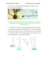
Illustration of hair follicle-like scaffold. There are
two types of cells which are necessary for hair
follicle generation. Blue dots represent
mesenchymal cells which can induce the
proliferation of epithelial cells (red dots). The scale
bar represents 100 μm. (Reproduced from David A.
Whiting. Histology of the Human Hair Follicle. In:
Ulrike Blume-Peytavi, Antonella Tosti, David A.
Whiting, Ralph M. Trüeb, editor. Hair Growth and
Disorders: Springer-Verlag Berlin Heidelberg;
2008. p 107–123. With kind permission of Springer
Science and Business Media.)
119
26
Figure 6
Schematic illustration for the whole process of the
microwell fabrication: i) silicon master
manufacturing, ii) PDMS stamp production and iii)
hydrogel wells fabrication.
34
Figure 7
Different dimensions of PDMS stamps and
corresponding hydrogel microwells. A:
Cross-sectional images of PDMS stamps (i-iv
35
XIII
microwell diameters, 56, 93, 180, 388µm) stained
by rhodamine B, where MD represents microwell
diameter; MH represents microwell height; IH
represents islet height. B: i-iv: images of microwells
with various diameters fabricated by 10% (w/v)
PEGDA. All scale bars represent 100 µm.
Figure 8
Microwell stability. Arrays were incubated in PBS
and their stability was analyzed by establishing two
methods, A: overall stability of microwell arrays
and B: stability of microwell arrays based on
counting. Overall stability was determined based on
whether each of the microwell arrays was intact
(100%) or detached (0%) from the glass slide (n =
9). Stability by counting was determined based on
the number of the undamaged microwells over total
number of microwells from each microwell array (n
= 9). Hydrated prepolymer solutions containing
10%, 20%, 50% and 80% (w/v) PEGDA were
analyzed over time, and stability of microwell
arrays decreased with increasing PEGDA
concentration. In the figure, diamond patterns (♦)
represent 10% (w/v) PEGDA; squares (□) represent
20% (w/v) PEGDA; triangles (▲) represent 40%
(w/v) PEGDA; crosses (×) represent 80% PEGDA.
37
Figure 9
Mechanical properties of PEGDA hydrogels with
varying gel percentage and thickness. A:
Representative nanoindentation curves from 10%
(w/v) PEGDA microwell bottom, 10% (w/v)
PEGDA hydrogel, 20% (w/v) PEGDA microwell
bottom, and 20% (w/v) PEGDA hydrogel. B:
Young’s modulus for 10% (w/v) PEGDA microwell
bottom, 10% (w/v) PEGDA hydrogel, 20% (w/v)
PEGDA microwell bottom, and 20% (w/v) PEGDA
hydrogel. Young’s modulus of 20% (w/v) PEGDA
was significantly higher than that of 10% (w/v)
PEGDA (***p < 0.001) while there were no
significant differences between Young’s modulus of
microwell bottoms and surfaces for both
concentrations of PEGDA.
39
Figure 10
Investigating HDF viability in PEGDA solution of
different MWs, PEGDA 575, 700 and 3500. A:
41
XIV
HDF viabilities increased with MWs of PEGDA.
HDF had highest viability in PEGDA MW 3500
solution over 2 hours. * and *** indicate p < 0.05
and p < 0.001 as compared to the viability of
corresponding control group (n=3). B: LIVE/DEAD
assay images at 2 hour for PEGDA of three
different MWs. Living cells were stained green and
dead cells were stained red. All scale bars represent
200μm.
Figure 11
Investigating HDF viability in cell suspension (2
million cells/mL) after exposure to UV of different
intensities. HDF viability was fairly consistent
across different UV intensities (n=3).
42
Figure 12
Investigating HDF viability in cell suspension (2
million cells/mL) after different length of exposure
time to photoinitiator. HDF viability is fairly
consistent across 2 hour period (n=3).
43
Figure 13
Investigating HDF viability in cell suspension after
UV exposure in the presence of photoinitiator.
There was a significant decrease in cell viability
after 2 hours. * indicates p < 0.05 as compared to
the viability of the control group (n=3).
44
Figure 14
A: Viability of HDF cells encapsulated in PEGDA
hydrogels photopolymerized at various conditions.
B: UV intensities and their corresponding minimum
UV exposure time for hydrogel formation.
45
Figure 15
Encapsulating HDF cells (2 × 10
6
cells/mL) within
microwell arrays. A-i: Cell viability (HDF) of the
control group before UV exposure. A-ii: Phase and
fluorescent superimposed image after applying a
Live/Dead assay to HDF cells which shows
relatively uniform cell distribution in the hydrogel.
A-iii: 3D cell-laden microstructures. A-iv: HDF
cells stained with Ethd (red) and calcein-AM
(green) in the 3D microstructure. B: Cell viability in
various MW PEGDA hydrogels. Cell viability of
PEGDA hydrogels after microwell fabrication
increased with increasing MW. PEGDA 575,
PEGDA 700 and PEGDA 3500 were highly
46
XV
hydrated polymers containing 10% PEGDA in PBS.
* and *** indicate p < 0.05 and p < 0.001 as
compared to the viability of corresponding control
group (n=3). All scale bars represent 100 µm.
Figure 16
Seeding HaCaT cells on the top of microwell
arrays. A-i: Cell viability (HaCaT) of the control
group before cell seeding. A-ii: Phase and
fluorescent superimposed image after applying a
Live/Dead assay to HaCaT cells demonstrates
HaCaT cells were located in microwells. B: Cell
viability before and after cell seeding showed no
significant difference. Initial cell concentrations
ranged from 1-12 million cells per mL (n=3).
47
Figure 17
Various densities of HaCaT cells seeded on the top
of microwell arrays. A-E: Representative images of
HaCaT cells stained with calcein-AM fluorescent
dye in the microwells with different cell seeding
densities. F: The average number of cells per well
increased with increasing initial cell concentration
(n=3). Scale bars represent 200 µm.
49
Figure 18
HaCaT cells growing in microwell arrays over 8
days. Live/Dead assay was performed to indicate
cell viability. A: Image of HaCaT cells on the top
just after cell seeding. B-C: Cell aggregates formed
on day 1 and day 3. D: Cell aggregates growing
bigger after 8 Days. All scale bars represent 100
µm.
50
Figure 19
A: Representative image of microstructure (200
μm). B1-B2: SEM images of hydrogels. B1: center
islet surface. B2: microwell bottom surface. C:
Upon cell seeding, HaCaT cells were spherical in
shape. D: After 3 days’ incubation, cell aggregates
formed.
51
Figure 20
HDF cell encapsulation in PEGDA (MW 3500)
hydrogel over 2 weeks. A: (i-iv) Phase contrast
images of HDF cells in microgels. After 72 h, cell
spreading was seen in the hydrogel and the
morphology of cells continued to change over 2
weeks (indicated by arrows). Except day 0, images
53
XVI
of day 3, day 7 and day 14 were from the same
location of the hydrogel. B: Quantification of cell
viability by Live/Dead assay over 2 weeks. Cell
viability decreased consecutively on first 7 days,
and then cell viability remained stable from day 7
onwards around 48%. All scale bars represent 100
µm.
Figure 21
A: Chemical synthesis of MeHA by reacting HA
and methacrylic anhydride. B:
1
H-NMR spectrum
of 75-kDa HA. C:
1
H-NMR spectrum of MeHA
(DM= 19.4%).
64
Figure 22
SEM images of 2.5%, 5%, 7.5% and 10% (w/v)
MeHA hydrogels (250 × magnification).
65
Figure 23
A: Contact angle of water measured on different
concentration MeHA hydrogels. Increasing trend
was observed with higher MeHA concentrations
(n=3); B: Effect of concentration of MeHA on
contact angle of water. As concentration of MeHA
of hydrogel increases, contact angle of water
increases (n=3).
66
Figure 24
Log-log plot of shear moduli (G’, G’’) vs. strain (n
= 3) for 2.5%, 5%, 7.5% and 10% (w/v) MeHA
hydrogels.
67
Figure 25
Images of HaCaT cells seeded in microwells taken
at the same location at different time points. A:
HaCaT cells growing in microwells (outer diameter
= 200 μm, inner diameter = 66 μm) over 14 days.
Green fluorescence represents live cells and red
represents dead cells. All scale bars represent 100
μm. B: Cell viability of HaCaT seeded on
microwells (n = 3). Cell viability fell gradually over
14 days, and remained high at 72.3 % at day 14.
69
Figure 26
A: Representative images of HDF cells
encapsulated in MeHA hydrogels (5.0% w/v) over
7-day incubation. B: HDF cell viability over 7-day
incubation in 2.5% and 5.0% (w/v) MeHA
hydrogels. Viability generally decreases over time.
All scale bars represent 100 μm.
70
XVII
Figure 27
A: Morphology of HaCaT and HDF cells in 2D
culture, respectively, as reference. B: Co-culturing
HDF and HaCaT cells in the microwells and 2D
culture dishes over time, respectively. On the day 0,
individual HaCaT cells were uniformly dispersed in
the microwells. Cell aggregates formed on day 3
and became bigger on the day 7 and day 14. On the
day 21, all centre inlets of microwells were covered
by cell aggregates. Live and dead assay on the day
21 showed that cell aggregates and most of HDF
cells were stained in green (live) and only a few of
cells in red (dead). For the control group, HDF and
HaCaT cells were dispersed in the culture dishes on
the day 0. Two types of cells attached and spread on
the day 3 and day 7. On the day 14 and 21, the
number of cells increased rapidly and it is difficult
to separate HDF or HaCaT cells from each other.
All scale bars represent 100 μm.
82&83
Figure 28
Co-culturing HDF and HaCaT cells for 14 days.
HDF cells were encapsulated in the thick hydrogel
and HaCaT cells seeded on the top. Live/Dead
assay was applied on the cell-incorporated hydrogel
on day 14. All scale bars represent 100 μm.
84
Figure 29
Confocal images of cell distribution and cell
development in the microstructural hydrogels on
day 1, day 3, day 7 and day 14.
85
Figure 30
3D confocal images on day 7 and day 14 and the
corresponding disassembled 2D images of
representative cell-incorporated microstructures. All
scale bars represent 100 μm.
86
Figure 31
Cell proliferation in A: 2D culture, B: non-patterned
hydrogels and C: 3D microstructures on day 3, day
7 and day 14. Similar to positive controls (2D
culture), most of HDF and HaCaT cells in and on
the microwells were positive in Ki-67 stain. For the
non-pattern hydrogels, HaCaT cells seeded on top
formed cell aggregates on day 3. Over time, HaCaT
cells behaved like in the culture dish and they
adhered, spread and grew into a large number of
88
XVIII
cells. All scale bars represent 100 μm.
Figure 32
Hair cortex keratin-specific AE-13 immunostaining
in the 2D culture, non-patterned hydrogels and 3D
microstructures on day 3, day 7 and day 14. AE-13
was expressed in fluorescent green color and cell
nuclei were counterstained with DAPI. Cell clumps
formed at day 14 in 2D culture and at day3, day 7
on the non-pattern hydrogels were positively
expressed AE-13 (indicated by arrows). HaCaT
cells in the microwells were positive in AE-13
staining. Views with higher magnification are also
shown for day 3, day 7, and day 14 3D culture,
showing the signals from representative microwells.
All scale bars represent 100 μm.
89
Figure 33
Quantitative real-time PCR results showing
differences of gene expression (Wnt10a, Wnt10b,
Shh, KGF and BDNF) between 2D culture and 3D
microwell system. *, ** and *** indicate p < 0.05,
p < 0.01 and p < 0.001 as compared to mRNA
concentration of corresponding control group (n=3).
91
Figure 34
Real-time PCR results showing Wnt10a, Wnt10b,
Shh, KGF and BDNF gene expression in 2D culture
and 3D microwell system supplied with high Ca
2+
DMEM culture medium. *, ** and *** indicate p <
0.05, p < 0.01 and p < 0.001 as compared to mRNA
concentration of corresponding control group (n=3).
92
XIX
LIST OF ABBREVIATIONS
Abbreviation
Full name
°C
degree Celsius
AGA
androgenetic alopecia
AMV
Avian Myeloblastosis Virus
ANOVA
analysis of variance
BDNF
brain-derived neurotrophic factor
BMP
bone morphogenic protein
calcein-AM
CLSM
calcein acetoxymethyl ester
confocal laser scanning microscopy
CTS
connective tissue sheath
DAPI
4',6-diamidino-2-phenylindole
DHT
dihydrotestosterone
DM
degree of methacrylation
DMEM
Dulbecco's modified Eagle's medium
DP
EBs
ECM
Edar
dermal papilla
embryoid bodies
extracellular matrix
ectodysplasin-A receptor
Ethd
ethidium homodimer
FDA
Food and Drug Administration
FE-SEM
field emission scanning electron microscope
FGF
fibroblast growth factor
FITC
fluorescein isothiocyanate
FUE
follicular unit extraction
G’
storage modulus
G”
loss modulus
XX
GFP
green fluorescent protein
GM
glassy membrane
HA
hyaluronic acid
HaCaT
human adult low calcium high temperature
HDF
HG
human dermal fibroblast
hair germ
HGF
hepatocyte growth factor
HHEMP
2-hydroxy-4’-(2-hydroxy-ethoxy)
-2-methylpropiophenone
HP
hair papilla
Hx
IGF1
Huxley's layer
insulin-like growth factor 1
IGFBP5
insulin-like growth factor binding protein 5
IL-1b
interleukin-1b
IRS
inner root sheath
kDa
KGF
kilo Daltons
keratinocyte growth factor
MC
matrix cells
MC1R
melanocortin-1 receptor
MD
microwell diameters
Me
melanocytes
MeHA
mL
MMP
methacrylated hyaluronic acid
milliliter
matrix metalloproteinase
MW
NaOH
molecular weight
sodium hydroxide
NMR
nuclear magnetic resonance
Ntf-3
neurotrophin-3
OCT
optimal cutting temperature compound
XXI
ORS
outer root sheath
PBS
phosphate-buffered saline
PDGF
platelet-derived growth factor
PDMS
polydimethylsiloxane
PEG
poly (ethylene glycol)
PEGDA
real-time PCR
poly (ethylene glycol) diacrylate
real-time polymerase chain reaction
RFP
red fluorescent protein
RGDS
Arg-Gly-Asp-Ser
SCID
severe combined immunodeficiency
SD
standard deviation
Shh
sonic hedgehog
TGF-α
transforming growth factor alpha
Tgfb1
transforming growth factor beta 1
TMS-PMA
3-(trimethoxysilyl) propyl methacrylate
Tnf
tumor necrosis factor
UV
ultraviolet
VEGF
vascular endothelial growth factor
α-SMA
α-smooth muscle actin
μg
micro gram
μL
micro liter
μm
micro meter
1
Chapter 1 Background
1.1. Introduction
Hair is one of the most important aspects of an individual’s appearance.
Physiologically, hair shaft serves as a protective device for maintaining body
heat and against the sunshine
1
. Besides, hair plays an essential role in
psychosocial communication. It works as a symbol of youth, health,
self-confidence and personal attractiveness, while hair loss often has adverse
impact on one’s self-esteem and interpersonal relationships
1
. Hair loss, termed
as alopecia in medical science, has been a widespread problem around the
world. There are several types of alopecia, including androgenetic alopecia,
alopecia areata, scarring alopecia, telogen effluvium and etc
2
. Alopecia may
occur as a natural part of aging, due to a disease or due to drugs and
medications
3
. In particular, androgenetic alopecia (AGA) is the most common
cause of hair loss in humans. According to a global report, AGA affects
approximately 50% of men and 20% to 53% of women by age 50 years
4
.
Currently, treatments of AGA include using anti hair loss drugs and/or surgical
implantation. Minoxidil and finasteride are two drugs for AGA
5
. However,
some side effects appear during the treatment and hair fall resumes upon
withdrawal of the drugs. On the other hand, using surgical procedure is
effective in hair regeneration by which grafts containing hair follicles were
transplanted onto the bald scalp of patients. Nevertheless, there are no other
alternatives of harvesting hair follicles other than from human donors
6
.
Therefore, it is important to explore a new method to generate hair follicles in
large quantity to meet the clinic needs. Recently, scientists attempt to
emphasize on the understanding of hair follicle morphogenesis, to investigate
the mechanism of hair follicle initiation and development, and to form hair
follicles from dissociated cells. This chapter will provide a brief account of
hair follicle morphogenesis, research progress of hair follicle generation and
the importance of cell compartmentalization in hair follicle bioengineering.