The characterization of soluble t cell receptors specific for the parasite toxoplasma gondii
Bạn đang xem bản rút gọn của tài liệu. Xem và tải ngay bản đầy đủ của tài liệu tại đây (4.66 MB, 164 trang )
THE CHARACTERIZATION OF SOLUBLE T CELL
RECEPTORS SPECIFIC FOR THE PARASITE
TOXOPLASMA GONDII
TAN ZHEN WEI
(B.Sc.(Hons.), NTU
A THESIS SUBMITTED
FOR THE DEGREE OF DOCTOR OF
PHILOSOPHY
NUS GRADUATE SCHOOL FOR INTEGRATIVE
SCIENCES AND ENGINEERING
NATIONAL UNIVERSITY OF SINGAPORE
2014
Declaration
I hereby declare that this thesis is my original work
and it has been written by me in its entirety. I have
duly acknowledged all the sources of information
which have been used in the thesis.
This thesis has also not been submitted for any
degree in any university previously.
Tan Zhen Wei
31 July 2014
I
Acknowledgements
Being able to write this page would mean the end of a four and a half year
journey, a journey which I will not be able to make alone. Here, I would like
to express my heartfelt thanks to these people. First and foremost, I would like
to thank my supervisor, Dr. Gijsbert Grotenbreg. This journey would not have
started without him. With his guidance, I could see myself grow over the
years, learning the skill sets expected of a researcher. He was a mentor but
never appeared too far to approach and always willing to provide advice.
Next, I would also like to thank the members of the lab, past and present.
Special thanks to Ming Yan and Kai Yee, both of whom showed me the ropes
when I first joined the lab. To Ling yun, for offering help and support as a
fellow post-grad. To Jia Wei, for being in charge of peptide synthesis, taking
care of the lab and making sure the lab is always in running order, not to
forget being a very good gym buddy. To Socks, for always willing to drive me
to NTU for experiments, teaching me on SPR, as well as providing advice
whenever needed. To Kenneth and Lionel, for offering advice on experiments
and football discussions. To Joanna, Gladys, Ping Ping and all other members
whom I did not list but have helped me in one way or the other in my journey.
Next, I would like to thank Dr. Johnathan Rapley and Dr. Rob Meijers
(European Molecular Biology Laboratory, Hamburg Germany) for solving the
crystal structure of the Rop7c1 TCR. This allowed me to do the supercharging
part of my project. I would also like to thank Dr. Lee Kim Swee and Dr. Hidde
Ploegh (Whitehead Institute of Biomedical Research, Cambridge USA) for
agreeing to share data on the effector function of the T cell clones, as well as
II
the chance to collaborate on a publication currently under review. I would also
like to thank Dr. David Thompson and Dr. David Liu (Howard Hughes
Medical Institute, Harvard University USA) for kindly sharing the AvNAPSA
program for our supercharging experiments. I would like to take this
opportunity to thank Dr. Markus Wenk and Dr. Cynthia He as well, both of
whom were ever so friendly and provided insightful advice during my TAC
meetings with them. I would like to thank my parents for their support during
this time, for trusting in me in my decision to do a Ph.D. Last, I would like to
thank my fiancée, Yi Zhen, for her support and her understanding that I was
always late for our dates whenever I was in lab.
III
Table of Contents
Summary………...………………………………………………………...VII
List of Tables……….…………………………………............................IX
List of Figures…………………………………………............................X
List of Illustrations…………………………………………………........XII
List of Abbreviations and symbols…………………………………..XIII
List of Publications……………………………………………………. XVII
Chapter 1: Introduction.......................................................................1
1.1 Adaptive immune system………………………………………………1
1.2 BCR recognition and diversity………………………………………....5
1.3 BCR therapeutics: Monoclonal Antibodies…………………………...8
1.4 TCR recognition and diversity………………………………………..14
1.5 TCR therapeutics: Adoptive T cell transfer…………………………18
1.6 TCR therapeutics: Soluble TCRs…………………………………….21
1.7 Supercharging to improve stabilities of soluble TCRs……………..27
Chapter 2: Production and characterization of 3 TCR clones with
identical antigen specificities
2.1 Introduction……………………………………………………………..31
2.2 Materials and Methods………………………………………………..35
2.2.1 Cloning of TCR α and β chains…………………………………….35
2.2.2 Expression and purification of TCR α and β chains.…………….36
2.2.3 Refolding and purification of soluble TCRs……………………….36
IV
2.2.4 TCR-tetramer binding assay……………………………………….37
2.2.5 Surface Plasmon Resonance………………………………………38
2.3 Results………………………………………………………………….38
2.3.1 Cloning, expression and purification of individual TCR chains…38
2.3.2 Refolding and purification of the 3 soluble TCR clones…………42
2.3.3 Binding specificities of refolded TCRs…………………………….48
2.3.4 Binding affinity between the three TCRs and Ld Rop7 MHC…...51
2.4 Discussion…………………………………………………………….. 55
Chapter 3: Production and characterization of supercharged TCRs
3.1 Introduction………………………………………………………….....60
3.2 Materials and Methods……………………………………….............65
3.2.1 Design of supercharged Rop7c1 TCR chains…………………....65
3.2.2 Production and purification of supercharged TCR chains………66
3.2.3 Refolding of supercharged TCRs………………………………….66
3.2.4 Assessing functional avidities of supercharged TCRs…………..66
3.2.5 RAW cells staining with TCR tetramers…………………………..67
3.2.6 Stability assay of supercharged TCRs……………………………67
3.3 Results……………………………………………………………….....68
3.3.1 Crystal Structure of Rop7c1 TCR………………………………….68
3.3.2 Production of supercharged Rop7c1 TCR α and β chains……..68
3.3.3 Production and purification of supercharged Rop7c1 TCRs……73
3.3.4 Functional avidity and specificity of supercharged TCRs……….84
3.3.5 Stabilities of supercharged Rop7c1 TCRs………………………..88
3.4 Discussion……………………………………………………………...98
V
Chapter 4: Comparing TCR binding affinity and effector function
4.1 Introduction……………………………………………………………105
4.2 Materials and Methods………………………………………………110
4.2.1 Surface Plasmon Resonance…………………………………….110
4.2.2 Structural modeling of Altered Peptide Ligands………………..110
4.2.3 Stability assays for pMHCs……………………………………….110
4.3 Results………………………………………………………………...111
4.3.1 Rop7c1, Rop7c2 and Rop7c3 recognize their cognate ligands
differently…………………………………………………………….111
4.3.2 Stabilities of the Ld MHC presenting APLs……………………...116
4.3.3 TCR binding affinity is not indicative of effector function………119
4.4 Discussion…………………………………………………………….122
Conclusion……………………………………………………………….130
Future Work………………………………………………………………131
Bibliography……………………………………………………………..132
Annex A………………………………………………………………….. 143
VI
Summary
CD8+ T cells are important for resolving attacks by pathogens such as viruses
and parasites. Recently, transnuclear mice monoclonal for each of three TCRs
recognizing the Rop7 antigenic peptide from the parasite Toxoplasma gondii
were generated. To better understand T cell immunity against the parasite, I
characterized the binding affinities of these three T Cell Receptors (TCRs)
against the Rop7 peptide MHC molecule. I observed a range of binding
affinities for these three TCRs. Moreover, binding kinetic studies also
revealed that they contact the peptide Major Histocompatibility Complex
(pMHC) for different periods of time. These data indicate that during an
infection by the parasite, T cells expressing TCRs with a range of binding
affinities are activated. Thus, T cell activation is not solely dependent on the
binding affinity of the TCR to its cognate ligand.
With the soluble TCRs generated from the binding affinity and kinetics
studies, I sought to increase the stabilities of soluble TCRs so as to increase
their attractiveness as a potential alternative immunoconjugate platform to
monoclonal antibodies. To this end, I selected the Rop7c1 TCR, which has the
highest refolding yield as well as binding affinity, to try and improve its
stability. Increasing the surface charges of a protein have been shown to
increase the thermostabilities of some proteins. Thus, I increased the surface
charges of the Rop7c1 TCR by selecting and mutating highly surface exposed
residues to either positively or negatively charged residues. However,
increasing the surface charges did not improve the stability of the TCR.
VII
Several factors such as free energy and surface charge-charge interactions play
a role in protein stability as well and thus, solely increasing the surface
charges of the TCR may not be sufficient to improve its stability.
With our collaborator’s data, I was also able to compare the binding affinities
of the three TCR clones with the effector function of the T cells expressing
them. I observed that the binding affinities of the TCR do not correlate
positively with the strength of their effector function. This implies T cell
effector function may not be dependent on TCR binding affinity but on other
possible intrinsic factors. More experiments will be required to identify these
factors.
VIII
List of Tables
Table 1: TCR gene usage...............................................................................40
Table 2: AvNAPSA scores for Rop7c1 TCR................................................70
Table 3: Attempted refolds of supercharged TCRs………………………81
Table 4: Buffer conditions for stability assays……………………………90
IX
List of figures
Figure 5: Construction of soluble TCR and expression of individual TCR
chains…...........................................................................................................41
Figure 6: Purification of soluble Rop7c1 TCRs…………………………...43
Figure 7: Purification of soluble Rop7c2 TCRs…………………………...45
Figure 8: Purification of soluble Rop7c3 TCRs…………………………...47
Figure 9: Functional avidity of the TCR tetramers to Ld Rop7 MHC…..49
Figure 10: Binding affinities of the TCR clones to Ld Rop7 MHC………52
Figure 11: Kinetic analysis of the TCR clones binding to the Ld Rop7
MHC…………………………………………………………………………53
Figure 12: Crystal structure of Rop7c1 TCR……………………………..69
Figure 13: Positions of mutations for negative supercharging of Rop7c1
TCR………………………………………………………………………….71
Figure 14: Positions of mutations for positive supercharging of Rop7c1
TCR………………………………………………………………………….72
Figure 15: Expression of individual supercharged TCR chains…………74
Figure 16: Purification of soluble Rop7c1α 1β (-11) TCR………………..76
Figure 17: Purification of soluble Rop7c1α 1β (-25) TCR………………..77
Figure 18: Purification of soluble Rop7c1α (-16) 1β (-11) TCR………….78
Figure 19: Purification of soluble Rop7c1α 1β (+5) TCR………………...79
Figure 20: Size exclusion chromatograms of positively charged TCRs…80
Figure 21: Surface charge distribution of the Rop7c1 TCR……………..82
Figure 22: Surface charge distribution of supercharged chains…………83
Figure 23: Functional avidity of the supercharged TCRs………………..85
X
Figure 24: Binding of supercharged TCR tetramers to RAW 264 cells...87
Figure 26: Stability assay for Rop7c1 TCR……………………………….92
Figure 27: Stability assay for Rop7c1α 1β (-11) TCR…………………….93
Figure 28: Stability assay for Rop7c1α 1β (-25) TCR…………………….94
Figure 29: Stability assay for Rop7c1α (-16) 1β (-11) TCR………………95
Figure 30: Stability assay for Rop7c1α 1β (+5) TCR……………………..96
Figure 31: Comparison of the Tms of the various TCR clones………….. 97
Figure 32: SPR sensorgrams of Rop7c1 and Rop7c2 TCRs binding to
APLs………………………………………………………………………..112
Figure 33: Equilibrium binding analysis of the TCR clones against the
various pMHC ligands…………………………………………………….113
Figure 34: Models for APLs in MHC peptide binding groove………….114
Figure 35: Stability assay for Ld IPAAAGRFF MHC…………………..117
Figure 36: Stability assay for Ld IPAFAGRFF MHC…………………..118
Figure 37: Stability assay for Ld IPANAGRFF MHC…………………..118
Figure 38: Comparison of Tms for all pMHCs…………………………..119
Figure 39: Effector function of the threeTCR clones upon activation…121
XI
List of illustrations
Figure 1: Recombination Mechanisms of T and B cell receptors…………7
Figure 2: Different types of monoclonal antibodies………………………11
Figure 3: Potential uses of soluble TCRs………………………………….23
Figure 4: Various life stages of the parasite Toxoplasma gondii…………33
Figure 25: Differential Scanning Fluorimetry…………………………….89
XII
List of Abbreviations and symbols
APC: Antigen Presenting Cell
AvNAPSA: Average number of neighboring atoms per side-chain atom
BCR: B Cell Receptor
BiTE: Bispecific T cell Engagers
BMDC: Bone marrow derived dendritic cell
BSA: Bovine Serum Albumin
CAR: Chimeric Antigen Receptor
CDR: Complementary Determining Region
CFSE: Carboxyfluorescein succinimidyl ester
CT: Congenital Toxoplasmosis
CTL: Cytotoxic T Lymphocyte
D: Diversity gene segment
DAG: Diacylglycerol
DALY: Disability-adjusted life year
Db: Diabody
DCs: Dendritic cells
DSF: Differential Scanning Fluorimetry
DTT: Dithiothreitol
EAE: Experimental Autoimmune Encephalitis
EBV: Epstein Barr Virus
EDC: Ethyl(dimethylaminopropyl) carbodiimide
EDTA: Ethylenediaminetetraacetic acid
ER: Endoplasmic Reticulum
XIII
FcRn: Neonatal Fc Receptor
FPLC: Fast Protein Liquid Chromatography
GFP: Green Fluorescent Protein
HIV: Human Immunodeficiency Virus
HSCs: Hematopoeitic Stem Cells
I.B.: Inclusion bodies
IEDB: Immune Epitope Database and Analysis Resource
IFN-γ: Interferon gamma
Ig: Immunoglobulin
IL-2: Interleukin 2
IL-4: Interleukin 4
IL-10: Interleukin 10
IL-13: Interleukin 13
IL-17: Interleukin 17
IL-22: Interleukin 22
IPTG: Isopropyl β-D-1-thiogalactopyranoside
IP3: Inositol trisphosphate
J: Joining gene segment
kDa: Kilodaltons
LCMV: Lymphocytic Choriomeningitis Virus
mAbs: Monoclonal Antibodies
MAPK: Mitogen-activated protein Kinase
MFI: Median Fluorescence Intensity
MHC: Major Histocompatibility Complex
MonoQ: Anion exchange column
XIV
NHS: (N-Hydroxysuccinimide)
NKT: Natural Killer T cells
O.D.: Optical Density
PBS: Phosphate Buffer Saline
PCR: Polymerase Chain Reaction
PE: Phycoerythrin
PFA: Paraformaldehyde
P.I.: Isoelectric point
PIPE: Polymerase Incomplete Primer Extension
pMHC: peptide loaded Major Histocompatibility Complex
PSMA: Prostate Specific Membrane Antigen
RUeqm: Equilibrium Response Unit
scFv: Single chain antibody
SDS-PAGE: Sodium Dodecyl Sulphate – Polyacrylamide Gel Electrophoresis
SHM: Somatic Hypermutation
Strep: Streptavidin
Strep-PE: Streptavidin conjugated phycoerythrin
S75: Superdex 75 column
S200: Superdex 200 column
TaFv: Tandem scFV
TAA: Tumour Associated Antigens
TAP: Transporter associated with Antigen Processing
TAPA: Tumour Associated Peptide Antigen
TCR: T Cell Receptor
TGF-β: Transforming Growth factor β
XV
TIL: Tumour Infiltrating Lymphocytes
Th cells: T helper cells
Tm: Melting temperature
Treg cells: T regulatory cells
V: Variable gene segment
XVI
List of Publications
1. Sources of diversity in T cell epitope discovery (2011) C.X.L.
Chang, L.Y. Dai, Zhen Wei Tan, J.A.L. Choo, A. Bertoletti, G.M.
Grotenbreg. Frontiers in Bioscience (vol 16 pg 3014-3035)
2. Affinity for self-MHC in the periphery, not affinity for antigen,
determines the function of antigen-specific CD8 T cells. Lee Kim
Swee, Zhen Wei Tan, Anna Sanecka-Duin, Gijsbert
Grotenbreg, Eva M. Frickel & Hidde L. Ploegh. PLOS Biology
(Under review)
XVII
Chapter 1: Introduction
Pathogens in our environment are constantly evolving, trying to evade host
defence mechanisms and looking for opportunities to infect us. The adaptive
immunity plays a huge role in protecting us against such microbial attacks. Its
arsenal primarily comprises of B cells and T cells. B cells recognize full and
unique structural patterns found on pathogens with their B cell receptors
(BCRs) whilst T cells recognize antigenic peptides, processed from
pathogenic proteins intra-cellularly and presented by Major Histocompatibility
Complexes (MHCs) on infected cell surfaces, with their T cell receptors
(TCRs). In this way, both the pathogen and infected cells can be cleared by the
immune system. As such, monoclonal antibodies, soluble forms of BCRs,
have been developed as a therapeutic tool against various diseases with much
success. However, a significant research gap exists for TCR therapeutics due
to the many challenges faced in developing soluble and stable TCRs.
1.1 Adaptive immune system
All jawed vertebrates possess the adaptive immune system, which is crucial to
protecting us against attacks by microbes and pathogens should our innate
immune system fail to do so 1. More importantly, the adaptive immunity is
coined as such due to the fact that it can adapt and confer long-term immunity
by allowing for a memory response against recurrent infection. The adaptive
immune system comprises mainly of two arms: B cells, which partake in the
1
humoural immune response, and T cells, which are responsible for cellmediated immune responses.
T cells originate from Hematopoietic Stem Cells (HSCs) in the bone marrow
and undergo development in the thymus where they undergo a selection
process before entering into the periphery as naïve T cells 2. These T cells
express a unique TCR each and are responsible for recognizing antigenic
peptides presented by MHCs found on infected cells 3,4. Naïve T cells then
patrol the periphery and lymph nodes searching for antigen. Dendritic cells
(DCs), also known as professional antigen presenting cells, pick up pathogenic
antigens and migrate to lymph nodes. T cells with TCRs specific for these
antigens then contact and bind to the DCs presenting these antigens at the
lymph nodes. These T cells are then activated, secreting cytokines such as
Interleukin-2 (IL-2) and Interferon-γ (IFN-γ). They then dissociate from the
DCs, proliferate rapidly and migrate to sites of infection 5. There are different
subsets of T cells and their effector functions are different upon activation.
CD8+ T cells, also known as cytotoxic T cells, recognize antigenic peptides of
8-9 amino acid residues, either cytosolic or nuclear in origin, presented by
class I MHC molecules 6. They kill off infected cells directly via cell-mediated
cytotoxicity. CD4+ T cells, or T helper cells (Th), recognize exogenously
derived antigens presented by class II MHC molecules 6. There are several
different subsets of CD4+ T cells as well. Th1 cells mediate responses against
intracellular pathogens, secreting IFN-γ to activate macrophages and IL-2 for
CD8+ memory formation 7. Th2 cells secrete a milleu of cytokines such as IL4, IL-5 and IL-9, effecting class switching in B cells, recruiting eosinophils
2
and activating mast cells in response to extracellular parasite invasions 7. Th17
cells secrete cytokines such as IL-17 and IL-22 as an immune response against
bacteria and fungi 8. Once the infection is cleared, most effector CD8+ T cells
will disappear with a small percentage becoming memory T cells, rapidly
activating and proliferating in response to a recurrent infection. T regulatory
(Treg) cells are also present at the end of an infection, secreting
immunosuppressive cytokines such as IL-10 and TGF-β to prevent
autoimmunity 9.
On the other hand, B cells undergo development and selection in the bone
marrow before entering the periphery. These naïve B cells express a unique
BCR each and recognize structural patterns on full and intact antigens
presented on cell surfaces or in soluble forms. Once bound to foreign antigens,
B cells ingest the antigen and digest it, subsequently expressing the fragments
on its class II MHC molecules. Activated CD4+ T cells that recognize the
same antigenic peptides presented by the B cells can then contact these B cells
and help them to proliferate and differentiate into plasma cells. Plasma cells
then secrete antibodies which can bind to the pathogenic antigens with high
affinity. Once bound, these secreted antibodies then carry out their effects via
several mechanisms. First, by binding to the antigens expressed on pathogens
or their toxic products, antibodies prevent their access, and thus harmful
effects, to the host’s cells. Second, phagocytes such as macrophages and
neutrophils recognize the antibody coated pathogens or toxins and ingest them
in a process known as opsonization. Last, antibodies serve as receptors for
complement proteins to be activated. The complement system, with several
3
different proteins such as c1q and c1s, can form an attacking complex to kill
extracellular pathogens.
There are five classes, commonly known as isotypes, of antibodies. IgM and
IgD are isotypes of BCRs found on mature naïve B cells. IgE antibodies target
allergens and parasitic worms including helminthes, and activate mast cells
and basophils. IgA targets pathogens found in the mucosal areas such as the
gut and respiratory tract whilst IgG is the major isotype that targets against
invading pathogens.
Other than targeting pathogens, the adaptive immunity also plays a part in
combating cancer. First, it combats cancer indirectly by killing viruses which
can cause cancer. Examples include the Epstein Barr virus, which is
responsible for Burkitt’s and Hodgkin’s lymphoma 10, and the Human
Papillomavirus, which is responsible for cervical cancer 11. Second, it can also
eliminate cancer cells directly by specifically identifying the tumour antigens
present on tumours and then killing them.
With such a huge arsenal of weapons, both B and T cells play a huge role in
protecting our body against invading pathogens and malignancies. Several
studies have since been done to try and utilize them and their effector
molecules as therapeutic tools against diseases and tumours 12-14.
4
1.2 BCR recognition and diversity
B cells are able to target antigens found on the pathogens themselves or
expressed on the surface of infected cells. Each B cell contains a unique BCR
and allows them to contact these antigens. In order to combat the huge
repertoire of possible foreign antigens, a large diversity in the BCRs expressed
by B cells is required.
The BCR, also known as an immunoglobulin (Ig), is made up of two heavy
chains and two light chains. Each chain contains a variable and a constant
region, with the variable regions responsible for antigen recognition. Within
the variable regions of each heavy and light chain are three hypervariable
regions i.e. regions that are highly diverse between each BCR. They are also
known as Complementary Determining Regions (CDRs) as they are the parts
that contact the antigen the most. There are three CDRs in each heavy and
light chain and they are responsible for the binding of antigens and thus, have
huge variabilities. With the presence of two heavy and light chains, each
antibody has two identical antigen binding sites. Thus, each BCR can
theoretically bind to two target antigens. The large diversity in the binding
regions of the BCR is generated primarily by the recombination of genes
encoding for the variable regions (Fig. 1). For the light chain, Variable (V)
gene segments and Joining (J) gene segments are recombined to produce the
variable region of a light chain. A total of two gene loci, κ and λ, encodes the
light chain of the BCR. Thus, each light chain could either be of the κ or λ
subtype. On the other hand, the variable regions of the heavy chain are
5
encoded by the recombination of the V, J and Diversity (D) gene segments.
Pairing of the recombined heavy and light chain for each BCR molecule adds
another level of diversity to their variable regions. In addition, more diversity
is generated as nucleotides are gained or lost randomly during joining of the
various gene segments. This is known as junctional diversification and
together with the recombination of the various gene segments and pairing of
the heavy and light chains, a large and diverse repertoire of BCRs is
generated. With such a huge diversity, it is not surprising that there will be
BCRs which can recognize self-antigens. If these BCRs were allowed to enter
the periphery, they will generate an autoimmune reaction. To prevent this
from happening, a selection process occurs in the bone marrow to ensure the
BCRs assembled are not reactive to self-antigens, before the B cells are
released into the periphery. If the B cells are reacting with self-antigens, they
are given a second chance to modify their BCRs by going for a second round
of recombination in a process known as receptor editing. B cells that still
produce self-reactive BCRs are then deleted.
In addition to diversity generated during B cell development, more diversity in
the BCR can be created upon contact with antigens and with T helper cell
stimulation, in a process known as affinity maturation. As B cells get
stimulated, they divide and accumulate point mutations in their BCRs’
variable regions. These mutations occur approximately once every cell cycle,
a rate many times faster than spontaneous mutation in other genes. Thus, this
process is referred to as somatic hypermutation (SHM). SHM generates a
small number of BCRs with increased affinities for the target antigen but
6
many more has either little or no effect on their binding affinities to the
antigen. B cells expressing BCRs with higher binding affinities are then
preferentially stimulated to survive and proliferate. After several rounds of
proliferation and SHM, a pool of B cells expressing BCRs with much higher
binding affinities to the antigen is generated. In this way, affinity maturation is
achieved with surviving B cells expressing BCRs which can bind to the target
antigens with high specificity. In the event of a recurrent infection, the
antibodies produced upon recall would also be of a high binding affinity,
shortening the time to resolve the insult.
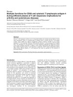
Figure 1. Recombination Mechanisms of T and B cell receptors. Diversity of the T cell
receptor (TCR) can be achieved from the (1) V(J) recombination of the α chain and V(D)J
recombination of the β chain, (2) nucleotides added or deleted in the joined segments and (3)
pairing of the re-arranged α and β chains. Similarly, diversity of the B cell receptor (BCR) can
be achieved from the (1) V(J) recombination of the light chain and V(D)J recombination of
the heavy chain, (2) nucleotides added or deleted in the joined segments and (3) pairing of the
re-arranged heavy chain with either the κ or the λ isotype of the light chain. Further diversity
is created when the re-arranged BCR undergoes somatic hypermutation upon contact with
antigen in a process known as affinity maturation. Not all J and D segments are shown in the
schematic.
7