A transmembrane mutation in FcgRIIb reveals the role of ceramide in phagocytosis and autoimmunity
Bạn đang xem bản rút gọn của tài liệu. Xem và tải ngay bản đầy đủ của tài liệu tại đây (13.18 MB, 260 trang )
!
A TRANSMEMBRANE MUTATION IN FcγRIIB REVEALS THE
ROLE OF CERAMIDE IN PHAGOCYTOSIS AND AUTOIMMUNITY
NURHUDA ABDUL AZIZ
NATIONAL UNIVERSITY OF SINGAPORE
2013
!
A TRANSMEMBRANE MUTATION IN FcγRIIB REVEALS THE ROLE OF
CERAMIDE IN PHAGOCYTOSIS AND AUTOIMMUNITY
NURHUDA ABDUL AZIZ
B.Sc (Forensic Science)(Hons), Curtin University of Technology, Australia
A THESIS SUBMITTED
FOR THE DEGREE OF DOCTOR OF PHILOSOPHY
NUS GRADUATE SCHOOL FOR INTEGRATIVE
SCIENCES AND ENGINEERING
NATIONAL UNIVERSITY OF SINGAPORE
2013
!
Declaration
I hereby declare that this thesis is my original work and it has been written by me
in its entirety. I have duly acknowledged all the sources of information which have
been used in the thesis.
This thesis has also not been submitted for any degree in any university
previously.
_________________________
Nurhuda Abdul Aziz
!
∀!
Acknowledgements
There are many people who were involved in the successful completion of this
project and production of this thesis:
I would like to thank Assoc. Prof Markus R. Wenk for supervising me. I am
grateful for the time and advice that he has so generously provided.
My gratitude also goes to Assoc. Prof Paul A. MacAry for being a great
supervisor. I have benefited tremendously from his expertise and experience in
cell biology. I could not have done this thesis work without the supervision and
encouragement from such a patient and understanding supervisor.
My great appreciation goes to Asst. Prof Gijsbert Grotenbreg and Asst. Prof
Brandon J. Hanson for their insightful comments and valuable suggestions.
Special thanks to Dr Olivia Oh for working closely with me to see through this
project well as to Dr Gan Shu Uin and Dr Paul Hutchinson helping me with
various technical issues related to this project. I would also like to extend my
appreciation to Dr Shui Guanghou for his help with the mass spectrometry, and
Ms Duan Xinrui for her assistance with statistical analysis.
Lastly, I would like to thank all my lab colleagues, past and present for your
friendship and for being a part of my research experience.
∀∀!
Table of Contents
CHAPTER 1 1
INTRODUCTION 1
1.1 Phagocytosis 2
1.1.1 The immune system and phagocytosis 2
1.1.2 Receptors involved in phagocytosis 4
1.1.3 Fcγ receptor mediated phagocytosis 5
1.1.3.1 Particle internalization and formation of phagocytic cup 5
1.1.3.2 Formation of Early Phagosomes 7
1.1.3.3 Formation of Late Phagosomes 8
1.1.3.4 Phagosome – lysosome fusion 9
1.2 Fragment Crystallizable γ Receptors (FcγRs) 11
1.2.1 Regulation of phagocytosis signaling by Fcγ receptors 15
1.2.2 The role of FcγRIIb in host defense and human autoimmunity 18
1.2.3 Mechanism for loss of FcγRIIb inhibitory function by Ile232Thr
polymorphism 20
1.3 The molecular biology of lipids 21
1.3.1 Lipid diversity, role and importance 21
1.3.2 Classification of the repertoire of lipids 24
1.3.3 The influence of lipids on membrane curvature 28
1.3.4 Lipid distribution and contribution in phagocytosis 32
1.3.5 Lipid rafts: Overview 45
1.3.5.1 Rafts in signal transduction 47
1.3.5.2 A role for ceramide in lipid rafts 48
1.3.6 Lipidomics: emerging lipid analytics 50
1.4 Objectives and thesis outline 53
CHAPTER 2 55
MATERIALS AND METHODS 55
2.1 Solutions and Buffers 56
2.1.1 Buffers for phagosome preparation 56
2.1.2 Buffers for plasma membrane isolation 57
2.1.3 Buffers for SDS – PAGE and western blotting 57
2.1.4 Buffers for flow cytometry 59
2.1.5 Buffers for confocal microscopy 59
2.1.6 Buffers for mycobacterial infection 59
2.2 Reagents 60
∀∀∀!
2.2.1 Latex beads 60
2.2.2 Antibodies 60
2.2.3 Plasmids and Cell lines 61
2.3 Cell culture 66
2.3.1 Cell culture and maintenance 66
2.3.2 Differentiation of U937 monocytes into macrophages 66
2.4 Detection of Protein kinase C activity assay 67
2.5 Preparation of plasma membrane isolates 67
2.6 Assessment of phagocytosis and phagosome maturation 68
2.6.1 Generation of IgG opsonized latex beads 68
2.6.2 Phagosome Formation and Isolation 69
2.6.3 Phagosome quantitation 70
2.6.4 Western blot analysis 70
2.6.5 Flow cytometry analysis 72
2.7 Confocal Microscopy 74
2.8 Mycobacteria infection assays 76
2.8.1 Culture of Mycobacteria 76
2.8.2 BCG infection and survival assays by U937 macrophages 76
2.8.3 Bioplex Cytokine Array 77
2.9 Lipid Analysis 78
2.9.1 Extraction of lipids from samples 78
2.9.2 Lipid fingerprinting by mass spectrometry 79
2.10 Statistical Analysis 80
CHAPTER 3 81
RESULTS I: GENERATION OF CELL LINES, REAGENTS AND MODEL
SYSTEMS FOR STUDYING Fcγ RECEPTOR MEDIATED PHAGOCYTOSIS . 81
3.1 Introduction 82
3.2 Characterization of Fcγ receptors on U937 cells 83
3.3 Establishment of conditions for phagocytosis in U937 cells 88
3.3 Use of latex beads for an in vitro phagosome model 92
3.4 Isolation of maturing phagosomes with step sucrose gradients 96
3.5 Extraction of plasma membrane 104
3.6 Discussion 108
CHAPTER 4 112
RESULTS II: ANALYSING THE EFFECTS OF FcγRIIB
232I
AND FcγRIIB
232T
ON
LATEX BEAD PHAGOCYTOSIS 112
4.1 Introduction 113
∀#!
4.2 Evaluation of phagocytic indexes of FcγRIIb
232I
and FcγRIIb
232T
macrophages 114
4.3 Assessment of phagosomal maturation 116
4.3 Assessment of phagosome acidification 119
4.4 Quantification of ROS produced in maturing phagosomes 122
4.5 Impact of FcγRIIb on calcium responses during phagocytosis 126
4.6 Discussion 129
CHAPTER 5 133
RESULTS III: INVESTIVGATING THE PHAGOCYTIC BACTERICIDAL ACTION
OF FcγRIIB
232I
AND FcγRIIB
232T
ON A PATHOGEN MODEL 133
5.1 Introduction 134
5.2 Ensuring Fc receptor mediated phagocytic uptake 135
5.3 Measurement of bacterial ingestion and killing 138
5.4 Assessment of inflammatory cytokines following phagocytosis 144
CHAPTER 6 153
RESULTS IV: Lipidomic Fingerprinting and Analysis 153
6.1 Introduction 154
6.2 Lipid composition of plasma membrane 154
6.3 Lipid composition in maturing phagosomes 162
6.4 Comparison of lipid profiles between plasma membrane and phagosomes
166
6.4 Discussion 172
CHAPTER 7 174
RESULTS V: INVESTIVGATING THE ROLE OF CERAMIDE IN
PHAGOCYTOSIS 174
7.1 Introduction 177
7.2 Generation and characterization of cell lines 179
7.3 Effect of ceramide on BCG killing and cytokine secretion 185
7.4 Discussion 195
CHAPTER 8 198
DISCUSSION 198
Discussion 199
#!
APPENDICES 208
Appendices 209
Appendix 1: Optimized MRM parameters for lipid species detected by LC-
MS/MS 209
Appendix 2: Trends of individual lipid species in maturing phagosomes 215
REFERENCE 225
References 226
!
#∀!
Summary
Receptor-mediated phagocytosis is a phylogenetically ancient biological process
employed for the protection of organisms from microbial infection and in the
maintenance of tissue homeostasis through clearance of cellular debris. The best
characterized cellular receptors that underlie this process are the receptors for
immunoglobulins-particularly IgG termed FcγRs and this form of phagocytosis is
termed opsonization. FcγRs can be broadly classified into activatory or inhibitory
receptors based on the presence of Immuno-Tyrosine Activatory Motifs (ITAM) or
Immuno-Tyrosine Inhibitory Motifs (ITIM) in their cytoplasmic domains. The
inhibitory receptor is proposed to regulate and dampen pro-inflammatory
signaling and hyper-aggressive phagocytic activity mediated by the activatory
receptors. The principle inhibitory receptor FcγRIIb also plays a role in controlling
autoimmunity for a single Isoleucine to Threonine substitution in its
transmembrane domain termed FcγRIIb
232T
renders the receptor non-functional
and confers susceptibility to systemic lupus erythematosus (SLE). The
FcγRIIb
232T
receptor is excluded from membrane microdomains where the WT
receptor regulates activatory FcγRs. In this study, we conduct a comprehensive
analysis of the lipid composition of phagosomes as these organelles invaginate,
internalize and mature through the endocytic pathway from the macrophage
plasma membrane. We demonstrate that maturing phagosomes captured at
different time points post phagocytosis, exhibit a distinct lipid composition from
the plasma membrane. Using cell lines stably transfected with either FcγRIIb
232I
!
#∀∀!
or FcγRIIb
232T
, we also demonstrate that FcγRIIb
232T
impacts upon cellular
ceramide expression/metabolism and this is linked to the observed
hyperaggressive phagocytic activity of these macrophages. These findings
represent the first comprehensive map of lipid composition and functionality in
FcR-mediated phagocytosis and highlight a novel role for ceramide in this vital
biological process.
!
#∀∀∀!
List of Figures
Introduction
Fig 1.1: IgG opsonized particles stimulates Fcγ receptor clustering in mediating
recognition of target particles for phagocytosis. 5!
Fig 1.2: Schematic representation of phagosome maturation highlighting the
molecules involved in relation to events along the endocytic pathway. 7!
Fig 1.3: Stages of phagosome formation and maturation. 10!
Fig 1.4: Schematic representation of human FcγRs. 14!
Fig 1.5: Diagrammatic representation of general FcγR signaling. 17!
Fig 1.6:
The basic structure of cell membrane is the lipid bilayer. 22!
Fig 1.7: Structure of phospholipid (specifically, phosphatidylcholine). 24!
Fig 1.8:
Basic structure of sphingolipid backbone and its various head groups. 26!
Fig 1.9: Sterols such as cholesterol are defined by their planar and rigid
tetracyclic ring 27!
Fig 1.10: Spontaneous curvature mediated by lipids depends on their molecular
geometry. 31!
Fig 1.11: Lipids are heterogeneously distributed between membranes and across
the membrane bilayer. 32!
Fig 1.12: PI serves as the basic building block for the synthesis of PIPS. 37!
Fig 1.13: Schematic illustration of PIP composition at different stages of a forming
phagosome. 39!
!
∀∃!
Fig 1.14: PIP species detected in maturing phagosomes. 40!
Fig 1.15: Cholesterol preferentially partitions into areas with sphingolipids.
43!
Fig 1.16: Lipid rafts are microdomains described as floating islands in a sea of
phospholipids. 46
Results I
Figure 3.1: U937 cells and the FcγRIIb knock –ins express FcγRI and FcγRII but
not FcγRIII. 85!
Figure 3.2: U937 knock – in cells express similar levels of FcγRII. 86!
Figure 3.3: FcγRIIb, the inhibitory receptor, is not expressed in U937. 87!
Fig 3.4: Effects of PMA stimulation on PKC activation. 90!
Fig 3.5: Cells differentiated with GM-CSF and PMA have increased phagocytic
capacity. 91!
Fig 3.6: Rabbit IgG coated latex beads are most efficiently taken up via Fc
receptors. 94!
Fig 3.7: IgG particles interact with Fcγ receptors and is internalized into the
phagolysosomal pathway. 95!
Figure 3.7: Latex bead phagosomes were isolated by flotation on step sucrose
gradients. 99!
Fig 3.8: Latex bead phagosomes were isolated at different stages of maturation.
100!
Fig 3.9: Phagosome isolates were devoid of major contamination from other
intracellular organelles. 101!
!
∃!
Fig 3.10: Phagosome concentration is normalized according to absorbance at
600nm. 102!
Fig 3.11: Equal loading of phagosomes was verified by silver stain. 103!
Fig 3.12: Coating of cells with cationic silica beads enables isolation of the
plasma membrane from internal membranes. 106!
Fig 3.13: Silver staining of proteins in plasma membrane extracts. 107
Results II
Fig 4.1: FcγRIIb
232T
macrophages exhibit enhanced phagocytosis. 115!
Fig 4.2: FcγRIIb
232T
polymorphism on the cell surface is sufficient to enhance the
rate of maturation of IgG opsonized beads. 118!
Fig 4.3: Phagosomes expressing FcγRIIb
232T
displayed a more rapid acidification
kinetic compared to FcγRIIb
232I
phagosomes. 121!
Fig 4.4: FcγRIIb
232T
phagosomes produced more ROS over time compared to
wild type FcγRIIb
232I
phagosomes. 125!
Fig 4.5: A more notable calcium response was observed during phagocytosis by
FcγRIIb
232T
macrophages. 128
Results III
Fig 5.1: Anti – 2F12 opsonized BCG activates Fcγ receptor mediated
phagocytosis. 137!
Fig 5.2: FcγRIIb
232T
expressing macrophages internalize more mycobacteria. 140!
Fig 5.3:
Macrophages expressing FcγRIIb
232T
have a much higher capacity to kill
ingested bacteria as
compared to FcγRIIb
232I
expressing macrophages.
142!
!
∃∀!
Fig 5.4: Killing capacities of macrophages were not affected by the increased
bacterial burden. 143!
Fig 5.5: Macrophages expressing FcγRIIb
232T
secrete higher levels of IL-1β and
TNFα 48h following BCG infection. 147!
Fig 5.6: Pro – inflammatory cytokine secretion was enhanced by macrophages
expressing FcγRIIb
232T
following 24 h incubation with BCG. 148!
Fig 5.7: Increased production of pro – inflammatory cytokines 48 h after Fcγ
receptor phagocytosis of BCG by FcγRIIb
232T
macrophages. 149
Results IV
Fig 6.1: Major lipid species in the plasma membrane. 156!
Fig 6.2: Individual lipid species in the plasma membrane revealed significant
differences between FcγRIIb
232I
and FcγRIIb
232T
macrophages. 157!
Fig 6.3: FcγRIIb232T resulted in increased levels of phospholipid species with
long acyl chains. 159!
Fig 6.4: Impact of FcγRIIb
232I
or FcγRIIb
232T
on saturation of plasma membrane
phospholipids. 161!
Fig 6.5: Comparison of phagosome lipids from FcγRIIb
232I
and FcγRIIb
232T
macrophages. 164!
Fig 6.6: Heatmap representation of changes in individual lipid species of
phagosome lipids. 165!
Fig 6.7: The lipid composition of the plasma membrane differed significantly from
that of phagosome membranes. 167!
Fig 6.8: Comparison of ceramide species in both the FcγRIIb232I and
FcγRIIb232T phagosomes. 170!
!
∃∀∀!
Fig 6.9: Raft lipids were enriched in cells expressing FcγRIIb
232I
. 171
Results V
Figure 7.1: Establishing the expression of SMPD1 gene in transduced U937 cell
lines. 181!
Figure 7.2: SMPD1 expression levels were confirmed by western blotting. 182!
Figure 7.3: Surface expression of ceramide was altered after over-expression or
silencing of SMPD1 gene. 183!
Figure 7.4: SMPD1 can modify the levels of ceramide on the plasma membrane.
184!
Fig 7.5: High level of ceramide retards the uptake of BCG into macrophages. 188!
Fig 7.6: Ceramide mediated raft modification is critical for pathogen survival. . 189!
Fig 7.7: Ceramide influences the secretion of pro-inflammatory cytokines. 192!
Fig 7.8: A low level of ceramide enhances excessive cytokine production after
48h FcγR phagocytosis. 193!
Fig 7.9: Production of IL-10 in culture supernatant after FcγR phagocytosis. 194!
!
!
∃∀∀∀!
List of Tables
Table 1: Molecular markers of endocytic organelles proposed to interact with
phagosomes. 10
Table 2: Fc- receptor polymorphisms in human autoimmune diseases. 18
Table 3: Classification system for phospholipids. 25
!
∃∀#!
Abbreviations
ASMase Acid sphingomyelinase
BCG Bacillus Calmette-Guérin
BSA Albumin from bovine serum
Cer Ceramide
CFU Colony forming units
Cho Cholesterol
EEA-1 Early endosome antigen 1
ESI-MS Electrospray ionization mass spectrometry
ER Endoplasmic reticulum
FcγR Fc receptor for immunoglobulin G
G-CSF Granulocyte colony stimulating factor
GFP Green fluorescent protein
GM-CSF Granulocyte-macrophage colony stimulating factor
IgG Immunoglobulin G
IFN-γ Inteferon gamma
IL Interleukin
!
∃#!
ITAM Immunoreceptor tyrosine activatory motif
ITIM Immunoreceptor tyrosine inhibitory motif
LAM Lipoarabinomannan
LAMP-1 Lysosome-associated membrane protein 1
LBPA lysobisphosphatidic acid
MHC Major histocompatibility complex
MIP-1α Macrophage inflammatory protein 1 alpha
MIP-1β Macrophage inflammatory protein 1 beta
MOI Multiplicity of infection
PA Phosphatidic acid
PAMP Pathogen-associated molecular pattern
PBS Phosphate buffered saline
PC Phosphatidylcholine
PI Phosphoinositol
PIPS Phosphoinositides
PE Phosphoethanolamine
PMA Phorbol 12-myristate 13-acetate
!
∃#∀!
PS Phosphatidylserine
ROS Reactive oxygen species
RT-PCR Reverse transcriptase-polymerase chain reaction
shRNA Short hairpin RNA
SLE Systemic lupus erythmatosus
SM Sphingomyelin
SMPD1 Sphingomyelin phosphodiesterase 1
SNARE SNAP and NSF attachment receptor
TNF-α Tumor necrosis factor alpha
!
CHAPTER 1
INTRODUCTION
Chapter 1| Introduction
!
!
%!
1.1 Phagocytosis
1.1.1 The immune system and phagocytosis
Phagocytosis is an essential component of our innate immune system. It is the
process by which foreign particles that are larger than 0.5 µm including microbial
pathogens, apoptotic bodies and cellular debris are internalized by phagocytic
cells and digested/eliminated. This process of recognition and engulfment of
pathogens or tissue debris that accumulate during infection, inflammation or
wound repair is essential for successful host defense. As such, phagocytosis
serves two vital functions: – (i) The removal of apoptotic cells or cellular debris
and (ii) the elimination of infectious agents [1-3].
Phagocytosis is an evolutionarily conserved process that was first observed by
the Russian biologist Elie Metchnikoff in the late 1800s and has been extensively
studied for over one hundred years [3-6]. Whilst the proteinaceous components
of this process have been characterized there remains a significant gap in our
knowledge about the role of lipids despite these being the major molecular
constituents of the phagosome membrane.
All eukaryotic organisms, with the exception of yeast, possess the ability to
phagocytose. In mammalian cells, phagocytosis is mediated primarily by a
specialized subset of immune cells termed “professional phagocytes”. This
includes monocytes, macrophages, neutrophils and dendritic cells. Professional
phagocytes are equipped to rapidly and efficiently ingest invading
Chapter 1| Introduction
!
!
&!
microorganisms in contrast to non-professional phagocytes, which are far less
efficient and are unable to eliminate as large a variety of targets. Non-phagocytic
cells include natural killer cells, basophils and eosinophils [7-10].
Phagocytosis is initiated by the interaction of specialized phagocytic receptors on
the plasma membrane of the phagocyte with ligands on the surface of the foreign
particle. The receptor-ligand interaction activates signal transduction pathways
that result in the internalization of the target particle. The internalized particle is
contained in a plasma membrane derived vacuole, termed a phagosome. The
phagosome subsequently undergoes maturation by interactions with endocytic
compartments, converting them into an effective microbicidal and degradative
compartment for the elimination/digestion of the internalized particle [7, 11].
Phagocytosis constitutes a mechanism in the first line of host defense through
the uptake and clearance of infectious targets and contributes to the
maintenance of tissue homeostasis, control of immune responses and the
resolution of inflammation. The understanding of the phagocytic process is
important as inappropriate clearance of apoptotic bodies can give rise to
autoimmune disorders, while a failure to engulf and kill pathogens can result in
deadly infections. Ingested pathogens are not only killed but are digested to
generate peptides that can be loaded onto class II major histocompatibility
complexes (MHC-II) for antigen presentation to cells of the adaptive immune
response. Hence, phagocytosis also serves to coordinate the link between the
innate and adaptive immune response [3, 11-14].
Chapter 1| Introduction
!
!
∋!
1.1.2 Receptors involved in phagocytosis
The surface of the phagocyte is adorned with a variety of phagocytic receptors
that are able to recognize and bind to invading microorganisms. The expression
of an array of specialized phagocytic receptors attributes to the cell’s unique
ability to efficiently internalize a variety of targets while also allowing for the
discrimination of pathogens from host self [15, 16].
Receptors involved in phagocytosis include pattern recognition receptors that
directly recognize the target pathogen through pathogen-associated molecular
patterns (PAMPs) such as surface carbohydrates, lipoproteins and
lipopolysaccharides that are present on bacteria, viruses or fungi; and receptors
that recognize targets coated in opsonic molecules [2, 10-13, 16].
Major opsonins include circulating serum immunoglobulin G (IgG) and
components of the complement cascade [2, 12]. Opsonization renders the target
particle more susceptible to engulfment by phagocytic cells. Complement
receptors (CR) recognize complement-opsonized pathogens, which get displaced
inwardly, gently “sinking” into the phagocytic cell without pseudopod extension. In
contrast, IgG opsonized particles are eliminated by Fc receptor (FcR) mediated
phagocytosis. The conserved Fc domain of IgG distributed over the surface of an
opsonized microbe is recognized by FcγRs present on the phagocytes and is
rapidly internalized by actin-dependent extension of the plasma membrane
around the target particle. The extending pseudopods eventually surround and
Chapter 1| Introduction
!
!
(!
internalize the target particle through a “zipper-like” process where the FcγRs
interact sequentially with IgG on the surface of the particle [3, 12, 13, 17].
1.1.3 Fcγ receptor mediated phagocytosis
1.1.3.1 Particle internalization and formation of phagocytic cup
The engagement of Fcγ receptors on the plasma membrane with IgG molecules
on the surface of the foreign particles triggers the formation of an actin-rich
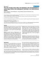
phagocytic cup as shown in Fig 1.1.
(adapted from Yeung et al, 2006)
Fig 1.1: IgG opsonized particles stimulates Fcγ receptor clustering in
mediating recognition of target particles for phagocytosis.
This leads to membrane extension and actin polymerization resulting in the
internalization of the target bound to the Fc receptor into the cell.
Chapter 1| Introduction
!
!
)!
The target particle is surrounded by the extending of pseudopods of the plasma
membrane that eventually engulfs the target particle. This ultimately results in the
delivery of the internalized particle into the cell interior within a plasma membrane
derived vacuole – the phagosome [17-19].
After internalization, actin is shed from the nascent phagosome. The phagosome,
derived from the plasma membrane does not initially possess microbicidal ability
and thus undergoes a coordinated maturation process similar to that observed for
early to late endosomes within the the endocytic pathway. The endocytic
pathway is organized as a continuum of organelles from early endosomes to
lysosomes. Phagosome maturation modifies the composition of the phagosomal
membrane and its luminal contents to endow the phagosome with an array of
microbicidal and digestive molecules needed to degrade the internalized particle.
Current phagosome maturation models imply the continuous removal and
addition of material from the endocytic compartments to the early phagosome to
convert it into a microbicidal phagolysosome (Fig1.2) [3, 10, 18]. The interplay
between the phagosomal and endosomal pathways has been described as a
“kiss and run” mechanism in which the partial and transient fusion of endosomes
and phagosomes (kiss) allows for the transfer of membrane and luminal contents
and is immediately followed by fission (run) which prevents complete mixing of
the two compartments. Therefore, in addition to the receptors and ligands that
participate in phagocytosis, other molecules provide unique characteristics to
each stage of phagosome maturation. These include molecules that regulate the