Coupling effects of NaYF4Yb,Er upconversion nanoshells and au ag metallic nanoshells 2
Bạn đang xem bản rút gọn của tài liệu. Xem và tải ngay bản đầy đủ của tài liệu tại đây (356.95 KB, 14 trang )
24
Chapter 2 Experimental Methods
2.1 Material synthesis: UC nanoshells
UC nanoparticles with hcp phase and diameter size ~10 nm were
synthesized using thermal decomposition.
71,72
A high UC emission was
achieved for the UC nanoparticles produced using this method.
73
In this
method, precursors of trifluoroacetates of metal ions were decomposed at high
temperature above 300
o
C in high boiling point solvents (e.g 1-octadecene)
and surfactants (e.g. oleylamine and oleic acid), leading to the formation of
UC nanoparticles stabilized by the surfactants.
Recent reports showed the hollow nanoparticles were synthesized via
Kirkendall effect.
74,75
The first Kirkendall effect was reported in 1942 and the
result was confirmed in 1947.
76,77
The Kirkendall effect was first studied for
the synthesis of hollow structures of cobalt sulfide nanoparticles from room
temperature to 182
o
C.
74,78
The formation mechanism of voids inside the
particles was dominated by outward diffusion of cobalt cations and balanced
by inward diffusion of vacancies. The small voids in each particle were
observed between the cobalt core and sulfide shell due to condensation of
vacancies at the boundary. These small voids coalesced into bigger ones,
followed by disappearance of the cobalt cores. Finally, a single void in the
center of the cobalt sulfide nanoparticles was formed.
More recently, the Kirkendall effect has been applied to synthesize
hollow UC nanoshells. For example, hollow NaYF
4
:Yb,Er UC nanoshells
were synthesized using a controlled ion exchange process from cubic-phase
Y
2
O
3
nanospheres
79
or thermal decomposition of a mixture of trifluoroacetate
25
precursors.
80
These hollow nanoshells were formed due to the Kirkendall
effect. In this thesis, NaYF
4
:Yb,Er nanoshells with hcp crystal structure were
synthesized using thermal decomposition of trifluoroacetate precursors in
oleylamine at 340 °C.
81
2.1.1 Preparation of trifluoroacetate precursors
Trifluoroacetate precursors of Y, Yb, and Er [(CF
3
COO)
3
M, M = Y,
Yb, and Er ions] were prepared by dissolving their respective oxides or
hydroxides in trifluoroacetic acid (CF
3
COOH), followed by drying in oven at
80
o
C.
73
Sodium trifluoroacetate (CF
3
COONa) was prepared by dissolving
sodium carbonate (Na
2
CO
3
) in trifluoroacetic acid, followed by drying in oven
at 80
o
C.
2.1.2 Synthesis of NaYF
4
:20%Yb,2%Er UC nanoshells
In the synthesis of NaYF
4
:20%Yb,2%Er nanoshells, 20 mL of
oleylamine was first reacted with 3 mL of trifluoroacetic acid in a 50-mL
three-neck flask under a continuous flow of Ar gas. A mixture of
(CF
3
COO)
3
Y (0.488 mmol), (CF
3
COO)
3
Yb (0.125 mmol), (CF
3
COO)
3
Er
(0.013 mmol), and CF
3
COONa (1.252 mmol) was then added and followed by
0.6 mL of deionized water under vigorous stirring at 60 °C for 5 min. This
mixture was heated to 340 °C using a heating mantle under refluxing condition
and in the presence of Ar gas for protection from oxidation. After 30 min, the
mixture was allowed to cool to 80 °C. The oleylamine-capped UC nanoshells
were isolated by centrifugation at 10000 rpm for 3 min, followed by washing
and redispersing in hexane for characterizations. To investigate the formation
26
mechanism of the UC nanoshells, the samples were collected at 300 °C, 5 min,
10 min, and 30 min at 340 °C for structure and microstructure analyses.
2.1.3 Synthesis of solid NaYF
4
core/NaYF
4
:20%Yb,2%Er shell
nanoparticles
In the synthesis of solid NaYF
4
core (~10 nm)/NaYF
4
:20%Yb,2%Er
UC shell (~3 nm), the solid NaYF
4
core was first prepared and then coated by
UC shell, following the reported methods.
32,35
In the preparation of solid
NaYF
4
core, CF
3
COONa (1.252 mmol) and (CF3COO)
3
Y (0.626 mmol) was
dissolved in oleylamine (20 ml) in a 50-mL three-neck flask at 90 ºC under Ar
until a clear solution was formed. The mixture was then heated to 340 °C
using a heating mantle and kept at such temperature for 30 min. The solution
was allowed to cool to 100 ºC. A mixture of (CF
3
COO)
3
Y (0.976 mmol),
(CF
3
COO)
3
Yb (0.250 mmol), (CF
3
COO)
3
Er (0.026 mmol), and CF
3
COONa
(2.504 mmol) were added to the solution. This mixture was heated to 340 °C
under refluxing condition and Ar. After 30 min, the mixture was allowed to
cool to 80 °C. The particles were isolated by centrifugation at 10000 rpm for
3 min, followed by washing and redispersing in hexane.
2.1.4 Synthesis of solid NaYF
4
:20%Yb,2%Er nanoparticles
In the typical synthesis of solid NaYF
4
:20%Yb,2%Er nanoparticles
(~15 nm), (CF
3
COO)
3
Y (0.976 mmol), (CF
3
COO)
3
Yb (0.250 mmol),
(CF
3
COO)
3
Er (0.026 mmol), and CF
3
COONa (2.504 mmol) were dissolved
in oleylamine (20 ml) in a 50-mL three-neck flask at 90 ºC under the presence
of Ar gas until a clear solution was formed. The mixture was then heated to
27
340 °C using a heating mantle and held at this temperature for 40 min. The
solution was allowed to cool to 80 ºC. The particles were isolated by
centrifugation at 10000 rpm for 3 min, followed by washing and redispersing
in hexane.
2.1.5 Surface coatings of NaYF
4
:20%Yb,2%Er UC nanoshells
Undoped NaYF
4
nanoshells were first synthesized using the synthesis
method of NaYF
4
:20%Yb,2%Er nanoshells (Sec. 2.1.2). For the preparation
of undoped NaYF
4
nanoshells, only (CF
3
COO)
3
Y (0.626 mmol) and
CF
3
COONa (1.252 mmol) were used in the first step without (CF
3
COO)
3
Yb
and (CF
3
COO)
3
Er. The surface coatings were done by coating the undoped
NaYF
4
nanoshells with Yb,Er doped NaYF
4
shell, followed by another
undoped NaYF
4
shell on top. The detailed procedure is given as follows. A
mixture of (CF
3
COO)
3
Y (0.976 mmol), (CF
3
COO)
3
Yb (0.250 mmol),
(CF
3
COO)
3
Er (0.026 mmol), and CF
3
COONa (2.504 mmol) were added to the
solution of as-synthesized undoped NaYF
4
nanoshells in a 50-mL three-neck
flask and then heated to 340 °C using a heating mantle under refluxing
condition and Ar. After 30 min, the mixture was allowed to cool to 100 °C.
Then (CF
3
COO)
3
Y (1.952 mmol) and CF
3
COONa (5.008 mmol) were added
and followed by heating to 340
o
C. After 30 min, the mixture was allowed to
cool to 80 °C. The particles were isolated by centrifugation at 10000 rpm for
3 min, followed by washing and redispersing in hexane.
28
2.2 Material synthesis: Au-Ag metallic nanoshells
Galvanic replacement reaction is a powerful method to produce hollow
metallic nanostructures.
52,82
In galvanic replacement reaction, Ag
nanoparticles are commonly used as the sacrificial metal template in synthesis
of hollow Au-Ag nanoshells. Au would be formed via the reduction of
HAuCl
4
and deposited on Ag templates being oxidized.
83
The deposition of
Au and oxidation of Ag solids lead to the formation of Au-Ag nanoshells with
interior cavity.
2.2.1 Synthesis of Ag nanoparticles
In a typical synthesis of Ag nanoparticles consisted of decahedral (~43
nm in size) and triangular prism (~53 nm in edge length) shapes, 5 mL of 1-
octadecene (ODE) and 3 mL of oleylamine were mixed using a magnetic
stirrer at a spin rate of 700 rpm in a 25-mL three-neck flask. ODE was
selected because of its high boiling point (~315
o
C) and good compatibility
with oleylamine, allowing the reaction at 160
o
C. The ODE-oleylamine
mixture was then heated to 160
o
C using a heating mantle under a continuous
flow of N
2
gas. A solution of 10 mg of AgNO
3
(58.9 mol) was immediately
injected to the mixture. The temperature decreased to ~155
o
C after injection
of the AgNO
3
solution, then increased to 160
o
C again within a few minutes.
After 30 min at 160
o
C, the solution was cooled to 60
o
C, followed by dilution
with 8 mL of toluene. The solution of Ag nanoparticles was kept in a vial
wrapped with aluminum foil and stored in the dark until further use. As-
synthesized Ag nanoparticles were used as a sacrificial template in synthesis
of Au-Ag nanoshell via the galvanic replacement reaction with HAuCl
4
.
29
2.2.2 Synthesis of Au–Ag nanoshells
As-synthesized Ag nanoparticles (16 mL in the solution) were added to
a 50-mL three-neck flask (with magnetic stirrer at a spin rate of ~700 rpm) and
then heated to 60
o
C in a water bath. A 5 mM HAuCl
4
solution was prepared
by dissolving 29.5 mg of HAuCl
4
•3H
2
O (75 mol) in 13.5 mL of toluene and
1.5 mL of oleylamine. Fresh HAuCl
4
solution was prepared and kept in the
dark before use. The 5 mM HAuCl
4
solution was added to the 50-mL reaction
flask at an injection rate of 0.25 mL/min. The samples were collected after
injection of various amounts of 5 mM HAuCl
4
solution as shown in Table 2.1.
The samples were isolated by centrifugation at 10,000 rpm for 3 min. The
obtained particles were washed three times with 8 mL of toluene, followed by
centrifugation, and re-dispersed in 8 mL of toluene for characterizations.
Table 2.1 Samples were collected after the injection of a variable amount of 5
mM HAuCl
4
solution for the measurement of microstructure, morphology,
chemical composition, and extinction spectra.
Sample
Volume of 5 mM HAuCl
4
solution (mL)
Amount of
HAuCl
4
(mol)
1 2.0 10
2 3.0 15
3 4.0 20
4 5.0 25
5 6.0 30
6 10.0 50
7 15.0 75
30
2.3 Assembly of Au-Ag nanoshells and UC nanoshells
To study the plasmonic effects of Au-Ag nanoshells on fluorescence
properties of UC nanoshells, single layer of Au-Ag nanoshells on glass
substrates were prepared using spin-coating, followed by the coating of silica
film using sputtering. UC nanoshells were further deposited on the silica film-
coated Au-Ag nanoshell layer to form an assembly of Au-Ag nanoshell
layer/silica film/UC nanoshell layer. In this assembly, the distance between
Au-Ag nanoshell layer and UC nanoshell layer was controlled by the thickness
of silica film. The assemblies with different surface coverage % of Au-Ag
nanoshell layer were also prepared. The surface coverage- and distance-
dependent fluorescence of the UC nanoshells was investigated.
2.3.1 Preparation of Au-Ag nanoshell layer
Glass substrate was prior soaked in aqua regia (3 parts concentrated
HCl (37%)/1 part concentrated HNO
3
(65%)) for removal of contamination at
the glass surface, followed by washing three times with ethanol. The washed
glass was then dried in oven at 80
o
C for 24 h. The glass was kept in vacuum
chamber before use.
Au-Ag nanoshell layer on glass substrate was prepared using spin-
coating. The Au-Ag nanoshell solution (25 L) of was dropped on a glass
substrate (1.5 cm x 1.5 cm), followed by spin-coating at a speed of 1500 rpm
for 30 seconds for each cycle. Samples were prepared by 5 – 40 cycles of spin
coating to obtain Au-Ag nanoshell layer with different surface coverage % on
the substrates. The samples were kept in the vacuum chamber before use.
Scanning electron microscopy (SEM) was performed for each sample. The
31
surface coverage % of the Au-Ag nanoshells on the substrate was calculated
from the SEM images using Java image processing and analysis program
(ImageJ). The surface coverage % is defined by the ratio of area occupied by
the nanoshells to the total analyzed area.
2.3.2 Assembly of Au-Ag nanoshell layer/silica film/UC nanoshell layer
Silica deposition was performed using a magnetron sputtering system
at a deposition rate of 15 nm per h at room temperature. The operating
conditions were 150 W (RF power), 0.5 Pa (chamber pressure), and 50
standard cubic centimeter per minute (sccm) (Ar flow rate). The silica
deposition rate was calibrated using surface profiler (KLA Tencor Alpha-Step
Q). The glass and silica have similar appearance and properties. For the
thickness measurement using the surface profiler, the glass substrate was first
coated with Ti film and followed by silica film. The silica thickness was
obtained from the total (Ti + silica) thickness normalized by the thickness of
Ti film. The average silica deposition rate was measured to be ~15 nm per h.
A similar silica deposition rate (~15 nm per h) was obtained from the silica
film sputtered on silicon substrates using the same operating conditions of the
magnetron sputtering system (Fig. 2.1). In this study, silica film was selected
due to its good chemical and thermal stability, and low thermal conductivity
(1.4 Wm
−1
K
−1
).
84
Its inertness to solvents (e.g. hexane and toluene) would
allow the deposition of hexane solution of UC nanoshells on the silica film-
coated Au-Ag nanoshells by solvent evaporation method, whereas the low
thermal conductivity would reduce the photothermal effects of Au-Ag
nanoshells to UC nanoshells.
32
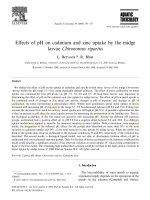
Fig. 2.1 (a) SEM image of cross-section of silica film sputtered on silicon
substrate for 4 hours. (b) Average thickness of silica films obtained at
different sputtering time. The average silica deposition rate was calculated to
be ~15 nm per h.
The Au-Ag nanoshell layer with different surface coverage % on the
glass was coated by 30-nm silica films using a magnetron sputtering system.
The silica film-coated Au-Ag nanoshell layer was then coated with UC
nanoshells. The procedure is given as follows. The sample of silica film-
coated Au-Ag nanoshell layer was placed in a 10-mL beaker glass. Then 2
mL of 0.07 wt% hexane solutions of UC nanoshells were added to the 10-mL
beaker glass. The UC nanoshells were then deposited on the Au-Ag nanoshell
layer/silica film by solvent evaporation in a vacuum chamber (Fig. 2.2). To
obtain the similar concentration of UC nanoshells deposited on the Au-Ag
nanoshell layer/silica film, same concentration and volume of hexane solutions
of UC nanoshells was used to fabricate all the assemblies in this study. The
effects of the Au-Ag surface coverage on the fluorescence of the UC
nanoshells were studied.
To investigate the distance-dependent fluorescence of UC nanoshells,
the Au-Ag nanoshell layer (prepared by 20 cycles of spin-coating) was coated
33
by silica films with different thickness (5 - 180 nm) using the magnetron
sputtering system. The UC nanoshells were then deposited on the Au-Ag
nanoshell layer/silica film to form an assembly of Au-Ag nanoshell layer/silica
film/UC nanoshell layer. The distance between the Au-Ag nanoshell layer and
UC nanoshell layer was controlled by the thickness of the silica film.
Fig. 2.2 Schematic of deposition of UC nanoshells (0.07 wt% in hexane) by
solvent evaporation in a vacuum chamber.
2.4 Material characterizations
2.4.1 Crystal structure
X-ray diffraction (XRD) is a non-destructive characterization
technique which can provide crystal structure information of materials. In this
technique, monochromated X-ray striking a sample is scattered by the atoms
in the sample. The scattered intensity of the X-ray is collected as a function of
the incident and scattered angle. In this thesis, the crystal structures of the
34
samples were investigated using powder XRD diffractometer system (Cu K
radiation) (Bruker AXS, Karlsruhe, Germany). XRD spectra of the samples
were compared with their corresponding standard XRD spectra [Joint
Committee on Powder Diffraction Standards (JCPDS), for example, file
number PDF 16-334 for hcp sodium yttrium fluoride and PDF 4-783 for fcc
silver].
2.4.2 Microstructure and surface morphology
Transmission electron microscopy (TEM) is a microscopy technique in
which an electron beam is transmitted through an ultra-thin sample, interacting
with the specimen as it passes through. An image is formed from the
interaction of the electrons transmitted through the sample. In this thesis, the
microstructure of the nanoparticle samples was studied using a JEOL JEM
2010F transmission electron microscope (JEOL, Tokyo, Japan) operated at
200 kV. Carbon-coated copper grids (400 meshes) were used to support the
nanoparticles. The average particle size was estimated by random
measurements of 100 – 200 particles from the TEM images. Elemental
composition of the samples were performed using energy dispersive X-ray
(EDX) in the transmission electron microscope. The average concentration of
the elements in the samples was determined from the EDX data randomly
collected at least 5 different selected area.
Different from the TEM which produces an image by detecting the
electrons transmitted through the sample, scanning electron microscopy
(SEM) produces an image by detecting the electrons such as secondary
electrons which are emitted from the sample surface due to excitation by the
35
primary electron beam. The SEM can produce high-resolution images of the
sample surface. In this thesis, the surface morphology of the samples was
investigated using a Zeiss Supra 40 field emission scanning electron
microscope (Carl Zeiss, Oberkochen, Germany). The samples were prepared
by depositing the particle solution on silicon substrate, followed by drying in
vacuum chamber.
2.4.3 Dynamic light scattering (DLS) analysis
DLS is a technique that can be used to determine the size distribution
profile of small particles in suspension. In DLS analysis, the motion related to
size of the particles is measured. The smaller particles move faster than the
larger ones in the solution. The particle size obtained by this technique is the
diameter of a sphere which is commonly referred to as the hydrodynamic
diameter. In this study, the DLS measurement was performed using a Malvern
Zetasizer Nano ZEN3600 (Malvern Instruments, Worcestershire, UK).
2.4.4 UC fluorescence
The room UC fluorescence spectra were measured using a LS-55
luminescence spectrometer (Perkin Elmer Instruments, Cambridge, UK) with
an external 980-nm laser diode (1 W, continuous wave with 1 m fiber, Beijing
Viasho Technology Co., Beijing, China) as the excitation source in place of
the xenon lamp in the spectrometer. The spectrometer was operated in the
bioluminescence mode, with a gate time of 1 ms, delay time of 10 ms, cycle of
20 ms, and a flash count of 1. The UC fluorescence photographs were taken
36
using Canon Powershot A620 and Canon EOS 60D cameras (Canon, Tokyo,
Japan).
2.4.5 UV–Vis–NIR extinction
UV–Vis–NIR spectrometer measures the intensity of light passing
through a sample and compares it to the intensity of incident light. For
example, when light passes through a particle, the light can be absorbed,
scattered, and/or transmitted by the particle (Fig. 2.3). The absorption and
scattering of the particle can be expressed by absorption and scattering cross
section, respectively. The sum of absorption and scattering cross section is
referred to as the extinction cross section. The extinction spectra can be
determined by recording the light passing through the sample using UV–Vis–
NIR spectrometer. In this thesis, the UV–Vis–NIR extinction spectra were
measured using a Cary-5000 UV–Vis–NIR spectrophotometer
(Varian, Palo
Alto, CA, USA). The extinction spectra of the samples were normalized to
that of their surrounding mediums (e.g. solvents, substrates).
Fig. 2.3 An illustration describes the transmission, absorption, and scattering
processes of lights passing through a particle.
Incident lights
Scattered
Transmitted
Absorbed
Particle
37
2.4.6 X-ray photoelectron spectroscopy (XPS)
Photoelectron spectroscopy is based upon a single photon in/electron
out process. In XPS, an X-ray photon of energy hv absorbed by an atom in a
molecule or solid leads to ionization and the emission of a core (inner shell)
electron. The kinetic energy (E
k
) of the emitted photoelectron is measured by
electron energy analyzer (electron spectrometer). Thus, the binding energy
(E
b
) of the electron can be calculated:
Φ2.1
where Φ is the spectrometer work function. From the XPS spectra, the
composition of the elements can be calculated from the ratio of integrated
peak areas normalized by respective sensitivity factors. The XPS analysis was
performed using Kratos Axis Ultra DLD (Kratos analytical, Manchester, UK)
with a monochromated Al K
α
source (1486.6 eV).
2.4.7 Inductively coupled plasma optical emission spectrometry (ICP-
OES)
ICP-OES is an analytical technique used for the detection of trace
metals. It is a type of emission spectroscopy that uses the inductively coupled
plasma to produce excited atoms or ions that emit electromagnetic radiation at
wavelengths characteristic of a particular element. The intensity of this
emission is indicative of the concentration of the element within the sample.
The composition of metal ions was performed using a Dual-view Optima 5300
DV ICP-OES system (PerkinElmer, Shelton, USA).