MODERN MORPHOLOGICAL TECHNIQUES AND THE EVOLUTIONARY BIOLOGY AND TAXONOMY OF SEPSIDAE (DIPTERA) 3
Bạn đang xem bản rút gọn của tài liệu. Xem và tải ngay bản đầy đủ của tài liệu tại đây (1.82 MB, 69 trang )
128
CHAPTER 6
__________
Using multiple data-sources to address taxonomic
uncertainties
Abstract
In this chapter, I use numerous data-sources in conjunction to resolve two issues
in sepsid taxonomy. In Part I, large differences between the genetic sequences of Sepsis
flavimana (Sepsidae) specimens from USA and Germany suggested the possible
presence of a 'cryptic' species. A follow-up investigation based on morphology, mating
behaviour data and reproductive isolation experiments revealed that there were indeed
two biological species. The American specimens were later revealed to be Sepsis
pyrrhosoma, a 'cryptic' species that was previously synonymised under S. flavimana.
For this study, I performed the morphological and taxonomic analysis, and contributed
to the phylogenetic analysis. In Part II, specimens resembling Themira leachi
(Sepsidae) were found in Neotropical Cuba, which is puzzling given that the species
has only been recorded in the Holarctic. A morphological and genetic comparison with
German specimens revealed that the Cuban specimens were indeed T. leachi, which
confirms a disjunctive distribution in the species that was likely brought about by
synantrophic processes. For this study, I similarly performed the morphological and
taxonomic analysis.
129
Part I: From ‘cryptic species’ to integrative taxonomy – an
iterative process involving DNA sequences, morphology, and
behaviour leads to the resurrection of Sepsis pyrrhosoma
(Sepsidae: Diptera)
3
DNA sequence data have recently gained much popularity in taxonomic
research and is generally acknowledged today that they provide important evidence for
delimiting species (Meier et al. 2008). DNA data can now be generated at a fast rate,
with relatively low cost, and by personnel uninitiated with taxon-specific knowledge
required for morphological research (Lee 2000, Scotland et al. 2003). However,
increasingly, the widespread use of DNA sequences has also created problems in the
form of so-called ‘cryptic species’ that are now routinely proposed when morphology
and DNA sequence evidence – at least initially – yield different inferences about
species boundaries (Bickford et al. 2007).The use of the term ‘cryptic species’ implies
that the unit is already properly diagnosed as a species. However, this is rarely so and
in most cases a resolution of the conflict between morphology and DNA sequence
information is not even attempted. As a consequence, ‘cryptic species’ are
accumulating in the literature and interfere with a proper classification and the
3
A version of this chapter has been published as Tan DS, Ang YC, Lim GS, Ismail MRB, Meier R
(2010) From ‘cryptic species’ to integrative taxonomy: an iterative process involving DNA
sequences, morphology, and behaviour leads to the resurrection of Sepsis pyrrhosoma (Sepsidae:
Diptera). Zoologica Scripta 39, 51-61, where I performed the morphological and taxonomic
analysis, and contributed to the phylogenetic analysis.
130
assessment of biodiversity (e.g. resulting in an undercount of species numbers). Here I
demonstrate how an iterative process based on multiple sources of data can move a
‘cryptic species’ from being only a putatively new species-level taxon to being
formally recognized as a species based on sufficient evidence (Petersen et al. 2007).
It is sometimes assumed that this process of iterative taxonomy only requires
enough data, but this not necessarily be the case, as the same data may yield different
species inferences under different species (Laamanen et al. 2003, Tan et al. 2008).
Many authors avoid this issue due to the vitriol related to species concept discussions.
However, it is precisely when data are in disagreement that it is important to be explicit
about species concepts, because in these cases species concepts can matter (Tan et al.
2008). Here I suggest that the best solution is applying a two-step process. One can first
evaluate the available data based on the species concept that is favoured by the authors.
Afterwards, the same data can be discussed under alternative species concepts
(Laamanen et al. 2003). This approach will ensure that species are clearly defined given
that the authors’ opinion based on their species concept will be binding under
nomenclatural rules. At the same time the treatment is transparent and allows
proponents of alternative species concept to draw their own conclusions.
Most species in entomology are recognized based on morphological characters.
Sepsid flies are no exception but the use of morphology for some species can be
problematic because of the bewildering amount of phenotypic variability present in this
family (Pont and Meier 2002). In sepsids most of this variability is related to
environmental factors, such as the amount of food available to the larvae (Meier
1995a). In these cases DNA sequences are particularly useful for clarifying species
131
boundaries, because the sequences are not affected by the environmental variables. In
other cases the observed intraspecific variability is at least partially genetic (Reusch
and Blanckenhorn 1998). Here, DNA sequences can still be used as additional
evidence, but any observed sequence variability across allopatric populations can be
difficult to interpret (Memon et al. 2006, Petersen et al. 2007, Ang et al. 2008a)
because recently diverged species can share barcodes and may thus be incorrectly
lumped into one species (Meier et al. 2006, Meier et al. 2008). Similarly, allopatric
populations within old species may have distinctly different sequences and DNA
evidence may erroneously suggest that they should be split into multiple species (Meier
2008, Meier et al. 2008).
Here I demonstrate the value of an iterative approach using multiple sources of
data by clarifying the species boundaries of Sepsis flavimana Meigen, 1826. As with
many similar cases in recent literature (Bickford et al. 2007), my taxonomic problem
started with finding unexpectedly high levels of COI divergence between what
appeared to be allopatric populations that were collected from various locations in
North America. Based on recently published identification keys (Ozerov 2000, Pont
and Meier 2002), these specimens all keyed out to one species, S. flavimana. This
particular species is one of the most morphologically variable sepsids, with much of its
variability related to size (Munari 1993, Pont and Meier 2002). Not surprisingly, this
species has spawned a large number of synonyms [eleven; Ozerov (2005)]. Among
others Ozerov (2000) synonymised four Nearctic species with S. flavimana when
revising the North American fauna (S. vicaria Walker, 1849, S. pyrrhosoma Melander
and Spuler, 1917, S. melanopoda Duda, 1926 and S. kertezsi Duda, 1926).
132
However, the unexpectedly high level of genetic variability that I found within
the North American populations of what appeared to be S. flavimana motivated me to
re-investigate the morphology in order to test whether these genetically distinct
populations may also be morphologically distinct. As additional sources of data, I was
also able to study the mating behaviour (under laboratory conditions) and test for
reproductive isolation based on cultures that had been established for two genetically
distinct populations from North America and Europe.
Materials and Methods
Collection, rearing and morphology. Sepsis ‘flavimana’ specimens were
collected from six American populations and stored in 100% ethanol for subsequent
morphological and genetic study (Fig. 5.1). In addition, live specimens from New
Orleans (LA, USA), Kevelaer (NRW, Germany), and Ahrensfelde (Schleswig-Holstein,
Germany) were reared in laboratory cultures using sucrose syrup as a carbohydrate
source and cow dung as a breeding substrate. Cow dung was initially frozen at -80˚C
for several days to kill any insects infesting the dung prior to collection. Fly cultures
were maintained at 25-28˚ C in 2l plastic containers. Compound microscopy and high-
fidelity microscopic photography (Visionary Digital ™ BK+ system using a Canon
EOS MkIII fitted with Infinity Optics K2 Long Distance Microscope on CF4P3
objective settings) were used to study the morphology of specimens from all eight
localities in detail.
DNA sequences. A 778bp fragment of cytochrome oxidase c subunit I (COI)
was amplified and sequenced, including the DNA barcoding region from 50 individuals
133
representing multiple populations of five nominal Sepsis species (S. biflexuosa, S.
duplicata, S. flavimana, S. fissa, and S. ‘pyrrhosoma’) with S. fissa designated as
outgroup based on Su et al. (2008). Genomic DNA was extracted from individuals via
a modified phenol-chloroform method described by Petersen et al. (2007a), and
amplification and sequencing protocols for COI followed Su and colleagues (2008). All
sequences were aligned with CLUSTALX 2.01 (Thompson et al. 1997) and the
alignment was free of indels.
Phylogenetic analyses. Maximum likelihood and maximum parsimony were
used to infer the gene-tree for the COI of S. ‘flavimana’ populations and related
species. A new technology parsimony search was implemented in TNT 1.1 (Goloboff
et al. 2008) with search level 55; the minimum tree length was found 10 times. Node
support was assessed through jackknife resampling, with absolute frequency
differences and 36% character deletion for 250 replicates. A maximum likelihood
bootstrap tree was obtained with GARLI 0.951 (Zwickl 2006). Using the Akaike
Information Criterion (AIC), MrModeltest (Nylander 2004) selected the GTR + Γ + I
model for COI. The analysis was automatically terminated if there was no improvement
of a log likelihood of 0.01 or more after 50,000 generations. Support was obtained as
maximum likelihood bootstrap with 250 replicates under the same settings used to
obtain the most likely tree.
Observations of mating behaviour. Virgin flies were obtained from each culture
by isolating a petri-dish of larvae-infested dung from the laboratory colony in an empty
container and segregating males and females within 6 hours of eclosion. Sepsid flies, at
least in the flavimana group, acquire sexual maturity after two to five days (personal
134
observation). Flies were thus assumed to be sexually mature after five days as adults.
To examine reproductive compatibility within and between populations, one virgin
male was introduced to a 3.5cm plastic petri-dish containing a single virgin female, and
the behaviour of both flies was recorded at 7X – 15X magnification with an analogue
video recorder attached to a trinocular Leica MZ16A microscope. Recordings began
upon introduction of both flies and ended either after successful copulation or after 60
minutes if copulation did not occur. The analogue recordings were then digitised and
analysed frame-by-frame (25 frames per second) using the non-linear video editing
software Final Cut Pro. Behavioural elements were then recorded to facilitate
comparisons among populations. 10 and 12 mating trials were recorded for the two
populations from North America and Europe respectively.
Observations of mating behaviour. To examine the reproductive compatibility
between populations of S. ‘flavimana’, we attempted to mate males and females from
different continents. Five sexually mature virgin flies of each sex were placed in
rearing containers under conditions identical to those in which cultures from individual
collection localities live and breed successfully. Male and female flies originated from
different continents, and the following reciprocal pairings were attempted: Ahrensfelde
♂ × New Orleans ♀; Ahrensfelde ♀ × New Orleans ♂. We also examined reproductive
compatibility between the two European populations: Ahrensfelde ♂ × Kevelaer ♀ and
Ahrensfelde ♀ × Kevelaer ♂. No flies died during the course of these trials. Each of
these five male × female pairings was thrice replicated. The breeding substrate in each
container (a 7cm petri-dish containing cow dung) was examined every other day for the
presence of fertilized eggs or larvae. Substrate with fertilized eggs or larvae was
removed and placed in separate containers for pupation of larvae. Where hybrid flies
135
were obtained, they were again segregated by sex within one day of eclosion to
maintain their virginity. We then attempted to back-cross these hybrids with virgin flies
from their parental cultures. To ascertain whether flies from failed back-crossing trials
were fertile, we attempted to mate them with other flies from their own respective
populations.
Results
Morphology
We found two discrete morphotypes from the North American specimens that
could be distinguished by a suite of morphological characters. One morphotype was
indistinguishable from all European specimens of Sepsis flavimana, while the other
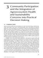
morphotype has the following distinguishing features (Fig. 5.2): (1) the flies are
consistently lighter in colour (especially on the thoracic pleura, face, gena and legs cf.
Fig 2. A, G vs. H); (2) the male fore-tibia lacks a distinct ventro-basal bump on the
tibia (C) as compared to the European morphotype (J); (3) the epandrium and base of
the surstylus of the male is light in colour and only the tip is dark (lateral view A,
dorsal view F); (4) the surstylus has a sub-medial tooth (D). Features 1 and 2 are
consistent with the description of S. pyrrhosoma by Melander and Spuler (1917) which
mentions that the species is “largely reddish along the sides” with “face and cheek
yellowish,” and with a male foretibia “slightly decreasing in size towards the tip and
bearing a very weak and setulose tubercle on the underside near the base.” Features 1–3
are also visible on the holotype of S. pyrrhosoma (W. Mathis, pers. comm.). For
convenience, I refer to this as the ‘pyrrhosoma’ morphotype. North American
136
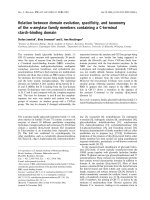
Figure 5.1: Consensus tree of Sepsis flavimana group. Parsimony jackknife percentiles are given above
branches and maximum likelihood bootstrap percentiles below.
137
Figure 5.2: A–G. Sepsis pyrrhosoma (♂ unless otherwise noted). A. Habitus, lateral view, showing
hypopygial capsule (hyp). B. Fore-femur, posterior view. C. Fore-tibia, posterior view. D. Surstylus,
dorsal view. E. Thorax, lateral view, showing pruinosity pattern on postprotonotum (ppn),
preepisternum (pest), anepisternum (aepst), ketepisternum (kepst), anepimeron (aepm), katatergite
(kat), meron (m) and metepimeron (mepm). F. Postabdomen, ventral view, showing 4
th
sternite (4
th
st.)
and hypopygial capsule. G. ♀ habitus, lateral view. H–K. Sepsis flavimana (♂). H. Habitus, lateral view,
showing hypopygial capsule. I. Fore-femur, posterior view. J. Fore-tibia, posterior view. K. Surstylus,
dorsal view. Scale bars (A, E, G, H): 1mm; (B, C, F, I, J): 0.5mm; (D, K): 0.1mm
138
specimens from Palmyra, VA, and Dyar Pasture, GA, are morphologically
indistinguishable from European S. flavimana, and are henceforth referred to as the
‘flavimana’ morphotype.
Molecular Data
We obtained COI barcode sequences of ca. 778 bp from 50 specimens
(GenBank accession numbers EU435804, EU435807, EU435808, EU435818,
GQ354410, GQ388730 – GQ388774). The parsimony analysis found 125 trees with a
length of 211 steps and the sequences for all species formed strongly supported
monophyletic clusters. The two morphotypes were monophyletic sistergroups with
strong support; this result was mirrored in the maximum likelihood analysis (see Fig.
5.1). Uncorrected pairwise distances between the two morphotypes from North
America ranged from 6.1–7.0%. However, distances within morphotypes were
considerably shorter: 0.0–0.5% in the ‘flavimana’ morphotype and 0.0–1.6% in the
‘pyrrhosoma’ morphotype. Distances between the European S. flavimana and
American ‘flavimana’ morphotypes were 1.7–2.9%, while distances between
‘pyrrhosoma’ morphotypes from North America and all other ‘flavimana’ morphotypes
(from both Europe and North America) were 6.2–7.7%. The distances between
European S. flavimana populations were 0.0–1.7% (Table 5.1).
139
Table 5.1: Uncorrected pairwise genetic distances between and within and between Sepsis flavimana
and S. pyrrhosoma morphotypes
S. flavimana
(North America)
S. flavimana
(Europe)
S. pyrrhosoma
(North America)
S. flavimana
(North America)
0.00-0.52%
S. flavimana
(Europe)
1.69-2.87% 0.00-1.70%
S. pyrrhosoma
(North America)
6.14-7.04% 6.17-7.65% 0.00-1.62%
Behavioural observations and reproductive isolation trials
The mating behaviour of S. pyrrhosoma differs from that of European S.
flavimana in several respects. In S. flavimana, the male proboscis only touches the
female on the dorsal region of the thorax, while in S. pyrrhosoma the male proboscis is
used to stimulate the female ocelli instead (Table 5.2; The video evidence for these
differences can be viewed from or Youtube at
and In addition, S. pyrrhosoma males were
observed to stimulate the postabdomen of females with their surstylus prior to
copulation, but this behaviour was absent in S. flavimana. During copulation, S.
flavimana females constantly shook their bodies, but this apparent resistance to mating
was not as violent or obvious in S. pyrrhosoma, where female body shakes were
sporadic and less energetic. Prolonged struggles lasting five or more seconds were
consistently observed during genital decoupling between S. flavimana individuals. In
contrast, separation was prompt in S. pyrrhosoma, with males typically requiring only
two quick about-turns to disengage from the female. Finally, the mating success rates
of virgin S. pyrrhosoma were much higher than in S. flavimana.
140
Table 5.2: Qualitative comparison of behavioural elements observed in S. flavimana and S. pyrrhosoma
(virgin) mating trials
Behavioural Elements
S. flavimana
S. pyrrhosoma
male mid-leg tarsi curl
observed in both species
male hind-leg tap of female abdomen
substance transfer (from male hind-leg to
female thorax)
male mid-leg rub of female thorax
degree of female resistance (shaking) violent and persistent mild and sporadic
separation after copulation
prolonged struggle to break
genital contact
rapid
precopulatory surstylus stimulation absent present and prolonged
location of male proboscis contact with
female
dorsal part of female thorax female ocelli
Mating success (virgin trials) 33.3 %
a
100%
b
a
n = 12;
b
n = 10
Table 5.3: Results of the hybridization experiments
Mass Crossings
Back Crossing
Ahrensfelde ♂ × New Orleans ♀
✕
(N/A)
Ahrensfelde ♀ × New Orleans ♂
✔ ✕
Ahrensfelde ♂ × Kevelaer ♀
✔ ✔
Ahrensfelde ♀ × Kevelaer ♂
✔ ✔
Mating trials reveal no potential for gene flow between the two species, since
hybrid offspring are produced in only one direction (Ahrensfelde S. flavimana ♀ × New
Orleans S. pyrrhosoma ♂) and these hybrids failed to produce viable eggs regardless of
whether they were mated with other hybrids or with either parent species (backcrosses;
Table 5.3). To demonstrate that laboratory conditions were sufficient to foster mating
between reproductively compatible flies, we successfully crossed S. flavimana from the
two European populations.
141
Taxonomic conclusions
Morphology, genetic data, behavioural differences, and reproductive isolation
support the resurrection of S. pyrrhosoma from synonymy, and I here re-describe the
species.
Species Re-description (Fig. 5.2)
Sepsis pyrrhosoma (Melander and Spuler 1917)(Family SEPSIDAE)
Sepsis pyrrhosoma (Melander and Spuler 1917): Fig. 14.
Holotype and allotype. 1 ♂ (Holotype) from Lafayette, IN (Melander and
Spuler 1917), 1 ♀ (Allotype) from Philadelphia, PN (Johnson, 1917), both in National
Museum of Natural History (NMNH), Washington, DC, USA.
Additional specimens. ♂♂♀♀ from ex culture established from ♀♀ from
grassland along Leake Avenue near Mississippi River, New Orleans, LA), ca. 5m ASL,
29˚ 55' 48.34" N 90˚ 8' 4.17" W 2008 (Coll. R. Meier); in Raffles Museum of
Biodiversity Research, Singapore (RMBR). Other samples from Raleigh, NC, New
York and Athens, GA.
Etymology. The specific name first given by Melander and Spuler in their
original description of the species (Melander and Spuler 1917), derived from the
combination of the Greek πυρο (pyro; fire) and σώμα (soma; body), an indication
towards the reddish hue of the fly’s body. The gender is neutral.
Distribution. Apparently limited to the South-eastern regions of North America,
142
Indiana, Louisiana, Georgia, North Carolina, Virginia, and Pennsylvania.
Diagnosis. Adult Sepsis pyrrhosoma resemble lightly coloured specimens of S.
flavimana. However, S. pyrrhosoma can be consistently distinguished from the latter by
the following characters. While S. flavimana (H) is always black to dark brown in
thorax and head colour, S. pyrrhosoma (A, G) is mostly reddish to yellow on the pleura
and abdominal sections as well as on the face and gena. Fore-femora of S. pyrrhosoma
are also consistently light yellow (B), while S. flavimana invariably retains a dark
brown region dorsally (I). Colour is the only way to distinguish ♀ S. pyrrhosoma (G)
from ♀ S. flavimana morphologically. Additional characters in the male are: (1) the
fore-tibial ventro-basal bump is always slight or non-existent in S. pyrrhosoma (C),
bearing small, weak bristles, while S. flavimana (J) has a distinct bump with longer and
thicker bristles; (2) The hypopygial capsule of S. flavimana (H) is entirely black with a
smooth, beak-like surstylus (K) while S. pyrrhosoma possesses a yellow hypopygial
capsule with only the surstylus darkened apically (A, F). (3) The S. pyrrhosoma
surstylus bears sub-medial inward-facing protrusions not present in S. flavimana (cf. D
& K).
The original description of Sepsis pyrrhosoma by Melander and Spuler (1917)
was brief. The following is a more detailed description of the adult based on the
specimens from New Orleans.
Colour (A–D, G). Similar in both sexes. Vertex and occipital region black,
frons and facial ridge dark brown. Parafacial, facial carina and gena light brown to
yellow. Pedicel and 1st flagellomere brown, arista black. Clypeal margins black.
143
Scutum and subscutellum black. Postprotonotum and pleural areas mostly yellowish
red, except for the dorsal margin of the anepisternum, pleural wing process, meron,
metepisternum and dorsal half of katepisternum, which are dark brown. All coxae and
trochanters always light yellow, as are fore-femora and tibiae. All tarsi are light yellow
until 4th and 5th tarsomeres, which are black. Mid and rear femora infuscate on the
dorsal and ventral side medio-distally, while tibiae are brown to dark brown basal-
medially. Abdomen with a cupreous tinge, yellowish red except for dorsal regions of
tergites and all sternites, which are dark brown. Epandria and cerci yellow, surstylus
yellow but black apically.
Macrotrichial pruinosity (E). Similar in both sexes. Head glossy except for
occipital region, gena and face, which is moderately pruinose with macrotrichia.
Scutum, pronotum and scutellum also moderately pruinose. Subscutellum and
anatergite glossy except for sparse microtrichia near margins. Proepisternum similarly
glossy with microtrichia limited to dorsal and ventral margins. Microtrichia also
present on posterior margin of anepisternum, anterior, dorsal and posterior areas of
anepimeron. Katepisternum and meron heavily pruinose, while katatergite medium
pruinose. Metepimeron with a clear ventrally.
Head (A, G). Similar in both sexes. Roundish, facial carina short and shallow.
Parafacial and gena narrow. With two subvibrissal bristles and numerous short setae
along lower genal margin. Numerous supracervical setae present. Eyes maroon,
roundish but posteriorly compressed on dorsal and ventral sides. Postcellar setae ¾ of
ocellar setae, both divergent. Outer vertical setae ½ of inner vertical setae. Pedicel
bearing setae along apical margin with 1 dorsal bristle. Flagellomere in profile long-
144
oval, rounded apically, almost twice as long as wide. Aristae dorsal and bare. Larger
specimens tend to have a disproportionately larger head compared to other specimens.
Thorax (A, E, G). With the following paired setae: 1 postprotonotal setae, 2
notopleural setae, 1 supraalar setae, 2 postsutural dorsocentral setae, 1 anepisternal
setae and 1 apical scutellar setae. Anepisternal setulae absent. Scutellum compressed,
more than twice as wide as is long.
Abdomen (A, D, F, G). Tergites (t) similar in both sexes; all with relatively long
setulae at discal and marginal regions. Synt1+2 with 1-2 pairs lateral marginal bristles,
proceeding t3 – 5 with 1 pair, t6 with none. Spiracles 1 & 2 in intersegmental
membrane, spiracles 3 – 5 on margin of tergite plate, spiracles 6 and 7 within t7.
Abdomen is slightly constricted after syntergite (synt) 1+2. Sternites (st) well defined,
with s4 bearing 2–3 rows of strong setae posteriorly (F). Bristles are more prominent in
males than females. ♂ terminalia – Symmetrical surstyli short and angulate,
decussating and fused to epandrium (D, F); with inward protrusion present medially
(D). Cercal lobes fused, each with 1 translucent apical seta.
Legs (A–C, G). ♂ fore-legs: slightly enlarged femur bearing one large ventral
(v) bristle at the middle and a slight tubercle bearing four to six shorter bristles on
posterio-ventral side (B). fore-tibia slim with a very slight basal bump bearing a row of
weak bristles (C). Mid-femur with one large and long anterior-ventral (av) bristle in
center. Mid-tibia with two smaller bristles av, centrally and preapically, one small
dorsal (d) bristle preapically; apex with bristles except on d region. Hind-femur without
distinct bristles; hind-tibia with one small d bristle preapically, one to two small av
145
bristles apically. Hind tibia bearing very faint region of osmoterium on anterior-dorsal
region medially. ♀ fore-legs simple and unmodified. Mid- and rear-femur without
bristles. Mid-tibia with one small v bristle on median, one small av bristle preapically,
with apice similar to ♂. Rear-tibia similar to ♂ but without osmoterial region.
Wing (A, G). Elongate, longer than abdomen. Veins bare except for a few
minute setulae on ventro-basal side of stem vein. Wing entirely covered with
microtrichia, with oblongish pterostigma at tip of R2+3. Anal lobe well developed, A2
not reaching wing margin. Upper calypter brown with long thin setae on margin. Lower
calypter absent. Halter creamy to yellow.
Discussion
Our study of the species boundaries of Sepsis pyrrhosoma demonstrates how
multiple sources of data can be used to resolve the status of so-called cryptic species
that are suggested by unexpectedly large genetic distances within a single nominal
species. my approach is iterative in that unexpected genetic variability prompted
renewed morphological evaluation. This re-evaluation uncovered consistent
morphological characters that distinguish S. flavimana and S. pyrrhosoma. However,
this morphological evidence initially appeared weak, because many Sepsis species
exhibit considerable size variability that is known to be correlated with differences in
body colour and other important diagnostic features such as fore-legs and claspers
(Pont and Meier 2002). Sepsis pyrrhosoma could therefore be easily mistaken as a S.
flavimana which probably explains why the former had been synonymised. In order to
further strengthen the morphological and genetic evidence for my hypothesis that S.
146
pyrrhosoma is a valid species, we studied the mating behaviour and reproductive
isolation and the morphological evidence corroborates with both. Overall, a case of
initial conflict between morphology and DNA sequences turned into a case of
concordance that was further strengthened with additional data. Note that I am not
proposing that such an extensive repertoire of data needs to be collected for all cases. I
believe that such detailed study will only be needed for the relatively small number of
cases where different data sources appear to be in conflict (Laamanen et al. 2003,
Petersen et al. 2007).
As explained earlier, the ultimate determination of species boundaries has to
depend on which species concept is used. And as pointed out by many authors, there
are a large number of species concepts. For example, Mayden (1997) lists 22 different
concepts, but fortunately this bewildering diversity can be pared down by either
grouping similar concepts into categories and/or only considering concepts that are
used regularly. I would argue that the four main categories of species concepts are
covered in Wheeler and Meier (2000): (1) concepts based on reproductive isolation or
cohesion typified by the Biological Species Concept (Mayr 2000) and the Hennigian
Species Concept (Meier and Willmann 2000); (2) concepts based on monophyly, as
represented by the Phylogenetic Species Concept sensu Mishler and Theriot (2000); (3)
concepts based on the diagnosability of populations, such as the Phylogenetic Species
Concept sensu Wheeler and Platnick (2000); and (4) concepts using a mixture of
criteria, such as the Evolutionary Species Concept sensu Wiley and Mayden (2000).
Reproductive isolation is the core criterion for both Biological and Hennigian
species concepts, and all evidence suggests that S. flavimana and S. pyrrhosoma are
147
reproductively isolated. Furthermore, these species are likely to be sympatric. Sepsis
flavimana were collected at Dyar Pasture, GA, which is only 28km south of Athens,
GA, where S. pyrrhosoma were collected, and it is likely that there is appropriate
breeding substrate (dung) between these two localities although the two species appear
to prefer different substrates. Sepsis flavimana is predominantly found on cow dung
(Pont and Meier 2002) while S. pyrrhosoma has only been collected on dog dung
(Raleigh, NC; Athens, GA) or in localities where dog dung is the most likely breeding
substrate (New Orleans, LA).
The phylogenetic species concept sensu Wheeler and Platnick (2000) defines
species as populations with a unique combination of characters. If S. pyrrhosoma and S.
flavimana are considered separate populations then each has a unique combination of
characters as well as distinct COI barcodes. I can thus defend S. pyrrhosoma and S.
flavimana as separate phylogenetic species although this conclusion is based on a
priori decisions on which taxa form populations (Laamanen et al. 2003, Tan et al.
2008), and such decisions have been criticized as being difficult to defend [see
discussion in Wheeler and Meier (2000)]. The phylogenetic species concept sensu
Mishler and Theriot (2000) is also likely to recognize S. pyrrhosoma as a separate
species because it forms a biologically distinct, reproductively isolated monophyletic
unit. These features — distinct biology and reproductive isolation — also likely render
S. pyrrhosoma a distinct species under the evolutionary concept. I believe that the
application of various species concepts to a dataset similar to ours will often support
the same conclusion. Furthermore, although I believe that all authors should have a
preferred species concept, proponents of different species concepts may often come to
the same conclusion; i.e., those authors that are afraid of criticism when applying a
148
particular species concept may have less to fear than they may think.
The only species concept that would have to come to a conflicting conclusion is
the recognition concept, which defines species as units that share a common
fertilization system (Paterson 1985). The decisive step in this species concept is the
recognition of the other specimens as being mating partners. As such S. pyrrhosoma
and S. flavimana would belong to the same species because they can successfully mate
and produce (albeit infertile) offspring. Please note that this species concept would also
lead to the synonymisation of numerous sepsid species, because males of many species
are known to initiate mating with all females of approximately right size.
Conclusion
We here demonstrate how ‘cryptic species’ proposed based on genetic evidence
can be resolved using multiple sources of data. I argue that these units either have to be
rejected or formally recognized, or else ‘cryptic species’ will overwhelm the systematic
literature. I also demonstrate that systematists can treat the ‘species-concept problem’
without having to fear the vitriol that is often related to discussing competing concepts.
I believe that for most species many concepts are likely to arrive at the same
conclusion. Finally, I have to acknowledge that in collecting the data for resurrecting S.
pyrrhosoma the North American S. flavimana emerged as a potential new ‘cryptic
species’ based on the genetic evidence. I believe that with the widespread use of DNA
sequences such cases will become very common. As one taxonomic problem is
resolved another appears based on the newly gathered data. In this sense, DNA
sequences will not speed-up taxonomic research, but will lead to the estimation of more
accurate species boundaries based on a more satisfactory amount of data.
149
Part II: Morphology and DNA sequences confirm the first
Neotropical record for the Holarctic sepsid species Themira
leachi (Meigen) (Diptera: Sepsidae)
4
Even for the most cosmopolitan of species, climate frequently presents effective
barriers for dispersal. Many eurytopic and synanthropic species go extinct when introduced
into a new climatic zone. For example, translocated ants remain in sheltered environments
reminiscent of their home climate (McGlynn 1999). Here I report the occurrence of a
primarily Holarctic dipteran species, Themira leachi (Meigen), in Neotropical Cuba. This
discovery suggests that the species may have a large disjunct distribution, as the next
closest record lies almost 3,500 km to the north in Nearctic Newfoundland, Canada
(Ozerov 1998).
The genus Themira comprises 35 species and belongs to the relatively small clade
of the cosmopolitan dung-fly family Sepsidae (Ozerov 2005). The genus is primarily
distributed in the Holarctic, with only four species bordering on other biogeographic
regions (Ozerov 1998, Pont and Meier 2002, Meier 2007). Themira leachi has been
recorded throughout Northern Europe, spanning eastwards through Asiatic Russia and
Mongolia. Ozerov (1998) added the species to the Nearctic fauna by reporting specimens
from Northern Canada.
4
A version of this chapter has been published as Ang YC, Lim GS, Meier, R (2008). Morphology
and DNA sequences confirm the first Neotropical record for the Holarctic sepsid species Themira
leachi (Meigen) (Diptera: Sepsidae). Zootaxa 1933, 63-65.
150
Materials and Methods
Recently, five specimens (four males, one female) were collected from dung in
Cuba (2002: Pinares de Mayarí pine forest, Sierra Cristal National Park, ca. 650m ASL).
The morphology of the males suggested that they are Themira leachi, but since this record
is so far beyond the known range of the species, I used detailed morphological study and
DNA sequencing to confirm this preliminary identification. Line drawings were prepared
for the Cuban specimen in order to compare them to drawings for European specimens. In
addition, I generated high-resolution color-photographs of the habitus and important
diagnostic structures for European and Cuban specimens using a Visionary Digital
TM
Plus
Lab System using a Canon EOS 40D with a mounted Infinite K2 Long Distance
Microscope (CF4 objective at position 1 and 3). For the images at the highest
magnification, a 10X Olympus objective was used (position 3).
Results and Discussion
Detailed morphological investigations reveal that the Cuban specimens are indeed
very similar to specimens from Europe and consistent with Ozerov's (1998) and Pont and
Meier's (2002) diagnoses. Forelegs, sternites and hypopygial capsules were used for
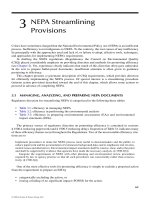
comparison; Cuban and European specimens are shown in Fig. 5.3. The fore femora and
tibiae of both specimens possess similar modifications whose function and co-evolution
with female wings have been discussed in the recent literature [on femur: c.f. B & H
(anterior view), C & I (posterior view); on tibia: c.f. D & J (Ang et al. 2008b, Ingram et al.
2008, Puniamoorthy et al. 2008)]. The 4
th
sternite and hypopygial capsule can be seen on
the abdomen and are also very similar in structure and diagnostic for T. leachi (lateral view:
c.f. E & K; ventral view c.f. F & L). Even more striking are the 2
nd
and 3
rd
sternites, which
151
are well developed and have been modified into a raised, anteriorly open crater on the 2
nd
sternite and a pronounced protrusion on the 3
rd
sternite. These sternite modifications are
unique to T. leachi and the only difference between the specimens is minor (more hook-like
protrusion on the European specimen). Overall, the foreleg, sternite, and hypopygial
capsule morphology are very similar between the European and Cuban specimens of T.
leachi and suggest the presence of only one species.
Recent studies of morphologically uniform species with wide distributions have
suggested that such species frequently contain ‘cryptic’ species that can be discovered once
DNA sequence data become available (Bickford et al. 2007). I thus compared the Cuban
and European specimens with regard to the mitochondrial gene COI. A ca. 660 bp piece of
the COI gene was sequenced for four Cuban specimens using the DNA extraction,
amplification, and sequencing protocols described in Su et al. (2008). These sequences
were submitted to Genbank (EU831274 – EU831277) and compared to a known sequence
of T. leachi from Europe (Genbank: EU435823) as well as COI sequences for ten other
Themira species (Su et al. 2008). Pairwise distances between the European and Cuban
sequences were 0.5% to 0.8%. Whether such distances are typical for inter- or intraspecific
variability can be judged when they are compared to a distribution of distances for closely
related species in Themira (Meier et al. 2006, Memon et al. 2006, Petersen et al. 2007).
Based on the known sequences for ten Themira species, the mean interspecific distance for
closest relatives is 6.2% and only one species pair [T. lucida (Staeger in Schiødte) vs. T.
flavicoxa Melander & Spuler] has distances below those observed between the Cuban and
European T. leachi. However, T. lucida and T. flavicoxa are morphologically distinct
(Ozerov 1998) while I did not find any significant morphological differences between the
Cuban and European specimens of T. leachi.
152
Figure 5.3: Morphology of Themira leachi from Cuba (photographed A-F; drawn M-R) and Europe
(photographed G-L). Habitus: A, G; fore-femoral modifications (anterior view): B, H; fore-femoral
modifications (posterior view): C, I; fore-tibial modifications (anterior view): D, J; abdomen (lateral view,
sternite bristles removed): E, K, N; abdomen (ventral view, sternite bristles removed): F, L, M; fore-
femur (anterior view): O; fore-tibia (anterior view): P; hypopygial capsule (dorsal view, setulation
omitted): Q; 4
th
sternite (dorsal view): R. Scale bars for A, G: 1mm; B-D and H-J: 0.1mm; E, F, K, L:
0.5mm.