Synthesis and structure investigation of stabilized aromatic oligoamides and their interaction with g quadruplex structures 2
Bạn đang xem bản rút gọn của tài liệu. Xem và tải ngay bản đầy đủ của tài liệu tại đây (990.69 KB, 72 trang )
17
Chapter 2
Snynthesis and Structure Investigation of Molecular Crescent
Aromatic Oligoamides
2.1 Introduction
Oligomers that adopt stable, compact conformations have been coined as
“foldamers”
1a
that are largely inspired by naturally occurring folding biomolecules
including proteins and DNAs. An effective strategy in the designs of foldamers
1,2
involves biasing the preferred conformations of synthetic oligomers by incorporating
a multitude of non-covalent interactions such as hydrogen-bonding (H-bonding),
solvophobic, π-π stacking and metal coordination bonds. Helical structures
3,4
appear
to occupy a privileged position among the folding patterns observed in reported
foldamers. The progress made so far in designing helical foldamers has allowed a
number of functionalities to be incorporated into various structural designs. As such,
folding helices endowed with diverse properties have been extensively investigated in
recent years that can 1) bind either neutral (saccharides,
4g,4h,5a-d
water
3f,5e-g
and other
small molecules
5h,5i
) or protein-membrane
7i-l
interactions, and 8) kill bacterial.
7m-q
A recently emerged concept in designing sophisticated helical foldamers explores
the proper use of multiply centered intramolecular H-bonds of varying types to
constrain the backbones of aromatic oligoamides and their analogs such as aromatic
oligohydrazides and oligoureas. Manipulating the folding of these aromatic oligomers
18
based on this strategy has allowed the creation of foldamers with a helically wrapped
interior cavity of as small as 1.4 Å
8
and as large as 30 Å in radius.
3d
This concept can
be traced back to the pioneering work on pyridine amide oligomers by
Hamilton,
3a,3b,9a
aromatic oligoamides/ureas/hydrazides/arylene ethynylenes by
Gong,
3d,10
aromatic oligoureas by Zimmerman,
9b
pyridine amide oligomers and
quinoline carboxamide oligomers by Lehn,
3c,11a-d
and Huc,
3c,3e,3f,3m,5e-g,6i,6k,11a,11b,11e-r
followed by the active explorations on aromatic amides/hydrazides by
Li,
4g,5b,5d,5m,5n,6a,6b,12a-c
Chen,
6f,12d-g
and others.
3g,12h-j
By including novel building
blocks and H-bonding patterns, we
8,13
and others
5n,10k,12a,12b,12i
have been interested in
further developing the corresponding field.
In this Chapter, we focus on shedding additional insights into the largely
unexplored structural features (backbone bending, columnar packing, and potential
channel formation) and on revealing the location-dependent strength of multiply
centered intramolecular H-bonds, which play a critical role in the folding of these
oligomers. Firstly,
1
H NMR and X-ray diffraction were used to establish the folding
of these conformationally rigid aromatic oligomers. Secondly, amide
hydrogen-deuterium (H-D) exchange studies were used to infer the strengths of
various intramolecular H-bonds placed at different locations along a backbone, results
from which has allowed us to pinpoint the local conformational weakness along the
oligoamide backbone. The conclusion from H-D exchange was confirmed by the
crystal structures of a series of oligomers, which allows a qualitative correlation
between the conformational stability of the H-bond enforced backbones and the
19
strength of individual H-bonds that are sensitive to local structural environments.
10e,13
The correlation derived from H-D studies and solid state investigations was
substantiated by results from ab initio calculations at the level of B3LYP/6-31G*.
Examining the assembly of the oligomers in the solid state revealed a columnar
packing shared by all the oligomers ranging from dimer to hexamer. The interplay of
π-π stacking and van der Waals’ interactions provide the driving forces for the
observed formation of columns. With their persistent shapes, tunable sizes and
tendency to aggregate into column- and channel-like structures, these folding
oligomers may serve as novel building blocks for constructing higher-order
supramolecular structures with non-collapsible pores and channels capable of
conducting ions and small molecules.
10m
2.2 Result and Discussion
2.2.1 Synthesis of Oligoamides
N
H
O
O
N
N
O
H
O
O O
O
H
O
O
O
2
N
3
R
1
R
2
6
5b: R
1
=OC
8
H
17
,R
2
=R
4
= OCH(CH
3
)
2
,R
3
=CH
3
5a: R
1
=R
2
=R
3
=H
5d: R
1
=R
2
=R
3
=H,R
4
=OCH(CH
3
)
2
5c:R
1
=OC
8
H
17
,R
2
=OCH(CH
3
)
2
,R
3
=R
4
=CH
3
N
H
O
O
NO
2
N
O
H
O
O O
O
R
2
R
3
N
H
O
O
N
N
O
H
O
O O
O
H
O
O
O
2
N
R
2
R
3
3
4
3b: R
1
=R
3
=OCH
3
,R
2
=H
3a:R
1
=R
2
=R
3
=H
4b: R
1
=OC
8
H
17
,R
2
= OCH(CH
3
)
2
,R
3
=CH
3
4a: R
1
=R
2
=R
3
=H
R
1
3d: R
1
=R
3
=OCH
3
,R
2
=OC
8
H
17
3c:R
1
=R
2
=R
3
=OCH
3
3f: R
1
=OCH
3
,R
2
= OCH(CH
3
)
2
,R
3
=OC
8
H
17
3e:R
1
=OCH
3
,R
2
=OC
8
H
17
,R
3
=OCH(CH
3
)
2
R
1
NO
2
N
O
H
O
O O
O
R
2
2
2b: R
1
=OCH
3
,R
2
=H
2a: R
1
=R
2
=H
R
1
2d: R
1
=R
2
=OCH
3
2c: R
1
=H,R
2
=OC
8
H
17
2g: R
1
=OCH
3
,R
2
=OCH(CH
3
)
2
2e: R
1
=OCH
3
,R
2
=OC
8
H
17
NO
2
N
O
H
O
O NH
O
OC
8
H
17
O
2f
1
5
6
10
11
15
16
20
21
25
N
H
O
O
N
N
O
H
O
O O
O
H
O
O
N
H
O
O
2
N
O
R
1
R
2
R
3
R
4
5
2
3
4
7
13
12
8
9
14
17
19
24 22
18
23
30
26
27
29
28
6
6
11
6
11
16
6
24
1
5
10
5
10
5
10
15
5
10
15
20
6a:R
1
=R
2
=H
6c: R
1
=OCH(CH
3
)
2
,R
2
=CH
3
6b: R
1
=OCH(CH
3
)
2
,R
2
=OC
8
H
17
1
5
6
8
2
3
4
7
9
20
All the aromatic oligoamides in Schemes 2-4 were synthesized from commercially
available salicylic acid, 2,5-dihydroxybenzoic acid and 2-hydroxy-5-methylbenzoic
acid in up to 18 steps.
Monomeric building blocks 1k, 1l, 1m, 1q and 1t were prepared according to
Scheme 2.1. These five building blocks differ from each other only by the remote
alkoxyl substituents meta to nitro group. Introducing of these side chains
prove critically important in conformational characterization in solution by 2D
NOESY study and in the solid state by X-ray diffraction method.
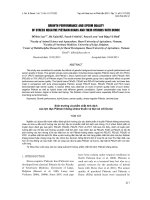
Scheme 2.1. Synthesis of Monomeric Building Blocks
a
a
a) conc. H
2
SO
4
, MeOH, reflux, 97%; b) K
2
CO
3
/RBr (or RI), anhydrous acetone, reflux, 51~65%; c)
Bi(NO
3
)
3
, MMT K10, THF, 58~67%; d) K
2
CO
3
/CH
3
I, DMF, 72~91%; e) NaOH, MeOH/H
2
O, reflux,
53~85%; f) conc. HNO
3
, Conc. H
2
SO
4,
80%.
Among the above five building blocks, 1k, 1l and 1m were prepared after five steps
starting form 2,5-dihydroxybenzoic acid. As shown in Scheme 2.1, esterification in
methanol provided methyl ester 1a in a high yield of ~90%. The second step
OH
OH
COOH
1a 1b
,
1c
,
1d
OH
COOCH
3
OH
OR
OCH
3
COOCH
3
O
2
N
OR
OH
COOCH
3
O
2
N
OR
OH
COOCH
3
1e
,
1f
,
1g
OR
OCH
3
COOHO
2
N
a
b
c
1h
,
1i
,
1j
1k, 1l, 1m
1b
,
1e
,
1h
,
1k
:R= CH
3
OH
COOH
OH
COOCH
3
O
2
N
1o, 1r
OCH
3
COOCH
3
O
2
N
1p, 1s
OCH
3
COOH
O
2
N
1q
,
1t
R
1
R
1
R
1 R
1
1o, 1p, 1q:R
1
=CH
3
1r, 1s, 1t:R
1
=H
d
e
f, a
d
e
1c
,
1f
,
1i
,
1l
:R=C
8
H
17
1d, 1g, 1j, 1m: R = CH(CH
3
)
2
21
involving chemoselective alkylation turned out to be quite sensitive to the solvents
used. While the use of dimethylformamide (DMF) produced dialkylated product in
both hydroxyl groups, a desirable shifting to the monoalkylation occurred almost
exclusively at the hydroxyl group meta to ester group with the use of alkyl
iodides/bromides under refluxing conditions in the presence of potassium carbonate
(K
2
CO
3
) in acetone. Since 1a has two hydroxyl groups on the same benzene ring, no
more than 1.1 equiv of the alkyl iodine (or bromide) was used. This led to a long
reaction time and moderate chemical yields (~ 60%) for 1b-1d. Nevertheless, simple
flash column chromatography allows the easy purification of the products and
recycling of the starting material. This chemoselective alkylation was unambiguously
confirmed by the determined crystal structure of 1c (Figure 2.3).
Attempted nitrations of 1b-1d by varying the ratio of conc. nitric acid and conc.
sulfuric acid in dichloromethane (CH
2
Cl
2
) at varying temperatures from -40
o
C to 45
o
C invariably led to a mixture of at least three products detectable by Thin Layer
Chromatography (TLC), from which the desired products 1e-1g were obtained in a
unacceptable low yield of less than 30%. After testing a few more other conditions
(i.e., conc. nitric acid (HNO
3
) in acetic acid (AcOH), or slow addition of conc.
sulfuric acid (H
2
SO
4
) into conc. nitric acid containing compounds to be nitrated),
the nitration method using montmorillonite (MMT) impregnated with bismuth nitrate
(Bi(NO
3
)
3
) was finally singled out. The condition was very mild, simply involving
mixing the compounds to be nitrated (1b-1d) with Montmorillonite K10 impregnated
with bismuth nitrate in Tetrahydrofuran (THF) at room temperature and stirring the
22
solution for 12 hrs. Under this condition, a clean reaction producing only 1e-1g was
obtained. The chemical yield was around 65%. It was later found out that a
considerable amount of nitrated products was absorbed into solid support
Montmorillonite K10, which can not be efficiently extracted out using CH
2
Cl
2
. This
issue was solved by adding a small amount of acid (1M hydrochloric acid (HCl)) to
the filtered Montmorillonite K10, followed by extraction with CH
2
Cl
2
to maximize
the chemical yield. The subsequent straightforward methylation of the second OH
group using iodomethane or dimethyl sulfate in DMF at 60 ˚C, following by the
NaOH-mediated saponification led to the production of monomeric acidic building
blocks 1k-1m.
During the synthesis of 1q from 2-hydroxy-5-methylbenzoic acid, bismuth
nitrate-mediated nitration at room temperature tends to give inconsistent low chemical
yields from time to time. It was finally realized that such nitration is highly sensitive
toward both reaction temperature and reaction time. By controlling reaction
temperature at -20 ˚C for 20 minutes, followed by immediate quenching with water,
desired product can be obtained in a yield of as good as 80%. This bismuth
nitrate-mediated nitration, surprisingly, did not work for 1r. Its mono-nitration,
however, can be accomplished using conc. HNO
3
and conc. H
2
SO
4
in CH
2
Cl
2
, under
which conditions, ironically the nitration of 1b-1d did not proceed at all. To facilitate
the separation of mono-nitrated acid product 1r from its isomer that contains a nitro
group ortho to hydroxyl group and minor product containing two nitro groups, the
reaction mixtures were converted to ester compounds. It is interesting to note that
23
saturated (Sodium hydrogen carbonate (NaHCO
3
) can dissolve dinitro compound, but
not mononitro compounds, into the aqueous layer. The two mono-nitrated isomers
thus can be efficiently separated by flash column chromatography using
hexane/CH
2
Cl
2
(v:v 4:1) as the eluent to give pure product 1r as a bright yellow solid.
Scheme 2.2. Synthesis of Trimers
a
a
a) H
2
, Pd/C, THF, 40 ˚C, 94%; b) ethyl chloroformate, 4-methylmorpholine, CH
2
Cl
2
, 1t (for 2b) or 1k
(for 2d) or 1l (for 2e), RT, 72~77%; c) ethyl chloroformate, 4-methylmorpholine, CH
2
Cl
2
, 1k, RT
67~83%.
Scheme 2.3. Synthesis of Hexamer 6a
a
a
a) H
2
, Pd/C, THF, 40 ˚C, 96%; b) ethyl chloroformate, 4-methylmorpholine, CH
2
Cl
2
, 1t, RT, 71%, c)
(COCl)
2
, DMF, CH
2
Cl
2
, 1t, then TEA/CH
2
Cl
2
,71~82%; d) KOH, KCl, MeOH/H
2
O, 2a, reflux, 83%; e)
ethyl chloroformate, 4-methylmorpholine, CH
2
Cl
2
, 2h, RT, 19%.
Following the elaboration of the synthetic routes for the efficient preparation of
various monomeric building blocks (Scheme 1: 1h-1m, 1p, 1s, 1q and 1t), a series of
oligoamides was prepared according to schemes 2-4. A convergent route was seldom
O
COOCH
3
O
2
N
1s
H
3
COOC N
H
O
O
NO
2
O
2a
:n=1;
3a
:n=2
n
a, b repeat
3a
HOOC N
H
O
O
NO
2
O
2h
H
3
COOC N
H
O
O
NO
2
O
5
6a
d
a, e
H
3
COOC N
H
O
O
NO
2
O
n
4a
:n=3;
5a
:n=4
a, c
repeat
4a
O
COOCH
3
O
2
N
1h
H
3
COOC N
H
O
O
NO
2
O
2b, 2d, 2e
OCH
3
OCH
3
N
H
O
O
NO
2
N
O
H
O
O O
O
R
2
H
3
CO
3b, 3c, 3d
R
1
R
1
2b: R
1
=H
2d:
R
1
=OCH
3
2e: R
1
=OC
8
H
17
3b: R
1
=H,R
2
=OCH
3
3c: R
1
=R
2
=OCH
3
3d: R
1
=OCH
3
,R
2
=OC
8
H
17
a, b
a, c
24
used here because it either did not give the expected product or gave a low coupling
yield (19% for 6a by coupling tetramer 4a with dimer 2h and 6% for 6b by coupling
trimer 3a with trimer 3g). Instead, backbone construction (C-to-N) of the oligoamides
1-6 in a unidirectional stepwise fashion proved to be a more efficient, time-saving
strategy by reacting monomeric active ester or acid chloride with amino-terminated
oligoamides. This stepwise construction can be exemplified by the preparation of
tetramer 4a (Scheme 2.4). The synthesis of 4a started from monomers 1s and 1t.
Reduction of 1s by Palladium on carbon (Pd/C)-mediated hydrogenation at 40
o
C in
THF converted 1s into amine intermediate that coupled with in situ generated active
ester produced from 1t (conditions: ethyl chloroformate, 4-methylmorpholine
(NMM), CH
2
Cl
2
, room temperature) to give nitro-terminated dimer 2a with a
chemical yield of 71%. Hydrogenation of 2a under the typical conditions (Pd/C,
Hydrogen (H
2
), THF, 40
o
C) produced amino-terminated intermediate that was
subjected to the next coupling reaction with the above in situ generated active ester
from 1t to afford trimer 3a with a chemical yield of 82%. Trimer 3a was further
hydrogenated (Pd/C, H
2
, THF, 40
o
C) to yield the corresponding amine intermediate
that reacted with the acid chloride, which was generated from 1t under the conditions
involving oxalyl chloride (COCl
2
) and a few drops of DMF in CH
2
Cl
2
at room
temperature, to produce 4a with a chemical yield of 61%.
Unfortunately, despite our numerous attempts, neither convergent nor stepwise
synthesis was able to produce oligoamides of higher than heptamer, a reason why
25
N
H
O
O
N
N
O
H
O
O O
O
H
O
O
O
2
N
3
O
OC
8
H
17
6b
O
COOCH
3
O
2
N
1t
H
3
COOC N
H
O
O
NO
2
O
2g
N
H
O
O
NO
2
N
O
H
O
O OH
O
3g
O
O
OC
8
H
17
b, d
b, c
N
H
O
O
NO
2
N
O
H
O
O O
O
3f
O
OC
8
H
17
g
h, i
3f
OCH
3
O
COOHO
2
N
OCH
3
O
O
2
N
H
N
1k
1u
C
8
H
17
O
NO
2
O
O
N
OCH
3
H O O
H
N
2f
O
COOCH
3
O
2
N
1s
H
3
COOC N
H
O
O
NO
2
O
2c
N
H
O
O
N
N
O
H
O
O O
O
H
O
O
O
2
N
4b
OC
8
H
17
O
N
H
O
O
N
N
O
H
O
O O
O
H
O
O
N
5b
,
5c
H
O
O
2
N
O
OC
8
H
17
O
R
1
OC
8
H
17
5b
:R
1
=OCH(CH
3
)
2
5c
:R
1
=CH
3
a
O
b, c
b, c
b, e
N
H
O
O
NO
2
N
O
H
O
O O
O
3e
OC
8
H
17
O
b, d
b, f
N
H
O
O
NO
2
N
O
H
O
O O
O
4a
b, d
b, e
N
H
O
O
N
N
O
H
O
O O
O
H
O
O
O
2
N
2
N
H
O
O
NO
2
N
O
H
O
O O
O
5d
3
O
3
O
6c
only the oligoamides of up to hexamers were presented and studied in the furture
work.
Scheme 2.4. Synthesis of Oligomers from Dimers to Hexamers
a
a
a) EDC, HOBT, Propan-2-amine, CH
2
Cl
2
, 95%; b) H
2
, Pd/C, THF, 40
o
C, 64%; c) ethyl
chloroformate, 4-methylmorpholine, CH
2
Cl
2
, 1l, 64%; d) ethyl chloroformate, 4-methylmorpholine,
CH
2
Cl
2
, 1m, 71%; e) (COCl)
2
, DMF, CH
2
Cl
2
, 1q, 83%; f) (COCl)
2
, DMF, CH
2
Cl
2
, 1m (for 5b) or 1q
(for 5c), 46~49%; g) KOH, KCl, MeOH/H
2
O, reflux, 93%; h) (COCl)
2
, DMF, CH
2
Cl
2
, 7%; i) H
2
,
Pd/C, THF, 40
o
C, 3a, then TEA/CH
2
Cl
2
, 90%.
26
2.2.2 One-Dimensional
1
H NMR Studies of Folding Oligoamides
The oligoamides 2-6 studied here contain three important sets of proton signals,
i.e., amide protons, aromatic protons and interior methoxy protons. Among them, the
chemical shift values of the amide protons are the simplest diagnostic of the existence
of intramolecular H-bonds when compared to other more advanced analytical
techniques (i.e., 2D NOESY and X-ray diffraction). In chloroform, upon forming
intramolecular H-bonds, amide protons typically exhibit a substantial downfield shift
due to the deshielding of amide protons by the adjacent electron-negative elements.
The degree of downfield shifting thus provides a good indication as to the occurrence
and strength of hydrogen bonds found in H-bond enforced aromatic foldamers. For
example, amide protons involved in two-center H-bonds have a typical chemical shift
of less than 9.6 ppm
4f
while those involved in three-center H-bonds most often
downfield shift to much larger than 10 ppm,
3d,4f,8,10a,10b,10e,13
suggesting that
three-center H-bonds have a higher stability than two-center H-bonds of similar
types.
10e
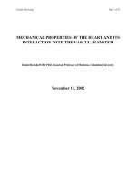
The representative
1
H NMR spectra containing the amide and aromatic signals
(Figure 2.1) for some selected oligomers were presented in Figure 2.1 with the
chemical shifts for all the amide protons of oligoamides 4-6 tabulated in Table 2.1.
The majority of these amide protons resonant at >10 ppm at 1 mM in CHCl
3
, a more
than 1 ppm downfield shift than the amide proton (8.70 ppm) in 2f and others
10b
that
are involved in the formation of two-center H-bonds. This experimental observation is
consistent with the expectation that these amide protons be engaged in a continuous
intramolecular H-bonding network as originally conceived. The formed H-bonding
27
network subsequently stabilizes the oligomers into a crescent-shaped well-defined
conformation rather than a random coiled structure, giving rise to the sharp proton
signals in all the spectra compiled in Figure 2.1.
Figure 2.1.
1
H NMR (500 MHz) spectra of some selected oligomers 4-6 in CDCl
3
at 298K: (a) dimer
2a (20 mM), (b) trimer 3a (10 mM), (c) tetramer 4a (20 mM), (d) pentamer 5a (5 mM), (e) pentamer
5c (25 mM), (f) hexamer 6a (20 mM), and (g) hexamer 6c (20 mM).
28
2.2.3 Two-Dimensional
1
H
-
1
H NMR Studies (NOESY) of Oligoamides
Since NOE intensity is proportional to the inverse sixth power of the distance, the
experimentally observed NOE intensity is largely determined by the shortest distance
between two interacting nuclei. As revealed in the crystal structures of oligoamides
2-4, the shortest inter-atomic distances between amide protons and the adjacent
interior methoxy protons measure from 2.28 to 2.97 Å, an indication that the two
NOE contacts between every amide proton and its adjacent methoxy methyl groups
should be seen in the 2D NOESY spectrum if a folded conformation induced by
intramolecular H-bonds does prevail for oligoamides 2-6 in solution.
15
Accordingly,
the crescent-shaped or helically folded conformations in oligoamides 2-6 were probed
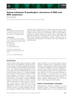
by 2D NOESY studies (Figures 2.2 and 2.3). Due to the highly repetitive nature of
oligoamides 2-6, extensive
1
H NMR signal overlaps among aromatic protons
were observed for an oligoamide as simple as trimer 3a. This prevents the accurate
and complete assignment involving the amide protons and adjacent interior methoxy
methyl protons and so hampers the elucidation of their folded structures in solution.
This issue was mostly solved by deliberately introducing linear and branched alkoxyl
side chains as well as a methyl group para to the interior methoxy groups into
oligoamides 2-6 (i.e., 2f, 3d, 4b, 5b, 5c, 6b and 6c). The introduction of these side
chains indeed led to the well-resolved amide protons, aromatic protons and internal
methoxy groups in oligoamides 2f (Figure 2.2a), 3d (Figure 2.2b) and 5b
8
that permit
us to detect the expected two NOE cross peaks for each amide protons. Additionally,
the majority of these NOE intensities between interior methoxy protons and amide
29
61116
10
5/15
1
20
(6,10) & (6,5)
(16,20)
(16,15)
(11,10) & (11,15)
10
5
(6,5)
(6, 10)
6
11 6
5
10
15
(11,15)
(11,10)
(6,10)
(6,5)
1
5
10
20
15
25
(6,1) & (6,5)
(11,10)
(21,25)
(11,15) & (21,20)
(16,15)
(16,20)
8
616
11/21
8
61116
10
5/15
1
20
(6,10) & (6,5)
(16,20)
(16,15)
(11,10) & (11,15)
10
5
(6,5)
(6, 10)
6
11 6
5
10
15
(11,15)
(11,10)
(6,10)
(6,5)
1
5
10
20
15
25
(6,1) & (6,5)
(11,10)
(6,1) & (6,5)
(11,10)
(21,25)
(11,15) & (21,20)
(21,25)
(11,15) & (21,20)
(16,15)
(16,20)
8
616
11/21
8
protons are much stronger than the weak NOE contacts between amide protons and
the neighboring aromatic protons ortho to the amide bonds. For example, the NOE
contact between protons 6 and 4 in pentamer 5c is much stronger than that between
protons 6 and 7 in the same molecule. This implies that the methoxy protons stay
much closer to the amide protons than to the aromatic protons, which is a direct
consequence resulting from the induced folding of the backbone by the internally
located H-bonds. Compared to 4a and 5a, better
1
HNMR signal dispersions are also
observed for 4b (Figure 2.2c) and 5c (Figure 2.2d), some
1
H NMR peaks still either
overlap substantially or display a small difference in chemical shift.
a) b)
c) d)
Figure 2.2. NOE contacts (NOESY, 500 MHZ, 298 K, 10 mM, 500 ms, 4 hrs) seen between amide
protons and their adjacent interior methoxy protons: (a) dimer 2f in DMSO-d
6
, (b) trimer 3d in 50%
CDCl
3
/50% DMSO-d
6
, (c) tetramer 4b in CDCl
3
and (d) pentamer 5c in CDCl
3
.
30
2.2.4 Solid State Structures of Oligoamides
Crystals of oligomers 2-4 suitable for X-ray structure determination were obtained
by slow evaporation of these oligomers in mixed solvents at room temperature (Table
2.1). The top and side views of the determined crystal structures for oligoamides 2-4
are presented in Figure 2.3. These crystal structures demonstrate that with the
stepwise addition of aromatic building blocks the elongated backbone becomes
increasingly curved in one direction. This is a result of the stabilizing forces from the
lengthened intramolecular H-bonding network that comprises up to six intramolecular
H-bonds (NH…OMe = 1.933-2.306 Å). As reported recently by us,
8
a longer
oligomer such as 5a or 6a with a long enough backbone eventually curve into a
helical conformation as a result of the stabilizing H-bonding interactions, which more
than compensate the unfavorable steric crowdedness involving the two end interior
methoxy groups.
Table 2.1. Crystal growth conditions for oligomers 2-4.
Solvent Pair
(1:1)
Solvent Pair
(1:1)
Solvent Pair
(1:1)
2a
CH
2
Cl
2
: MeOH
2d
CHCl
3
: MeOH
3c
C
CHCl
3
: Hexane
2b
CH
2
Cl
2
: Hexane
3a
CHCl
3
: MeOH
3d
DMF : CH
3
CN
2c
CHCl
3
: MeOH
3b
CH
2
Cl
2
: Hexane
4a
CH
2
Cl
2
: MeOH
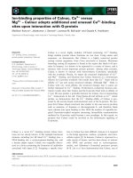
A closer look into the crystal structures of oligoamides 2 & 3 reveals a quite
surprising structural feature: while the aromatic rings in all the four dimer molecules
2a-2d are always coplanar, the nature of exterior side chains has an influential
distorting effect on the planarity of the trimeric backbone in trimers 3a-3d (Figure
31
2.4). This type of distortional behavior involving aromatic backbones is quite unusual
and not seen in other H-bonded short aromatic oligomers of similar
types.
3d,5m,10a,10l,12a,16
As illustrated in Figure 6, the distortion angels (i.e., the dihedral
angle formed between the plane defined by the first two benzene rings at the nitro end
and the plane defined by the first benzene ring at the ester end) are 55 º in 3a, 30 º in
3b, 26 º in 3c, and 10 º in 3d, respectively. Interestingly, while the first two benzene
rings at the nitro end stay coplanar in all the four trimer molecules 3a-3d, all the
distortions if any are fully concentrated around the amide linkage at the ester end
whose NH proton 6 seems to form a much weaker six-membered H-bond than amide
proton 11 at the nitro end. This weakness in H-bond strength can be inferred from
amide hydrogen-deuterium exchange studies on protons 6 and 11 of trimers 3a-3f (see
Table 2.2, Figure 2.9 and the corresponding text) and can be further substantiated by
theoretical calculations on the strength of intramolecular H-bond in 3a at the level of
B3LYP/6-31G* (see Figure 2.10 and corresponding text). A need to maximize the
favorable aromatic π-π stacking interactions during the crystal packing likely is
another decisive factor that contributes to such a deviation from the planarity (Figure
2.7). In other words, the intermolecular π-π stacking interactions may override to a
good extent the planarizing forces coming from the weaker H-bonds at the ester end,
thereby causing the plane involving the weaker H-bonds to deviate significantly from
the plane involving stronger H-bonds at the nitro end. A combination of π-π
interactions, the presence of amide proton 11 forming weak intramolecular H-bonds,
repulsive interactions between end nitro and ester groups and steric hindrance among
32
interior methyl groups shall account for the non-planar aromatic backbone observed
in tetramer 4a (Figure 2.5a). Contrasting with both trimer and tetramer and as
evidenced from their crystal structures, all the dimers 2a-2d contain a strong
three-center H-bond that forces the dimeric backbone defined by two aromatic rings
and one amide bond into a perfectly coplanar geometry.
Table 2.2. Chemical shifts (ppm)
a
and the half-lives (hrs, in parenthesis) of H-D exchange
b
of amide
protons for oligomers.
c
oligomer H
6
H
11
H
16
H
21
H
26
2a
10.36
(0.13)
2b
10.37
(0.20)
2c
10.47
(0.90)
2d
10.25
(0.24)
2e
10.49
(0.56)
2f
10.24
(0.30)
8.70
(0.21)
2g
10.49
(0.83)
3a
10.21
(0.04)
10.23
(0.27)
3b
10.24
(0.05)
10.37
(0.24)
3c
10.35
(0.08)
10.38
(1.34)
3d
10.36
(0.11)
10.38
(0.65)
3e
10.35
(0.07)
10.36
(0.90)
3f
10.33
(0.05)
10.35
(0.72)
4a
10.20
(0.63)
10.10
(0.03)
10.00
(0.06)
33
c)
4b
10.33
(0.92)
10.14
(0.11)
10.10
(0.08)
5a
10.25
(0.33)
9.85
(0.05)
9.85
(0.20)
10.25
(0.27)
5b
10.35
(1.90)
9.85
(0.07)
9.65
(0.28)
10.25
(0.37)
5c
10.35
(1.25)
10.10
(0.08)
9.85
(0.24)
10.10
(0.32)
5d
10.28
(0.14)
10.21
(0.09)
9.95
(0.09)
9.84
(0.21)
6a
10.19
(0.07)
10.12
(0.09)
9.84
(0.06)
10.28
(0.11)
10.15
(0.38)
6b
10.12
(0.42)
10.08
(0.29)
9.81
(0.27)
10.21
(0.40)
10.09
(5.79)
6c
10.28
(0.21)
10.25
(0.17)
10.15
(0.21)
10.15
(0.40)
9.85
(1.20)
a
Chemical shifts were measured at 1 mM in CDCl
3
(500MHz) at room temperature.
b
Half-lives of
H-D exchange data in parenthesis were measured at 5 mM in 5% D
2
O/47.5% DMSO-d
6
(v:v) in CDCl
3
at room temperature.
c
Figure 2.3. Crystal structure of intermediate 1c as well as top and side views of crystal structure of
2a-2d. In 2c, the dummy atom represents the octyl side chain. In top views, all the interior methoxy
methyl groups were removed for clarity of view. In side views, all the nitro groups, ester groups and
side chains were removed for clarity of view.
1c 2a 2b 2c 2d
34
Figure 2.4. Top and side views of crystal structure of 3a-3d. The side views were generated by placing
the nitro end in the back. In 3d, the dummy atom represents the octyl side chain. All the interior
methoxy methyl groups were removed for clarity of view. Solid arrows highlight the twisted
six-membered H-bonds.
Figure 2.5. Top and side views of crystal structure of (a) 4a and (b) 6a.
8
The calculated structure of 6a
is shown in (c). All the interior methoxy methyl groups were removed for clarity of view.
35
a) b) c) d)
Figure 2.6. Side and top views of columnar assemblies observed in the solid state structures of (a) 2a
along axis a, (b) 2b along axis ac, (c) 2c along axis a and (d) 2d along axis.
a) b) c) d)
Figure 2.7. Side and top views of column formation and further association observed in the solid state
structures of (a) 3a, (b) 3b, (c) 3c and (d) 3d. Dotted rectangles highlight the inter-columnar.
36
a) b) c)
Figure 2.8. Top: channel formation observed in the solid state structures of (a) 4a, (b) 5a, and (c) 6a.
Bottom: side views illustrating π-π interactions that lead to the channel formation in 4a-6a. Dotted oval
shapes or circle in red highlight one repeating channel unit. For clarity of view, some methoxy methyl
groups inside the channels were removed.
Examining the solid state structures of dimers 2 (Figure 2.6) and trimers 3 (Figure
2.7) reveals a one dimensional (1D) columnar assembly consisting of molecules
packed in an anti-parallel fashion via aromatic π-π stacking interactions (inter-plane
distances along packing axis = 3.3–3.6 Å).
10l
For dimers, except for 2b that packs along diagonal axis ac forming an angle of 63º
between axis ac and plane of the molecules, all the other three dimers (2a, 2c and 2d)
are stacked along axis a with angles of 90º, 53º and 70º, respectively, between axis a
and plane of the molecules by virtue of aromatic π-π interactions (Figure 2.6). These
columns further assemble into 2D sheets and 3D structures via van der Waals
interactions among aromatic C-H bonds, nitro groups, ester groups and methoxy
groups.
Three packing patterns can be observed for trimers. As shown in Figures 2.7a-c, the
stacking of 3a-3c along axis a is mediated by aromatic π-π interactions involving the
first two benzene rings at the nitro end remaining coplanar. In 3d (Figure 2.7d), the
37
π-π interactions between the central benzene ring and the end benzene ring close to
the nitro group are responsible for associating the molecules into 1D columns along
axis a. While the assembly of 1D columns into 2D/3D structures in both 3a and 3d
(see dotted rectangles in Figures 2.7a and 2.7d) does not involve π-π stacking, π-π
interactions do play an important role in driving the inter-columnar assemblies in the
solid-state assembly of both 3b and 3c (see dotted rectangles in Figures 2.7b and
2.7c). Additionally, among the eight oligomers 2a-2d and 3a-3d, only 3d packs in a
parallel fashion along the stacking axis.
The consistent columnar assembly of the above short oligomers 2-3 in solid states
suggests that, with their cavity-containing backbones, oligomers longer than trimer
may be capable of stacking on top of one another into channel-like structures. This
possibility was first examined by the solid-state structure of 4a. As expected, the
molecules of 4a stack into a channel that is stabilized by π-π interactions along the c
axis of the unit cell (see dotted red oval shape in Figure 2.8a). Re-examining the
crystal structures of 5a and 6a reveals similar channels in the solid state (see dotted
red circle or oval shape in Figures 2.8b and 2.8c). The side views of these channels
along axis b for 4a, approximately along axis b for 5a and along axis c for 6a show
that all the channels are stabilized by π-π interactions through partial overlap of
aromatic backbones. Although the interior of the channels formed by 4a-6a are
decorated by oxygen atoms, the presence of hydrophobic methoxy methyl groups
must obstruct the channels’ ability to conduct inorganic species such as Na
+
and K
+
.
Nevertheless, given the fact that 4a-6a all enclose a small cavity radius of ~2.8 Å
38
from the center of the cavity to the nucleus of the interior oxygen atom and that
coordination distances between oxygen atom and majority of metal cations falls
below 2.8 Å, we hypothesize that, replacing the interior methoxy groups with
phenolic hydroxyl groups and subsequent deprotonation of hydroxyl group will
restore the oxygen’s ability to bind metal ions. The resultant channels will be able to
bind/stabilize partially or fully dehydrated metal ions and subsequently allow the flow
of ions under applied electrochemical gradients (i.e., concentration or voltage
gradient).
10m
This object is being pursued and will be reported in due course.
2.2.5 Amide Hydrogen-Deuterium (H-D) Exchange Studies
The H-D exchange of amide protons can be detected by
1
H NMR and has been used
as a general method to distinguish between intramolecularly H-bonded and
solvent-exposed amide moieties in biological settings such as α-helices and
β-sheets. In H-D exchange experiments, the solvent-exposed amide protons are
continually exchanging with the solvent molecules such as D
2
O and usually are
replaced much faster by deuterium atoms than their H-bonded counterparts, leading to
the considerably shorter H-D exchange half life. Similarly, protons that are involved
in weaker H-bonds are exchanged faster with a shorter half life than those forming
stronger H-bonds if these protons are accessible equally well by solvent molecules.
Consequently, amide H-D exchange experiments offer a sensitive reflection of the
H-bond strength of amide protons as well as their solvent exposure degree. Very
recently, this method was applied to both H-bond detection and quantitative
39
measurement of H-bond strength in some aromatic foldamers by Gong
10e
and us.
13
A
H-D experiment can be initiated by adding a 50 l Dμ
2
O into 0.95 ml 5 mM oligomer
in deuterated solvents containing CDCl
3
: DMSO-d
6
(v:v 1:1), followed by monitoring
a change in the integration of amide proton signals at the appropriate time intervals
and fitting the obtained time-dependant integration change into a pseudo-first-order
rate equation to derive the half-life of the amide protons.
Under the presently used H-D exchange conditions, the half-lives of all the amide
protons in oligoamides 2-6 have been determined at 5 mM and selectively compiled
in Table 2.1. Given the overall dimensionality of 2-6, sticking-out orientation of
interior methyl groups and a low concentration of 5 mM used for amide H-D
exchange experiment, the intermolecular aggregation for all the studied oligoamides
is a highly unlikely event in solution. Thus, for short oligoamides including dimers
2a-2g, trimers 3a-3g and tetramers 4a-4b, the relative values of H-D half-lives should
reflect the relative stabilities of H-bonds; for longer oligomers such as pentamers and
hexamers that take up a helical conformation, amide H-D exchange shall be
additionally affected by a steric factor arising from the end-to-end overlapping as
observed in helical structures 5a and 6a.
8
In this regard, comparison of H-D exchange
values among the short oligomers of same length such as dimers 2a-2g, trimers 3a-3g,
or tetramers 4a-4b shows that the exteriorly located electron-donating side chains 1)
cause a large variation in H-bond strength and 2) result in intramolecular H-bonds in
oligomers modified with exterior side chains stronger than those found in the
oligomers 2a-4a that carry no side chains. Although an increase in H-bond strength
40
involving proton 6 in 5b and 5c cannot be excluded, the observation of much larger
H-D exchange half-lives for proton 6 in 5b (t
1/2
=1.90 hrs) and 5c (t
1/2
= 1.25 hrs) than
that in 5a (t
1/2
= 0.33 hrs) may indicate that the presence of exterior side chains make
proton 6 in 5b and 5c less accessible by D
2
O molecules than the same proton in 5a.
Similarly, comparison of H-D exchange values for proton 26 in 6a (t
1/2
= 0.38 hrs), 6b
(t
1/2
= 5.79 hrs) and 6c (t
1/2
= 1.20 hrs) shall allow us to surmise that proton 26 in6b
and 6c that both contain two exterior side chains on the nitro end is much less
solvent-exposed than proton 26 in 6a that contains no exterior side chains.
Figure 2.9. The half-lives of hydrogen-deuterium exchange rate for the amide protons 6 and 11 in
trimer 3. See Table 1 for the conditions and Supporting Information for the half-lives of all the other
amide protons in oligomers 2-6. Depending on R
1
-R
3
, plane A is twisted by varying degrees from the
plane defined by planes B and C that remain coplanar.
Of particularly interesting to note are the amide H-D exchange behaviors of the two
amide protons found in trimers 3a-3g (Figure 2.9): the half-lives of H-D exchange
rate for the amide proton 6 in all the studied trimers fall within a narrow range of 0.04
to 0.11 hrs that is significantly much smaller than those for proton 11 (0.27 to 1.34
hrs) in the same molecules. This suggests to us that the amide group involving proton
0
0.2
0.4
0.6
0.8
1
1.2
1.4
1.6
3a 3b 3c 3d 3e 3f
half‐lives(hrs )
H6
H11
41
6 shall constitute a conformationally weak point along the H-bonded aromatic
backbone of these trimer molecules. This reasoning is in an excellent agreement with
the backbone distortion fully centered around proton 6 as observed in the
solid state structures of 3a-3d (Figure 2.4) and can be further supported by the
theoretically determined relative H-bond strengths for all the four intramolecular
H-bonds found in trimer 3a (Figure 2.10). A further examination on H-D exchange
data (Figure 2.9) shows that the H-bonding strength involving proton 6 increases in
the order of 3a, 3b, 3c and 3d, which is surprisingly consistent with the observation
that the distortional dihedral angle decreases in the order of 3a, 3b, 3c and 3d (Figure
2.4).
2.2.6 Computational Studies on Oligoamides
Ab initio molecular modeling with the B3LYP/6-31G* basis set has consistently
allowed us to reliably predict the 3D topography of a circular pentamer
13
and an
acyclic helical pentamer 5a
8
and many others that are to be reported. As discussed
above, amide proton 6 in trimers 3a-3d is involved in the relatively weaker H-bonding
interactions than proton 11 and becomes the “battle of the bulge” where the backbone
readily gets twisted out of the planarity. To understand this focused twisting, we
carried out the ab initio calculation at the level of B3LYP/6-31G* on a total of eight
conformers (1A-4A & 1B-4B in Figure 2.10) generated by alternatively
flipping/rotating one benzene ring to disrupt one H-bond while keeping the rest three
H-bonds intact and by a further subtle adjustment on the interior methoxy side chain