Dissecting transcriptional network in mouse embryonic stem cells
Bạn đang xem bản rút gọn của tài liệu. Xem và tải ngay bản đầy đủ của tài liệu tại đây (6.81 MB, 257 trang )
DISSECTING TRANSCRIPTIONAL NETWORK
IN MOUSE EMBRYONIC STEM CELLS
FANG FANG
(M.Sci., Wuhan University)
(B.Sci., Wuhan University)
A THESIS SUBMITTED
FOR THE DEGREE OF DOCTOR OF PHILOSOPHY
IN COMPUTATION AND SYSTEMS BIOLOGY (CSB)
SINGAPORE-MIT ALLIANCE
NATIONAL UNIVERSITY OF SINGAPORE
2011
i
ACKNOWLEDGEMENTS
I am indebted to many people for their constant support during my PhD
training. I can’t even imagine myself write this part of thesis without their
helps along this exciting but strenuous journey.
Special thanks to my advisor, Paul Matsudaira, who has been a tremendous
supportive, encouraging and inspiring advisor. I would have quitted my PhD
career if he was not so helpful and supportive when I was in my career crash.
I am also grateful for his guidance and patience for ensuring the continuous
progress of my research progress.
I would like to thank my co-advisor, Harvey Lodish, who is, although, for
most of time, thousands of miles away from Singapore, for his encouragement
and instruction for my research and future career planning.
I have also received a lot of critical and helpful advice from my thesis
committee members, consisting of Gong Zhiyuan, Yu Hao, Chan Woon
Khiong and Neil Digby Clarke.
Thanks also go to my labmates, in particular Chen Xi and Xu Yifeng, who
have been inspiring mentors and good friends and contributed a lot to the work
described here.
My PhD training is supported by funding and a graduate fellowship from the
Singapore-MIT Alliance and I am grateful for all the administrative support
during last four and half years. Especially the independent research funding
for PhD students provide us a lot of freedom to carry out our own ideas.
ii
Last but not the least; I am deeply grateful to my family, who has
unconditionally given me invaluable love and support for me to overcome all
the difficulties during PhD trainings. I can only say no success in life would
have been possible without them.
iii
TABLE OF CONTENTS
Summary……………………………………………………………….…… iv
List of tables………………………………………………… v
List of figures………………………………………………… vi
Chapter I. Literature review………………………………………………… 1
1.1 Derivation and culture of pluripotent stem cells……………………… 1
1.2 Characteristics of mouse embryonic stem (ES) cells……………………8
1.3 Application of ES cells……………………………………………… 10
1.4 Molecular characteristics of ES cells……………………………… …12
Chapter II. Zfp143 regulates Nanog through modulation of Oct4 binding… 29
2.1 Summary of chapter II……………………………………………… 30
2.2 Introduction of chapter II………………………………………………31
2.3 Material and methods for chapter II……………………………………33
2.4 Results for chapter II…………………………………………… ……40
2.5 Discussion for chapter II……………………………………………….59
Chapter III. Dissecting early differentially expressed genes in a mixture of
differentiating embryonic stem cells………………………… 65
3.1 Summary of chapter III………………………………………… … 66
3.2 Introduction of chapter III……………………………………… ……67
3.3 Material and methods for chapter III…………………………… ……68
3.4 Results for chapter III………………………………………… ……77
3.5 Discussion for chapter III………………………………………… ….96
Chapter IV. Coactivators p300/CBP regulate self-renewal of mouse embryonic
stem cells by mediating long-range chromatin structure………100
iv
4.1 Summary of chapter IV……………………………….…………… 101
4.2 Introduction of chapter IV…………………………………………….102
4.3 Material and methods for chapter IV…………………………………104
4.4 Results for chapter IV………………………………………… … 117
4.5 Discussion for chapter IV………………………………………… …144
Chapter V. Conclusion and perspectives……………………………………152
Bibliography……………………………………………… ………………156
Appendix I. Integration of external signaling pathways with the core
transcriptional network through transcription factor
colocalization hotspots in embryonic stem cells…………… 173
Appendix II. A biophysical model for analysis of transcription factor
interaction and binding site arrangement from genome-wide
binding data………………………………………………… 204
v
SUMMARY
Embryonic stem (ES) cells are featured by their ability of self-renewal and
pluripotency. Although external signalling pathways as well as epigenetic
signatures have been shown necessary for ES cells maintenance, considerable
evidence indicates that naïve pluripotency of ES cells is dependent on their
specific transcription network that regulate the gene expression programs in a
spatially and temporally orchestrated and precise pattern. Delineating the
transcription network within ES cell system should be a fascinating science
challenge that would provide new insights into the fundamental nature of
pluripotency as well as advance its application in regenerative medicine. My
thesis project has applied computational and systems biology tools to dissect
transcriptional network of mouse ES cells, and has extensively expanded our
knowledge of the network by introducing novel self-renewal and pluripotency
associated transcription factors into the known core regulatory circuit.
Furthermore, I looked into coactivators that facilitate the functions of
transcription factors and further linked coactivator regulation to higher-order
chromatin structure. This is the first study of in vivo higher-order chromatin
organization that is unique to pluripotent cells based on the binding sites of
transcription factors and coactivators, adding a new content to the list of
unusual findings regarding the chromatin structure in ES cells as well as a new
layer to the ES cell specific transcriptional network.
vi
LIST OF TABLES
Table 1.1 Comparison of mouse and human ES cells 3
Table 2.1 Known transcription activator and repressor binding sites
at the Nanog regulatory regions.……………………………… 64
Table 3.1 Fisher’s Exact Tests between top-ranked genes of the
Differentiation-Test and benchmark gene list…………………… 84
Table 3.2 200 top-ranked differentially expressed transcription regulators
from the Differentiation-Test in 4-day EBs……………………….85
Table 3.3 Two sample comparison methods…………………………….… 96
Table 4.1 Sequences of primers used in this study………………… …… 111
vii
LIST OF FIGURES
Figure 1.1 Origin of stem cells during mammalian embryogenesis……… 5
Figure 1.2 Differentiation of mouse ES cells by EB formation.……… 9
Figure 1.3 The cell cycle of ES cells…………………………………………14
Figure 1.4 Bivalent chromatin domains in ES cells…………………….……19
Figure 1.5 Blocking FGF4/ERK and GSK3 signaling pathways are able to
maintain ES cells……………………………………………… 23
Figure 1.6 Model of core ES cell regulatory circuit………………………….27
Figure 2.1 Zfp143 expression is downregulated in both human and mouse ES
cells upon RA induced differentiation…………………………. .41
Figure 2.2 Zfp143 is required for the maintenance of undifferentiated
state of ES cells………………………………………………… 42
Figure 2.3 Zfp143 is required for the maintenance of undifferentiated
state of D3 ES cells………………………………………………43
Figure 2.4 Zfp143 knockdown reduced ES cell capacity to form
colonies in replating assay……………………………………… 44
Figure 2.5 Rescue of differentiation phenotype induced by Zfp143 RNAi… 45
Figure 2.6 Global gene expression changes after knockdown of Zfp143……47
Figure 2.7 Zfp143 and Oct4 co-occupy Nanog proximal promoter………….48
Figure 2.8 Zfp143 regulates Nanog proximal promoter…………………… 51
Figure 2.9 Nanog is a key downstream effector of Zfp143 for maintaining
ES cells……………………………………………………………52
Figure 2.10 Zfp143 is an Oct4 interacting protein………………….……… 54
Figure 2.11 The binding of Oct4 to chromatin is dependent on Zfp143….…57
Figure 2.12 Zfp143 and Oct4 co-occupy other targets that are important
for ES cells………………………………….………………… 58
Figure 2.13 A model depicting the different transcriptional regulators that
interact with Nanog cis-regulatory regions…………………… 63
Figure 3.1 A toy example of gene expression levels during a cellular
viii
differentiation process………………………………………….….77
Figure 3.2 An illustration of the inter-replicate variations of the average
expressions of a gene……………………… ………………… 79
Figure 3.3 Phase contrast micrographs of differentiating mouse
ES cells on gelatin………………………………….………… 82
Figure 3.4 Scatter plots of standard deviation vs. mean………………….….82
Figure 3.5 Variance comparison………………………………………… …83
Figure 3.6 Significance calibration from 10,000 random gene lists… ……84
Figure 3.7 Depletion of candidate genes by RNAi for two days…….………89
Figure 3.8 Depletion of candidate genes by RNAi for four days………… 90
Figure 3.9 A regulatory network in differentiating ES cells…………………92
Figure 3.10 Enrichment of the RBP-J motif in the upstreams of the
differentiation module…………………………….…………… 95
Figure 3.11 Average motif counts……………………………………………95
Figure 4.1 p300 and CBP are dispensable for the maintenance of ES cells 118
Figure 4.2 p300 and CBP are required and playing redundant roles for the
maintenance of ES cells………………………………………….119
Figure 4.3 Over-expression of p300 or CBP is able to rescue the double
knockdown effect………………… ……………………………121
Figure 4.4 p300 and CBP are recruited to Nanog-Oct4 -Sox2 cluster loci in
mouse genome………………………………………………… 123
Figure 4.5 Mapping the interaction domains of p300/CBP and Nanog…….125
Figure 4.6 KIX and Histone acetylation (HAT) domain of p300 and CBP are
important for their function in the maintenance of ES cells…… 126
Figure 4.7 p300 and CBP mediate intragenic looping interactions
among colocalization loci……………………………………….130
Figure 4.8 Intragenic looping interactions are specific to the
pluripotent state………………………………………………….132
Figure 4.9 RNAi samples for 3C assays……………………………… ….133
ix
Figure 4.10 p300 and CBP mediate intergenic looping interactions
among colocalization loci………………………… ………….136
Figure 4.11 Intergenic looping interactions are specific to the
pluripotent state…………………………… …………………138
Figure 4.12 The intragenic and intergenic looping interactions are
conserved in human ES cells………………………………… 139
Figure 4.13 Characterization of the DNA fragments involved in looping
interactions…………………………………………………….142
Figure 4.14 Model showing the three-dimensional organization of
Dppa3-Nanog-Slc2a3 loci…………………….……………… 150
1
CHAPTER I: Literature review
1.1 Derivation and culture of pluripotent stem cells
Although the era of embryonic stem (ES) cells is considered to begin officially
in 1981, when mouse ES cells were first isolated and successfully cultured in
vitro as self-renewal and pluripotent cell lines by two groups (Evans and
Kaufman, 1981; Martin, 1981), the adventure to search for exogenous cells
that are capable of recapitulating early embryogenesis had started earlier. At
the beginning, researchers had tried to manipulate early mouse embryogenesis
by embryonal carcinoma cells (EC) cells. EC cells are the pluripotent stem
cells from teratocarcinomas, which are highly malignant tumors that
occasionally occur in a gonad of a fetus and are comprised of a mixture of a
large population of undifferentiated cells and differentiated cells of multiple
lineages. EC cells could be maintained indefinitely with mitotically inactivated
embryonic fibroblast in vitro, and is able to give rise to cells of multiple
lineages (Finch and Ephrussi, 1967). However, further studies have found out
that EC cells were karyotypically abnormal or unable to differentiate normally
(Berstine et al., 1973; Papaioannou et al., 1975), which led to the efforts to
isolate a new type of stem cells, embryonic stem cells, from the mouse
embryo.
Embryonic stem cell lines are derived from the inner cell mass (ICM) of the
mouse blastocyst at embryonic day 3.5 (E3.5). These cells were initially
maintained in culture as self-renewal and pluripotent cell lines in either EC
cell-conditioned medium (Martin, 1981), or in a co-culture system in which
cells were grown on a layer of mitotically inactivated mouse embryonic
2
fibroblast (MEF) feeder population in the presence of blood serum (Evans and
Kaufman, 1981). Since medium conditioned by feeder cells is sufficient to
sustain the self-renewal and pluripotent state of mouse ES cells, the presence
of a diffusible factor has been postulated. Further research has found out that
under serum-free culture conditions, specific cytokines promoted the
maintenance of ES cells. Leukaemia inhibitory factor (LIF) and bone
morphogenetic protein 4 (BMP4), a member of the transforming growth factor
(TGF) β family, were required to sustain ES cells indefinitely in culture
(Chambers and Smith, 2004; Ying et al., 2003), as in the absence of them, ES
cells identity cannot be preserved, which led to profound differentiation.
Similar to mice, ES cells have also been established from other primates
(Thomson and Marshall, 1998), and the extensive studies and characterization
of these ES cells finally led to the derivation of human ES cells, which hold
tremendous potential for the development of cell transplantation therapies for
regenerative medicine and the treatment of various human diseases. The first
successful human ES cell line was derived by Thomson group (Thomson et
al., 1998). They isolated human ES cells from the blastocyst derived from day
5 to day 8 blastocysts after in vitro fertilization (IVF) and plated them onto
mitotically inactivated MEF cells. Under in vitro conditions, they exhibit the
prolonged undifferentiated proliferation and differentiation potential, which
are the two basic characteristics of ES cells. Two years later, Reubinoff et al.
(Reubinoff et al., 2000) confirmed that human ES cells could be efficiently
derived from surplus embryos. Since then, rapid progress has been achieved
and numerous studies have described the derivation of new human ES cell
lines and optimized the methods of growing undifferentiated human ES cells.
3
Similar to mouse ES cells, human ES cells can also be cultured under feeder
free conditions, however, instead the requirement of LIF and BMP4, human
ES cells rely on Activin and FGF2 for the maintenance, suggesting that mouse
ES cells may not be equivalent to human ES cells in the developmental stage.
In fact, besides culture conditions, human and mouse ES cells differ in a few
other aspects, such as morphology, gene expression profile and epigenetic
landscapes, as shown in Table 1.
Table 1.1: Comparison of mouse and human ES cells.
4
Recently, Brons et al., (2007) demonstrated that pluripotent stem cells may be
derived from the late epiblast layer (embryonic day 5.5–7.5) of post
implantation mouse embryos, and these cells are called EpiSCs (post-
implantation epiblast derived stem cells) (Brons et al., 2007). These cells
display profound differences from mouse ES cells in the combination of
growth factors that maintain their pluripotent states. They can only be cultured
using chemically defined media supplemented with FGF2 and Activin, and
they display flat colony morphology, which resemble the culture conditions
and morphology of human ES cells. More importantly, upon stimulation by
Activin and Fgf2, mouse ES cells can develop to EpiSCs, indicating that
EpiSCs are in a more advanced developmental stage than are ES cells and it
may be at the same developmental stage as human ES cells. Although EpiSCs
are able to form teratomas, they contribute very little to the germline in
chimeric mice.
FAB-SCs, another form of pluripotent stem cells, can be derived from mouse
blastocysts in the presence of bFGF, activin, BIO (which is a GSK3 kinase
inhibitor) and an anti-LIF antibody (Chou et al., 2008). These cells cannot
differentiate as mouse ES cells unless stimulated by LIF and BMP4 or force
expression of E-cadherin, suggesting that these cells are in a latent state of
pluripotency.
5
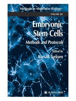
Figure 1.1. Origin of stem cells during mammalian embryogenesis. In this
figure, the pluripotent cells of the embryo are tracked in green. From left to
right, the morula-stage mouse embryo (embryonic day 2.5; E2.5) holds a core
of pre-ICM cells that turn into ICM cells at cavitation/blastulation (E3–E4). At
this stage, ES cell and Trophoblast stem (TS) cell lines can be derived in vitro,
and implantation occurs in vivo. FAB-SCs can be derived from mouse
blastocysts in combination of bFGF, activin, BIO and an anti-LIF antibody.
As the blastocyst fully expands (and undergoes implantation in vivo), the ICM
delaminates giving rise to a primitive ectoderm and a primitive endoderm
layer. At this stage, pluripotent cell lines that are known as embryonal
carcinoma (EC) cells can be derived from the primitive ectoderm. EpiSCs are
derived from E5.5–E5.75 post-implantation epiblasts in the presence of activin
and bFGF. At E6 and subsequent stages, the experimental ability to derive ES
Cells, TS cells and EC cells from the mouse embryo is progressively lost, and
the in vivo embryo will start gastrulating. (Adapted from Bioani and Sholer et
al. 2005)
Another source of pluripotent stem cells is provided by induced pluripotent
stem cells (iPSCs) from somatic cells by enforced expression of a few
pluripotency-associated transcription factors. The discovery of induced
pluripotency can be traced back to the work of somatic cell nuclear transfer
(SCNT) that first established by Briggs and King (Briggs and King, 1952;
King and Briggs, 1955). The cloning of Dolly sheep further showed that the
genome of even terminally differentiated cells preserve the potential to
6
develop into an entire organism (McLaren, 2000; Wilmut et al., 1997).
However, SCNT is technically challenging and the cloned animals always
exhibit abnormalities in gene expression and phenotype. An alternative
approach is developed by in vitro hybridization between somatic and
pluripotent cells. The hybrid cells by fusion of EC cells with somatic cells,
such as thymocytes, resemble EC cells in terms of biochemical properties and
differentiation potential, while lose the features of somatic cells (Miller and
Ruddle, 1976, 1977), indicating that some soluble regulatory factors in EC
cells confer a pluripotent state to somatic cells. However, hybrid cells lack
therapeutic potential because of their abnormal ploidy and the presence of
nonautologous genes from the pluripotent parent. A great breakthrough was
achieved by Yamanaka and Takahashi in 2006 (Takahashi and Yamanaka,
2006). The original idea was to induce pluripotency from somatic cells by
enforced expression of specific transcription factors, which was based on the
observation that lineage-associated transcription factors were able to change
the cell fate when ectopically expressed in certain heterologous cells (Davis et
al., 1987; Laiosa et al., 2006; Xie et al., 2004; Zhou et al., 2008). To induce
pluripotency, they performed an elegant screen for factors within a pool of 24
pluripotency-associated candidate genes and came out a core set of four genes,
Oct4, Sox2, Klf4 and c-Myc, called “Yamanaka genes”, which are minimally
required to be enforced expressed for reprogram mouse fibroblasts to iPSCs.
The resultant mouse iPSCs have passed the most stringent test of pluripotency,
tetraploid complementation, a technique in which iPSCs are injected into a
tetraploid blastocyst and are shown to contribute to the generation of an entire
living mouse (Kang et al., 2009; Zhao et al., 2009). The iPSCs field has
7
progressed at a breathtaking pace in the last 5 years, including derivation of
iPSCs from other species, such as human; optimization of the efficiency of
iPSCs generation; development of virus-free factors delivery system and
establishment of disease-specific iPSCs. In addition to being an exciting
academic research model to study cellular development, iPSCs hold
significant therapeutic potential for regenerative medicine, disease modeling
and drug development. Notwithstanding these achievements, iPSCs
technology remains in its infancy and a better understanding of the
reprogramming process is required in order to develop more efficient
strategies for pluripotency induction and a careful analysis of the genomic and
epigenomic characteristics of iPSCs, as well as the development of a robust
protocol for directed differentiation are required for future utilities of iPSCs in
clinic medicine.
Although different types of pluripotent stem cells have been generated and
broadly expand our knowledge for pluripotency, the biggest challenge remains
to produce mature, functional and pure derivatives of cell types that can be
utilized for transplantation purposes. To facilitate these developments, a large
amount of efforts is put to get a comprehensive understanding of the biology
of ES cells including genes that are important for the maintenance of ES cells,
especially human ES cells. However, due to the ethical challenge of the source
of human ES cells and the inability to test pluripotency of human ES cells by
chimera formation, extensive work has been carried out initially on mouse ES
cells. Mouse ES cells are easier to manipulate and have been extensively
characterized for 20 more years than human ES cells; therefore the discovery
on mouse ES cells will eventually shed light on the understanding of human
8
ES cells. In my thesis work, I focus all my studies on mouse ES cells, and
particularly on the transcriptional regulation of these cells, to understand the
molecular mechanisms underlying pluripotency.
1.2 Characteristics of mouse embryonic stem (ES) cells
Mouse ES cells are well known for two distinguished properties: self-renewal
and pluripotency. Self-renewal is the ability of ES cells to proliferate
continuously in culture in undifferentiated state (Smith and Benchimol, 1988).
More importantly, unlike EC cells and other primary cell lines that can only be
passaged for several times before senesce, these cells can be passaged for
years while maintaining normal karyotypes (Keller, 2005).
The second property of ES cells is that they recapitulate full developmental
potential when injected into mouse blastocysts, contributing cells to all three
germ layers and to the germline of chimeric animals. It is known as
pluripotency, which has attracted huge interest of numerous researchers
because of its promising applications in regenerative medicine. The golden
rule to judge pluripotency of ES cells is by their ability to integrate into the
ICM of the blastocysts and contribution to germline formation. So far,
pluripotency has only be proven conclusively in mouse ES cells, as they can
completely integrate into the blastocyst, after transplantation, and exhibit high
efficiency of chimera formation and germline transmission. ES cells can also
be induced to differentiate in vitro by a number of strategies. By cultivation in
vitro as 3D aggregates called embryoid bodies (EBs),
ES cells can differentiate
into derivatives of endoderm, mesoderm, and ectoderm. Removal from the
9
self-renewing environment by taking out cytokines, such as LIF or BMP4,
from culture medium triggers intrinsic differentiation programs that resembles
a developmental course that was interrupted when the ICM was extracted from
the blastocyst. Moreover, adding in soluble molecules, such as retinoic acid,
will stimulate ES cell differentiation as well.
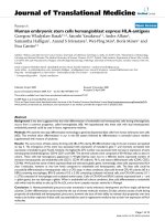
Figure 1.2. Differentiation of mouse ES cells by EB formation. (A) Mouse
ES cells cultured under feeder free condition; (B) Embryoid bodies (EB) are
formed 8 days after suspension culture. (C-E) Examples of mesoderm lineage:
Cardiomyocytes (C), Skeletal muscles (D) and Smooth muscles (E); (F-H)
Examples of ectoderm lineage: Neurons (F), Glial (G) and Epithelial (H); (I-L)
Examples of endoderm lineage: Pancreatic cells (I), Hepatocytes (K-L).
However, the therapeutic use of ES cells will require more precise control
over this process in order to make these cells differentiate efficiently and
strictly to a specific lineage. Intensive work has been conducted to the field of
directed differentiation to influence the lineage commitment of ES cells in
vitro. Various strategies involving supplementation of growth factor cocktail,
10
cell co-cultures, conditioned medium and specific gene transfection are used
to drive lineage specific emergence (Fair et al., 2003; Ogawa et al., 2005;
Wells and Melton, 1999; Zhou et al., 2007c). Nevertheless, the improved
knowledge of the molecular mechanisms governing ES cell maintenance and
differentiation towards specific lineage are desired to better facilitate direct
differentiation of ES cells for therapeutic applications.
1.3 Application of ES cells
As we have discussed above, the most extraordinary property of ES cells is
their ability to re-enter embryogenesis. Indeed, a major interest of ES cells to
the scientific community is their utility as cellular vehicles for engineering of
the mouse genome. Mouse ES cells can be injected into the blastocysts and
integrate into the ICM cells to produce viable chimeras. The derivation of
transgenic mice from genetically modified ES cells was first reported in 1984
(Bradley et al., 1984). Afterwards, ES cell technology has been most often
used to produce null mutants (gene knockouts) through homologous
recombination (Thomas and Capecchi, 1986) for the in vivo study of gene
function during development and this can even be achieved in a conditional
knockout manner. Moreover, they can also be used to introduce subtle genetic
modifications down to the level of single nucleotide mutation in endogenous
mouse genes. Transgenic mice derived from ES cells has not only
revolutionized basic biological research through the creation of genetically
altered animals, but also permits the evaluation of therapeutic strategies in
models of human disease, as well as the investigation of disease progression in
11
a manner not possible in human subjects.
The discovery of human ES cells has been considered as the key tool for
understanding most of the fundamental questions in both basic and clinical
human biology. Human ES cells may allow scientists to investigate how early
human cells become committed to specific lineages and differentiated into the
myriad functional cell types that build up tissues and organs of the entire body.
The knowledge gained will greatly accelerate our understanding of the causes
of birth defects and thus lead directly to their possible prevention. Human ES
cells can also be applied as a valuable in vitro model system to study diseases
that only occur in human or have significant difference between human and
other species, such as HIV, HCV. In the clinic trail, they could be used to
create an unlimited supply of cells, tissues, or even organs that could be used
to restore function. Human ES cell-derived progeny have been successfully
exploited in animal models of spinal cord injury (Keirstead et al., 2005; Sharp
et al., 2010), retinopathies (Lamba et al., 2009), and Parkinson‟s disease
(Yang et al., 2008). And this idea is greatly promoted by the generation of
patient-specific iPSCs. Disease-specific iPSCs have already been created from
patients suffering from amyotrophic lateral sclerosis (Dimos et al., 2008),
juvenile onset type 1 diabetes mellitus (Park et al., 2008a), Parkinson‟s disease
and spinal muscular atrophy (SMA) (Ebert et al., 2009). Critically, the
pathophysiology of SMA could be recapitulated in motor neurones derived
from patient-specific iPSCs. In the long run, these patient-specific iPSCs may
be ideally suited for cellular therapy, given that they are derived from the
patient to be treated, thus minimizing the risk of immune rejection. However,
it is noteworthy that these iPSCs, however, are only the starting point for the
12
preparation of cells for clinic trials, as therapeutic cells should be
differentiated cell lines with the characteristics proper of the various tissues
(muscle, neural, epithelial, haematic, germinal, etc.). Methods for obtaining
therapeutic cells from human ES cells or iPSCs are still being studied and
even if successful for some specific cell types, a testing assay to certain that
the inoculation or therapeutic implant was free of stem cells is also crucial, as
the remnant stem cells may result in tumors.
1.4. Molecular characteristics of ES cells
The maintenance of ES cells engages complex and precisely controlled
molecular and cellular regulatory machinery. While self-renewal and
pluripotency associated genes are up-regulated to maintain the undifferentiated
state of ES cells, genes that induce differentiation are suppressed but poised
for subsequent expression during cellular differentiation. Tremendous effort
has been applied to uncover the molecular mechanisms governing self-renewal
and pluripotency in ES cells, and based on our current knowledge, the
balanced state of ES cells is achieved through the complex interplay of cell
cycle regulation, signaling pathways, epigenetic modification, small regulatory
RNAs as well as ES-specific transcriptional network.
1.4.1. Cell cycle regulation
Cell cycle program of mouse ES cells is characterized by extraordinarily rapid
proliferation rate and a pluripotent cell specific cell cycle structure, which is
controlled by an unusual mode of cell cycle regulation. The work from the last
13
few years has revealed the importance of cell cycle regulation to the
maintenance of ES cells, as the process of self-renewal requires the
coordination of cell cycle progression and cell-fate determination (self-
renewal versus commitment). A few transcription factors as well as cell cycle
regulators appear to be critical to this regulation.
Mouse ES cells have relatively short cell cycle period compared with
differentiated cells, with ~8 to 10 hours total generation time, and an unusual
cell cycle structure, with a reduction in the duration of G1 phase. Although
human ES cells share a similar cell cycle structure, their generation time is
significantly slower (~32-38 hours; (Dalton, 2009; Ohtsuka and Dalton, 2008)
indicating that a short division may not be a pre-requisite for pluripotency.
This is supported by the study showing that slowing cell cycle of mouse ES
cells with chemical inhibitors has no measurable impact on the maintenance of
ES cells (Stead et al., 2002). Instead, other observations suggest that
mechanisms making up the specific cell cycle structure are more crucial to the
ES cell maintenance. The short G1 phase allows ES cells to be less responsive
to the differentiation signals sent by certain mitogenic signaling pathways that
are active and act as potent differentiation inducer during G1 phase in somatic
cells. It has been shown that mitogenic signaling pathways inhibit mouse ES
cells self-renewal and promote their differentiation, while self-renewal of
mouse ES cells is enhanced by the addition of inhibitors of mitogenic
signaling pathways to the culture medium (Burdon et al., 2002; Burdon et al.,
1999). Furthermore, the extended S phase may also shield cells from extrinsic
differentiation signals by maintain chromatin in an “open” euchromatic state
to facilitate rapid activation or repression of genes (Filipczyk et al., 2007;
14
Herrera et al., 1996).
Figure 1.3. The cell cycle of ES cells. The cell cycle of ES cells is shortened
relative to that of most other cells, which is due to an abbreviated G1 phase.
For most cells, the transition through early G1 phase requires the accumulation
of cyclin D, resulting in the hyperphosphorylation of the retinoblastoma
tumour suppressor protein (RB) by cyclin D–CDK4 or cyclin D–CDK6
complexes (D/4,6). Inactivation of RB by hyperphosphorylation results in the
mitogen-independent activity of cyclin E–CDK2 complexes, the defining
characteristic of late G1 phase. In ES cells, cyclin E–CDK2 (E/2) is
constitutively active throughout the cell cycle, which allows the transition of
ES cells from M phase directly to late G1. The resulting absence of the cyclin
D-dependent early G1 phase shortens the G1 phase and the entire cell cycle. +
refers to cyclin–CDK activity: +/-, negligible; +, low; ++, intermediate; +++,
high (Adapted from Orford and Scadden et al., (Orford and Scadden, 2008)).
A direct relationship between cell cycle regulation and master regulators of ES
cells has recently been described. Oct4 and Sox2 are shown to regulate miR-
15
302, which targets cyclin D1, Rb, E2F1 and p130 (Card et al., 2008) and
Nanog is suggested to be a regulator of G1 to S transition in ES cells through
regulation of CDK6 and CDC25A, which are key players in the G1 cell cycle
(Zhang et al., 2009).
The role of cell cycle regulation in maintaining ES cell identity is further
emphasized by the study of reprogramming and iPSCs derivation. Myc is one
of the four “Yamanaka factors” for iPSCs generation. Although subsequent
studies have demonstrated that Myc is dispensable for the iPSCs recipe, it is
shown to be critical for the early stages and high efficiency of reprogramming
as it maintains the cells in a proliferative state in which they respond better to
the other exogenous factors (Knoepfler, 2008; Zhao and Daley, 2008). Unlike
other transcription factors in the reprogramming recipe, Oct4, Sox2 and Klf4,
which have significant functions for maintaining self-renewal and
pluripotency in ES cells, there is no much evidence indicating the direct
relationship between the expression level of Myc to the state of ES cells, as no
developmental defects have been observed in c/N-Myc knockout mice.
However, there is considerable evidence linking Myc to the cell cycle
regulation in ES cells. Elevated c-Myc expression accelerates progression
through G1 by positively regulating cyclin-Cdk activity, whereas ES cells lost
its specific cell cycle structure during differentiation while the expression of
Myc is downregulated (Cartwright et al., 2005; White and Dalton, 2005). All
these data place Myc at the center of a regulatory network linking fundamental
self-renewal and pluripotency mechanisms to the cell cycle machinery in ES
cells.
1.4.2. Small regulatory RNAs