Mechanism of protein quality control in the cytosol in budding yeast
Bạn đang xem bản rút gọn của tài liệu. Xem và tải ngay bản đầy đủ của tài liệu tại đây (8.29 MB, 165 trang )
MECHANISM OF PROTEIN QUALITY CONTROL IN
THE CYTOSOL IN BUDDING YEAST
RUPALI PRASAD
(M.Sc, IIT Bombay)
A THESIS SUBMITTED
FOR THE DEGREE OF DOCTOR OF PHILOSOPHY
DEPARTMENT OF BIOLOGICAL SCIENCES
NATIONAL UNIVERSITY OF SINGAPORE
2011
i
ACKNOWLEDGEMENTS
This research work is by far one of the most significant scientific accomplishments in
my life and it would have been impossible without the following people, who
supported me and had belief in me.
First and foremost, I want to express my wholehearted gratitude to my mentor and
research advisor Associate Professor Davis Ng, for his expert guidance and
motivation throughout my research work. I am grateful to him for his invaluable
support and also for introducing me to the wonderful and interesting field of protein
quality control.
I would also like to express my sincere thanks to Dr. Shinichi Kawaguchi and Ms
Alisha for assisting me and being a part in the projects. I am also thankful to Dr
Kazue Kanehara, Dr Guillaume Thibault, and Songyu Wang for fruitful discussions
and suggestions. I owe very special thanks to all current and previous members of
Cell Stress and Homeostasis Group, especially Dr Nurzian Ismail, Dr. Chia Ling Hsu
and Dr. Xie Wei and to all my friends at TLL. I want to thank them for all their help,
support, interest and valuable hints.
I gratefully acknowledge the financial support rendered by the National University of
Singapore in the form of Research Scholarship. I am also grateful to the academic and
technical staffs at the Temasek Life Sciences Laboratory who have helped me in one
way or the other in my research work.
ii
Above all, I want to thank my family, which continuously supported me at all times. I
thank my parents for teaching me the value of education at a young age and instilled
in me a desire for higher education. I wish to thank my sisters for their love and
support. Words cannot express the love, encouragement and unequivocal support I
received from my beloved husband Anil without whose constant help and support, my
PhD. research work would have remained a daydream. The loving family
environment and support I enjoyed from all my family members was greatly
instrumental in providing me the tranquility and enthusiasm to pursue my research
with a piece of mind.
iii
TABLE OF CONTENTS
Acknowledgements i
Table of contents iii
Summary vii
List of tables ix
List of figures x
List of abbreviations xii
List of publications xiv
CHAPTER 1: Introduction 1
1.1 Protein quality control (PQC) 2
1.2 ER quality control 3
1.2.1 Role of ER-lumenal chaperones 3
1.2.2 Recognition of ERAD substrate 4
1.2.3 ERAD Complex 6
1.2.4 Retrotranslocation and the Cdc48 complex 8
1.3 Cytosolic protein quality control 10
1.3.1 Recognition of damaged proteins and repair mechanism 10
1.3.2 Transcriptional regulation 13
1.3.3 Autophagy-lysosome system 14
1.3.4 Sequestration into large aggregates 15
1.3.5 Degradation of misfolded proteins by UPS 16
1.4 Ubiquitin-proteasome system 18
1.5 26S proteasome 20
1.5.1 Substrate recognition by 26S proteasome 20
1.5.2 Substrate unfolding, translocation and proteolysis 21
1.5.3 Cellular localization of 26S proteasome 21
1.6 Structure and function of selected cytosolic chaperones and
cochaperones 23
1.6.1 Hsp70/Hsc70 23
1.6.2 Hsp40 26
1.6.3 Nucleotide Exchange Factors (NEFs) 28
iv
1.6.4 Hsp90 29
1.7 Objectives of the thesis 31
CHAPTER 2: Materials and methods 33
2.1 S. cerevisiae strains, growth media and genetic techniques 33
2.1.1 List of S. cerevisiae strains 33
2.1.2 Media for culturing yeast 36
2.1.3 Mating, sporulation and tetrad dissection 36
2.1.4 Yeast transformation 37
2.1.4.1 Low efficiency plasmid transformation via simple and
rapid way 37
2.1.4.2 High efficiency transformation using lithium acetate 37
2.1.5 Serial dilution-spotting growth assay 38
2.2 Molecular biology techniques 38
2.2.1 List of plasmids used in this study 38
2.2.2 List of oligonucleotide primers used in this study 39
2.2.3 Plasmids construction 40
2.2.4 Yeast genomic DNA extraction 43
2.2.5 Plasmid DNA extraction from yeast 44
2.3 Biochemical and immunological techniques 44
2.3.1 Antibodies used in this study 44
2.3.2 TCA precipitation of yeast whole Cell Lysate 44
2.3.3 SDS-PAGE and western blot analysis 45
2.3.4 Cycloheximide chase assay 46
2.3.5 Cytosol/membrane fractionation 46
2.3.6 Ubiquitination assay 46
2.3.7 Trypsin sensitivity assay 47
2.3.8 Co-immunoprecipitation Assays 47
2.3.9 TAP-tagged Hsp70 pulldown assay 48
2.3.10 Cell labeling and Immunoprecipitation analysis 48
2.3.10.1 Pulse chase assay 48
2.3.10.2 Immunoprecipitation 49
v
2.4 Microscopy techniques 50
2.4.1 Indirect immunofluorescence 50
2.4.2 Live cell imaging 51
2.5 Genetic screening method used in this study 51
2.5.1 UV mutagenesis 51
2.5.2 Primary selection 52
2.5.3 Secondary screen for CytoQC defect 53
2.5.4 Cloning of QCC genes 54
CHAPTER 3: A nuclear-based quality control mechanism for cytosolic
proteins 55
3.1 Introduction 55
3.2 The model cytosolic substrate ΔssCPY* is degraded by CytoQC and ERAD 57
3.3 DssPrA and D2GFP are misfolded proteins and degraded by proteasome 61
3.3.1 Novel CytoQC substrates are highly unstable 61
3.3.2 DssPrA and D2GFP are bona fide substrates for CytoQC 63
3.4 Misfolded cytosolic proteins traffic into the nucleus for degradation
65
3.5 San1p-dependent pathway is a general mechanism of CytoQC substrates 67
3.5.1 Nuclear E3 ligase San1p is required for degradation 67
3.5.2 CytoQC substrates polyubiquitination is dependent on E3 ligase San1p 69
3.6 San1p can interact with CytoQC substrate in vivo 70
3.7 San1p pathway is a constitutive mechanism of CytoQC 74
3.8 CytoQC substrates are polyubiquitinated and degraded inside the nucleus 76
3.8.1 Substrate degradation is independent of nuclear export 76
3.8.2 Nucleus is the site for CytoQC substrates degradation 77
3.9 E3 ligase Doa10p is not required for degradation of DssPrA and D2GFP 78
3.10 Ubr1p augments, but is not required for, DssPrA and D2GFP degradation 79
CHAPTER 4: Roles of molecular chaperones in the cytosolic quality control 85
4.1 Introduction 85
4.2 The Hsp70 chaperone machinery is essential for the degradation of
cytosolic misfolded proteins 87
vi
4.3 The Hsp70 chaperone can interact directly with cytosolic misfolded
proteins in vivo 90
4.4 The Hsp70 chaperone system is required for efficient nuclear transport of
cytosolic misfolded proteins 92
4.5 Effect of temperature on CytoQC substrate localization 93
4.6 The Hsp70 co-chaperone Ydj1p is directly involved in nuclear import of
cytosolic misfolded proteins 94
4.7 Nucleotide exchange factor Sse1p is essential for CytoQC pathway 97
4.8 Lack of Hsp90 inhibit substrate degradation and has paltry effect on nuclear
import 99
4.9 Discussion 101
CHAPTER 5: A genetic screen to identify genes required for cytosolic quality
control 106
5.1 Introduction 106
5.2 Folding state of proteins in the cytosol or membrane tethered forms, are
monitored by different PQC pathways 107
5.3 Ste6C requires all the factors of cytosolic quality control 112
5.4 Genetic screen to identify new components required for cytosolic quality
control pathway 116
5.5 The qcc mutants are defective in cytosolic quality control pathway 121
5.6 Cloning of QCC genes by complementation 123
5.7 Identification of new candidates in CytoQC pathway 123
5.8 Discussion 128
CHAPTER 6: Conclusions and future directions 132
References 135
vii
SUMMARY
Intracellular quality control systems monitor protein conformational states.
Irreversibly misfolded proteins are cleared through specialized degradation pathways.
Their importance is underscored by numerous pathologies caused by aberrant
proteins. In the cytosol, where most proteins are synthesized, quality control remains
poorly understood. Stress-inducible chaperones and the 26S proteasome are known
mediators but how their activities are linked is unclear. In this thesis, I have used
Saccharomyces cerevisiae as a model organism to study the quality control of
cytosolic misfolded proteins. To better understand quality control of cytosolic
proteins in chapter 3 and 4 of this thesis, a panel of model misfolded substrates was
analyzed in detail. Surprisingly, their degradation occurs not in the cytosol but in the
nucleus (Prasad et al., 2010). Degradation is dependent on the E3 ubiquitin ligase
San1p, known previously to direct the turnover of damaged nuclear proteins (Gardner
et al., 2005). San1p, however, is not required for nuclear import of substrates. Two
reasons can account for nuclear trafficking of misfolded cytosolic proteins. First, in S.
cerevisie, nucleus accounts for over 80 % of proteasomes at steady state throughout
the cell cycle, suggesting the requirement of nuclear import of misfolded cytosolic
proteins. Second, by trafficking misfolded proteins in the nucleus, cells provide
enough time for newly synthesized proteins to fold in proper conformation. One view
asserts that a key strategy of protein quality control is the integration of timing
devices to permit folding (Helenius and Aebi, 2004). As such, proteins failing to fold
within a set window are targeted for degradation. Experimental precedence comes
from ERAD studies where a sophisticated timing mechanism utilizes a series of
glycosidases to set a time limit for folding (Clerc et al., 2009). Proteins still unfolded
viii
after the final trimming step by Htm1p are detected by the Yos9p ERAD factor (OS-9
in mammals), which binds the resulting glycan signal (Quan et al., 2008). In CytoQC,
nuclear import of substrate can provide an analogous function. The detailed analyses
of cytosolic substrates have provided a clue that the Hsp70 family proteins Ssa1p and
Ssa2p and its co-chaperone Ydj1p are needed for efficient import and degradation. In
chapter 5 of this thesis, I have described a genome wide genetic screen to identify the
genes involved and to decipher the mechanism for quality control of cytosolic protein.
Among the genes identified, there are genes that encodes for proteasomal subunits
(RPN7 and RPN11) and UMP1, a chaperone required for assembly of 26S proteasome.
Together all our data reveal a new function of the nucleus as a compartment central to
the quality control of cytosolic proteins.
ix
LIST OF TABLES
Table 2.1 List of yeast strains used in this study 33
Table 2.2 List of plasmids used in this study 38
Table 2.3 List of oligonucleotide primers used in this study 39
x
LIST OF FIGURES
Figure 1.1
ERAD recognition of misfolded protein by N-glycan signal 6
Figure 1.2 Distinct ERAD complexes in yeast 8
Figure 1.3 Role of molecular chaperones in the balance of folding,
degradation and aggregation 12
Figure 1.4 Cell stress response 14
Figure 1.5 The ubiquitin-proteasome system for intracellular protein
degradation 19
Figure 1.6 The 26S complex of Saccharomyces cerevisiae 22
Figure 1.7 The Hsp70 ATPase cycle 26
Figure 3.1 A fraction of the misfolded protein ΔssCPY* translocates
into the ER without a signal sequence and ΔssCPY* degrades
via ERAD and CytoQC 60
Figure 3.2 DssPrA and D2GFP are substrates of CytoQC 62
Figure 3.3 Turnover of DssPrA and D2GFP requires cytosolic quality
control chaperones 64
Figure 3.4 DssPrA and D2GFP are localized in the cytosol and nucleus 66
Figure 3.5 Intracellular localization and degradation of ΔssPrA-3A 66
Figure 3.6 San1p is required for DssPrA and D2GFP degradation 68
Figure 3.7 San1p is required for DssPrA and D2GFP ubiquitination 70
Figure 3.8 San1p overexpression enhances substrate degradation 72
Figure 3.9 Effect of San1p-V5 overexpression on its localization and
substrate degradation 73
Figure 3.10 Effect of substrate expression level on degradation 75
Figure 3.11 Substrate degradation in nuclear export defective
xpo1-1 cells 77
Figure 3.12 Visualization of intracellular substrate decay 78
Figure 3.13 Doa10p is not required for ΔssPrA and Δ2GFP degradation 79
Figure 3.14 Ubr1p augments the San1p system 80
Figure 4.1 Analysis of SSA1-4 single and double mutants in
cytosolic quality control 89
Figure 4.2 Ssa1p and Ssa2p specifically bind substrate proteins in vivo 91
xi
Figure 4.3 Ssa1p and Ssa2p are required for the nuclear localization
of CytoQC substrates 93
Figure 4.4 Temperature effect on substrate localization 94
Figure 4.5 Substrate loses its ability to be transported into the nucleus
when Ydj1p function is eliminated 96
Figure 4.6 Sse1p is required for substrate degradation 98
Figure 4.7 Hsp90 is required for substrate degradation and partly for nuclear
Impot 100
Figure 4.8 Model of CytoQC-degradation pathway for the transport of
misfolded cytosolic proteins into nucleus 104
Figure 5.1 Ste6C degradation does not depend on the Doa10p E3
ubiquitin ligase 110
Figure 5.2 Cdc48 ATPase complex is required for membrane anchored
version of Ste6C 111
Figure 5.3 Ste6C is present both in cytosolic and membrane fraction 113
Figure 5.4 Ste6C is a cytosolic quality control substrate 115
Figure 5.5 A schematic diagram representing the strategy taken to obtain
genes involved in cytosolic quality control 118
Figure 5.6 Stabilization of Ste6C-Ura3p fusion protein in qcc mutant strains 119
Figure 5.7 The qcc mutant strains can also stabilize other cytosolic
quality control substrates 122
Figure 5.8 Introduction of complementary gene restores the cytosolic
quality control function 125
Figure 5.9 Introduction of complementary gene suppresses the
thermosensitive growth effect of temperature sensitive
qcc mutant strains 126
xii
LIST OF ABBREVIATIONS
ATP Adenosine-5’-triphosphate
CFTR Cystic fibrosis transconductance regulator
CHX Cycloheximide
CPY Carboxypeptidase Y
CytoQC Cytosolic quality control
DAPI 4’,6-diamidino-2-phenylindole
DMSO Dimethyl sulfoxide
DTT Dithiothreitol
ER Endoplasmic reticulum
ERAD ER-associated protein degradation
ERQC ER quality control
Endo H Endoglycosidase H
HRD HMG-CoA reductase degradation
Hsp Heat shock protein
Htm1 Homologous to mannosidase I
IPODs Insoluble protein deposits
JUNCs Juxtanuclear quality controls
Man Mannose
Mns α-mannosidase
NEF Nucleotide exchange factor
NES Nuclear export signal
NLS Nuclear import signal
OD Optical density
OST Oligosaccharyltransferase
xiii
PBS Phosphate buffered saline
PCR Polymerase chain reaction
PDI Protein disulfide isomerase
PMSF Phenylmethylsulphonylfluoride
PrA Proteinase A
QCC Quality control in the cytosol
RING Really Interesting New Gene
RPM Round per minute
SC Synthetic complete
SBD Substrate binding domain
SDS-PAGE Sodium dodecyl sulphate-polyacrylamide gel electrophoresis
SEM standard error of the mean
TAP Tandem affinity purification
TCA Trichloroacetic acid
TPR Tetratricopeptide repeats
UPS Ubiquitin-proteasome system
VHL von Hippel-Lindau
Yos9 Yeast osteosarcoma 9
YPD Yeast peptone dextrose
xiv
LIST OF PUBLICATIONS RELATED TO THIS STUDY
Prasad, R., Kawaguchi, S. and Ng, D.T. (2010). A nucleus-based quality control
mechanism for cytosolic proteins. Molecular Biology of the Cell. 21, 2117-2127.
Prasad, R., Kawaguchi, S. and Ng, D.T. Nuclear import of misfolded cytosolic
protein is mediated by Hsp70-Hsp40 chaperone system. Manuscript in preparation.
1
CHAPTER 1
Introduction
The central dogma of molecular biology—DNA to RNA to protein—concisely
describes the information flow of protein synthesis. Errors arising at any step can
disrupt protein folding and lead to potentially toxic products. Although DNA
replication is highly accurate, transcriptional and translational error rates can be as
high as 10
-4
and 10
-3
, respectively (Zaher and Green, 2009). Even with a correct
sequence, the need for chaperones and, in some cases, modifying enzymes for folding
makes an already complex process even more precarious. The consequences of
accumulating aberrant proteins are so serious that numerous sophisticated protein
quality control mechanisms (PQC) have evolved to protect cells.
Efficient protein folding is very important for proper function and viability and to do
so cells contain elaborate enzymatic machinery called molecular chaperones. Some
bind to non-native polypeptide and promote their folding in an ATP-dependent
manner (Hartl and Hartl, 2002). Protein misfolding can occur by genetic mutation,
biosynthetic errors and cellular stress such as chemical and temperature perturbation.
If a cell fails to eliminate these misfolded non-native proteins, it can lead to formation
of toxic aggregates, non-functional proteins and ultimately cell death. A number of
diseases associated with aberrant protein conformation highlight the importance of
protein quality control for cell survival (Dobson, 2004).
To repair or remove damaged or unfolded proteins, both prokaryotes and eukaryotes
use protein quality control system that includes numerous molecular chaperones and
proteases. In general intracellular misfolded proteins have three fates: they either get
2
rescued by molecular chaperones or destroyed via proteases or form aggregates.
Chaperones are a diverse group of proteins defined by size, cellular compartment and
function. They work together to prevent protein aggregation and facilitate the correct
folding of nascent polypeptide into native conformation. Misfolded proteins are
degraded by a variety of proteases; however, the main intracellular degradation site is
the proteasome.
1.1 Protein quality control (PQC)
As there is a high degree of compartmentalization in eukaryotic cells, proteins are
more prone to damage in specific organelles. For example, in the endoplasmic
reticulum (ER), proteins are vulnerable to errors in glycosylation, disulfide bond
formation and membrane insertion (Alexander et al., 2010). In contrast, proteins in
the cytosol are more sensitive to stress, while ER proteins are more stable because of
the effect of disulfide bonds and glycosylation. The protein quality system is adapted
to handle any situation that results in a change in protein conformation from its native
state. Protein quality control degradation systems have been identified in several
organelles such as cytosol, ER, mitochondria and nucleus. In the cytosol and ER,
degradation is mainly brought about by polyubiquitination of misfolded proteins by
protein-ubiquitin-complexes that mark the substrate for proteasomal degradation
(Hampton, 2002; McDonough and Patterson, 2003; Trombetta and Parodi, 2003). In
these two compartments, PQC can also occur via transport to the lysosome/vacuole
(Trombetta and Parodi, 2003). In the mitochondria, localized proteases function in
PQC and degradation (Arnold and Langer, 2002). Although, PQC is found
everywhere proteins are made or mature, the best understood PQC mechanisms are in
the endoplasmic reticulum (ER).
3
1.2 ER quality control
The ER is a protein factory, where secretory and membrane proteins enter and are
folded, which account for one-third of total cellular proteins. ER has a quality control
system that monitors the correctly folded proteins and sends them to their final
destination. Proteins which misfold or unfold, are recognized by the quality control
system in the ER which retain and refold them (Ellgaard and Helenius, 2003).
Accordingly, ER quality control mechanisms have the added responsibility to control
trafficking, to prevent the premature exit of folding intermediates (Vembar and
Brodsky, 2008). For proteins that fail to fold, the integration of ER associated
degradation (ERAD) pathways removes and destroys aberrant products. ERAD
includes the involvement of general folding factors like Kar2p and protein disulfide
isomerase as well as specialized factors that recognize and target misfolded proteins
to ERAD processing sites. These sites, made up of factors organized by E3 ubiquitin
ligases, function to translocate and ubiquitinate substrates before they are degraded by
the 26S proteasome (Carvalho et al., 2006; Denic et al., 2006; Gauss et al., 2006).
1.2.1 Role of ER-lumenal chaperones
Misfolded glycosylated proteins in the ER lumen are first recognized by Hsp70
homologue, Kar2p. Kar2p is also required for posttranslational protein translocation
into the ER and protein folding in the ER lumen (Kabani et al., 2003, Vashist and Ng,
2004). Kar2p binds to hydrophobic patches of misfolded or unfolded proteins to
prevent them from forming aggregates (Kabani et al., 2003; Flynn et al., 1991). Two
mutants of Kar2p have been identified; kar2-1 and kar2-133 and both of them showed
reduced affinity for its substrate and ERAD defect. In these mutant strains, ERAD
4
substrates get aggregated in the ER lumen and this defect can be rescued by
coexpression of wild type Kar2p.
Another abundantly present ER chaperone, protein disulfide isomerase (PDI) is a thiol
oxidoreductase. This chaperone is involved in disulfide bond formation, protein
folding and recognition and targeting of misfolded protein for ERAD (Nishikawa et
al., 2005; Tu and Weissman, 2004). Yeast PDI contains a characteristic CXXC motif
that is located in the active site of the enzyme (Tian et al., 2006). PDI binds to
hydrophobic patches of proteins in the folding process in its groove, breaks the wrong
disulfide-bonds and catalyzes the formation of correct ones.
1.2.2 Recognition of ERAD substrate
The best understood mechanism for recognition of ERAD substrate is the
glycan-dependent pathway. Majority of polypeptides, which are synthesized in the ER
are modified by N-linked glycans. The oligosaccharyltransferase covalently add
Glc
3
Man
9
GlcNAc
2
glycan to asparagines in Asn-X-Ser/Thr motif of polypeptides.
Additions of glycans to polypeptides make them more hydrophilic and direct them for
folding. Moreover, it provides information about the folding status of glycoproteins.
The outermost glucose residues are further removed by Gls1p and Gls2p to label
polypeptide as in the process of folding (Figure 1.1). The resulting glycoproteins
which bear, Glc
1
Man
9
GlcNAc
2
glycan, can associate with glycan-dependent
chaperones, which promote their maturation. Once liberated from these chaperones,
Gls2p trims the final glucose residue from polypeptide to attain their native structure.
If a polypeptide fails to mature within its folding window, further trimming of glycans
trigger its destruction. ER resident Mns1p (α1,2-mannosidase 1) trims the outermost
5
glycan residue from the B branch of N-glycan of a polypeptide, resulting in
Man
8
GlcNAc
2
glycan. However, Mns1p can process glycoproteins regardless of their
folding status, thus Man
8
itself is not sufficient to be a degradation signal (Jakob et al.,
1998; Jelinek-Kelly and Herscovics, 1988). Therefore additional signature glycan is
needed to distinguish defective polypeptides from the mature protein. Htm1p
(mannosidase 1) a lectin, further removes the C branch from the glycan, resulting in
Man
7
GlcNAc
2
structure which flags potential ERAD substrates for degradation (Clerc
et al., 2009). Man
7
GlcNAc
2
structure, in turn is recognized by glycan binding protein
Yos9p (yeast osteosarcoma 9) (Quan et al., 2008). Recent studies have shown that
Yos9p binds misfolded proteins but is unable to interact with their folded counterparts.
Yos9p is part of the Hrd1 complex, as it directly interacts with the large luminal
domain of Hrd3p, indicating that it can links the recognition of misfolded
glycoproteins to the ubiquitin-proteasome system.
6
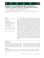
Figure 1.1.
ERAD recognition of misfolded protein by N-glycan signal.
In yeast,
glucosidases 1 and 2 trim the two outermost glucose residues to generate
Glc
1
Man
9
GlcNAc
2
. Final sugar is removed by
glucosidases 2, generating Man
9
GlcNAc
2
and thus protect the glycoprotein from degradation. Next, Mns1 cleaves the middle
α1,2-linked mannose to generate the Man
8
GlcNAc
2
glycan. Htm1 then processes the
terminal mannose rsidue from the C-branch yielding the terminal α1,6 mannose
residue as the Yos9p ligand.
1.2.3 ERAD Complex
ERAD components form multi-protein complexes on the ER membrane to ensure
efficient recognition, translocation and polyubiquitination of misfolded proteins
(Hoseki et al., 2010). There are two distinct membrane protein complexes that define
different ERAD pathways in the Saccharomyces cerevisiae. If a protein is misfolded
in the ER luminal domain, ERAD-L pathway degrades it. When misfolded region or
lesion is present in transmembrane domain or in the cytosolic domain, ERAD-M and
ERAD-C pathways degrade them respectively (Carvalho et al., 2006; Vashist and Ng,
2004). Thus, the site of the lesion is an important determinants for the pathway used
for degradation of misfolded proteins.
E3 ubiquitin ligase Hrd1p forms a complex with Hrd3p, Yos9p and Kar2p (Figure 1.2).
This complex is involved in the degradation of luminal misfolded proteins (ERAD-L
7
pathway). Yos9p, a lectin, interact with Hrd3p and Kar2p and recruits misfolded
proteins to this complex for ubiquitination (Bhamidipati et al., 2005; Kim et al., 2005;
Quan et al., 2008; Szathmary et al., 2005). In addition, there are extra components
found in the Hrd1p complex that function to recognize and deliver misfolded proteins
to the ERAD machinery (Carvalho et al., 2006; Denic et al., 2006) which include
Ubc7p/Cue1p (E2 complex), Der1p, Ubx2p and Usa1p. Misfolded proteins that have
lesions present within transmembrane domain are also polyubiquitinated via Hrd1p
complex (ERAD-M pathway). Proteins with misfolded cytosolic regions are degraded
via ERAD-C pathway, which requires the Doa10 ubiquitin-ligase. All the three
pathways share common factors such as the E2 ubiquitin conjugating enzyme, Ubc7p
and Cdc48 complex (Carvalho et al., 2006; Denic et al., 2006; Vashist and Ng, 2004).
8
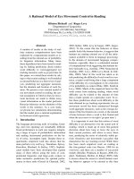
Figure 1.2. Distinct ERAD complexes in yeast. (A) Dao10 complex recognizes ERAD-C
substrates which contain a misfolded cytosolic domain. (B) The Hrd1p complex recognizes
proteins with a misfolded luminal or TM domain and defines ERAD-L and ERAD-M
pathway. Figure taken from Ismail and Ng, 2006.
1.2.4 Retrotranslocation and the Cdc48 complex
Once selected and polyubiquitinated, misfolded proteins need be to retrotranslocated
from the ER to the cytosol for proteasomal degradation. Membrane proteins must
translocate either prior to or during degradation from the lipid bilayer. To do so,
ERAD substrates require a retrotranslocon channel. The proposed translocon
complexes are Sec61 (Plemper et al., 1997) and the Derlins (Knop et al., 1997 and Ye
9
et al., 2004) and these protein complexes are bound to factors linked to substrate
selection, ubiquitination and translocation. E3 ubiquitin ligases such as Hrd1
(Hampton et al., 1996 and Plemper et al., 1999) and Doa10 (Swanson et al., 2001)
have also been proposed to be protein-conducting channels, as they are made up of
multispanning transmembrane domains, which can from oligomers to form channels.
Whether retrotranslocation occurs through the Sec61 translocon or require Der1p or
ubiquitin ligases is still a matter of debate.
In nearly all cases, ERAD substrates are ubiquitinated and released from the ER by a
ubiquitin-specific chaperone complex consisting of Cdc48p, Ufd1p and Npl4p (Ye et
al., 2001; Braun et al., 2002; Jarosch et al., 2002). This complex binds to
polyubiquitinated substrates and couples ATP hydrolysis with retrotranslocation. ATP
hydrolysis couples force on the substrate protein to extracts it from the translocation
channel. Cdc48p is an AAA-ATPase, which binds to the proteasome and has a high
affinity for ubiquitin chain (Dreveny et al., 2004). Ubx2p is an integral ER membrane
protein, which recruits Cdc48p-Ufd1p-Npl4p complex to E3 ubiquitin ligase complex
and to ERAD substrates (Neuber et al., 2005; Schuberth et al., 2005). Once the
substrates are polyubiquitinated and extracted from the membrane, they are further
targeted to the proteasome via the action of Rad23p and Dsk2p. Both proteins have a
C-terminal UBA (Ubiquitin associated) motif that recognize the polyubiquitinated
substrates and target them to proteasome with the help of subunits Rpn10p and
Rpn13p. Before proteasomal degradation, substrates are deubiquitinated by
deubiuinating enzymes to remove the polyubiquitin chain from the substrates
(Hirsch et al., 2009).
10
1.3 Cytosolic quality control
Protein misfolding in cytosol is toxic to cells and the accumulated toxic proteins can
lead to protein misfolding diseases (For examples, Parkinson’s and Alzheimer’s
diseases). Misfolding of proteins can expose hydrophobic surfaces that result in
unnecessary binding to normal proteins which disrupt the essential interactions
between cellular proteins. To avoid this situation, quality control systems present in
the cytosol monitor protein folding and remove misfolded proteins in the cytosol.
Hydrophobic patches of misfolded proteins are recognized by molecular chaperones
that mask them and transfer the misfolded species to the ubiquitin-proteasome system
and chaperone-mediated autophagy to eliminate them. The entire quality control
systems in cytosol are regulated by stress-inducible transcription factors, molecular
chaperones and other factors for the effective elimination of toxic proteins.
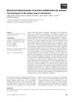
1.3.1 Recognition of damaged proteins and repair mechanism
Recognition of non-native cytosolic proteins is the first step towards their elimination,
which rely mainly on the interaction between chaperones and non-native folding
intermediates. Once recognized, cell can respond to their presence in three ways
(Figure 1.3). First, cytosolic factors may attempt to rescue the non-native folding
intermediates by folding them to a functional native state. Second, in order to prevent
toxic interactions, cells can sequester misfolded proteins. Finally, proteins that cannot
be folded into native conformation must be eliminated by ubiquitin-proteasome
pathway.
There are different chaperones systems present in the cytosol, which possess specific
mode of substrate binding that determine substrate specificity and range. The highly