A systems biology approach to elucidating the frequency decoding mechanism governing differential mammalian gonadotropin subunit gene expression
Bạn đang xem bản rút gọn của tài liệu. Xem và tải ngay bản đầy đủ của tài liệu tại đây (4.91 MB, 233 trang )
A SYSTEM BIOLOGY APPROACH TO
ELUCIDATING THE GnRH FREQUENCY
DECODING MECHANISM THAT GOVERNS
DIFFERENTIAL EXPRESSION OF THE
GONADOTROPIN-SUBUNIT GENES
STEFAN LIM
B.Sc(Hons.), Edin. U
A THESIS SUBMITTED
IN ACCORDANCE WITH THE REQUIREMENTS OF
THE NATIONAL UNIVERSITY OF SINGAPORE
FOR THE DEGREE OF DOCTOR OF PHILOSOPHY
Acknowledgments
It would be hard to envision myself completing this arduous journey of half-a-decade
without the tremendous help and encouragement of the following people:
My deceased father, who before he passed on secretly told my mother that I would se-
cure this scholarship to commence graduate studies, who believed I could embark on this
treacherous journey and make it.
My mother, who upheld and continued the convictions of my father, and encouraged me
throughout this period; if nothing else, silently praying for strength and perseverance for
me.
My wife, who has remained patient and understanding throughout this time, enduring
lengthy periods of loneliness when through the force of circumstances, I have had to de-
vote more time to research than to her.
Dr. Guna, who made it possible for me to do this PhD, by first accepting me into the
M.Sc in Bioinformatics programme, and then recommending me to A-Star for the award
of the Ph.D scholarship. If the former hadn’t happen, I would never have entered the
beautiful world of Biology
i
Dr. Philippa, whom I will always maintain as the best person who could ever have su-
pervised me, who took every risk imaginable in accepting me into her lab as an ignorant
intern at first, and then later, as her student. Moreover, for the last half-a-year of my can-
didature, when my stipend had dried up, she gave me employment in the lab, so that I
would never have to go hungry even for a day. I will never cease to respect and marvel
at her trust in my non-abilities, which she constantly sees as opportunities for personal
growth and fulfillment, and to be grateful to her for the one memorable visit to Israel, the
most beautiful country on earth. She is truly God-sent.
Prof. Zvi Naor, who has inspired me a great deal not only through his published work
in this field of gonadotropin gene regulation, but also through active discussions with him
during his visits to Singapore, as well as during my visit to Israel. He embodies all of
what great scientists ought to have - intelligence, drive, fantasy and an aura of humanity,
humility and congeniality.
Mingshi, who mentored me and taught me so patiently every aspect of experimental Bi-
ology, who taught me the beauty of life, and who is the sole reason why I have chosen to
pursue a Ph.D in this field and in this lab.
Stella, who was my dearest friend and god sister, and had been the constant inspiration in
my life, however hard and trying times might have been. She taught me the simple truths
of selfless love and friendship, and that it was not shameful nor cowardly to cry when
things surrounding me became overwhelmingly difficult to bear. In more ways than one,
and as only she would comprehend, I owe my continued existence to her.
ii
Kathy, who became my friend very late on in my PhD career, and when she was about to
leave Singapore for France to pursue her own academic dreams. She epitomizes every-
thing of a great scientist-to-be, and is probably one of the very few people in my life who
wouldn’t mind talking science with me on the subway, all the way home. She re-kindled
my interest in the French language - good or bad - it is not a worthless skill, at the very
least.
Andrea and Serena, who have been inseparable in their friendship and inseparable in
working their good deeds and charm. Thank you for the little card you gave me before
you left our lab, bearing a message that reminded me for the remainder of my time in this
lab that clearing trash and dirty bottles every so often was not a thankless task after all.
Sue Yuan, who was someone I tried to encourage all through her period of sorrow, but
ended up being encouraged by her fortitude and experiences. Thank you for being such a
dear friend, and for the mince pies you brought back from England.
Members of Philippa’s Lab, some of whom have out-stayed me, while others haven’t.
Regardless, each one of them has contributed no small part to my reaching the end, and
has made the pain of each experimental failure a little less.
Liu Ping, who helped me much with all the experiments involving FCCS and live cell
imaging.
Keng Hwee, who has at times played the role of devil’s advocate, and at other times,
the author’s advocate. Whichever role he assumed, he did it better than anyone else.
A*star, who funded this research project and also my studies.
iii
NGS, who supported me administratively throughout the course of my studies.
Celine, who came into my life rather unexpectedly, but most timely. Her extraordinary
blend of teenage innocence and youthful exuberance worked wonders for an aching heart,
tormented by the mistrust of others and the despair of a rejected thesis. She acted as an
angel commissioned by God, who appeared, and then disappeared - but who in the few
weeks that we shared life together, became my wonderfully adorable child, my sweet and
doting kid sister, my most precious friend, and everything else I could and would ever
wish for in life. Her charmingly facetious tendencies and insatiable appetite for food and
knowledge, were a joy to behold and a pleasure to oblige. She ran alongside me, encour-
aged me and infused me with just enough strength to complete this final mile. Without
her, I most certainly would have given up short of the finishing-line. It is thus only appro-
priate to reserve my final and most needful word of thanks to an earthly being for her, with
whom I was not acquainted when this thesis was first submitted, but fully and endearingly
so, by the time it was eventually re-done.
God, who is the One I will have to reserve most gratitude and honor for, without whom
nothing would have been possible. It was He, who created our amazing universe, and all
the science that undergirds the functionality of it all. The pursuit of scientific study is but
only a God-given opportunity to try and understand the beauty and wonder of creation.
iv
Abstract
The synthesis of the gonadotropin-subunits is directed by pulsatile gonadotropin-releasing
hormone (GnRH) from the hypothalamus, with the frequency of GnRH pulses governing
the differential expression of the common α-subunit (αGSU), luteinizing hormone β-
subunit (LHβ) and follicle-stimulating hormone β-subunit (FSHβ). In many vertebrate
species, levels of these hormones vary quite dramatically throughout their life cycles ow-
ing to low levels of GnRH secretion that occur during the juvenile stage, suggesting a na-
tive state of gene repression. Preliminary findings point to the actions of histone deacety-
lases (HDACs) in repressing the gonadotropins. In this study, a system biology approach
is taken to unravel the mechanisms for GnRH-frequency decoding and GnRH-induced
de-repression of the gonadotropin-subunit genes. Three mitogen-activated protein kinases
(MAPKs), ERK1/2, JNK and p38, are known to be contributing uniquely and combinato-
rially to the expression of each of these subunit genes. Using mathematical modeling and
computer simulations, it was found that dual specificity phosphatase (DUSP) regulation of
the activity of these MAPKs through negative feedback, forms the basis for decoding the
frequency of pulsatile GnRH. Furthermore, a fourth MAPK, ERK5, whose activation ki-
netics and role in FSHβ gene expression are shown, was found to enhance the preference
of FSHβ for low GnRH pulse frequencies. Evidence is presented for ERK5-activation of
FSHβ gene expression through Nur77-dependent and independent mechanisms, through
interactions with MEF2D. This involves the Ca
2+
-activated calcineurin both in activating
Nur77 transcription, as well as possibly dephosphorylating Nur77, which is required for
its activity. Having established that distinct sets of HDACs repress the two β-subunits, a
role for GnRH-activated Ca
2+
/calmodulin-dependent protein kinase I (CaMKI) is eluci-
v
dated in the de-repression of the FSHβ gene, which primarily involves phosphorylating
certain class IIa HDACs, critical for their nuclear export. Finally, Gem, a negative reg-
ulator of calcium L-type channels, is shown to be involved in regulating αGSU expres-
sion through influencing ERK1/2 activation in both a Ca
2+
-dependent and independent
way. These rely on Gem’s ability both to be re-localized to the cytosol upon CaM bind-
ing, and to effect cytoskeletal remodeling upon 14-3-3 binding. These findings reveal a
complex interplay of signal transducers, transcription factors, and both chromatin- and
cytoskeletal-remodeling proteins at different levels to orchestrate the expression of vari-
ous gonadotropin-subunit genes under the diverse actions of GnRH.
vi
Contents
Acknowledgments i
Abstract v
Contents xii
List of Tables xiii
List of Figures xviii
Nomenclature xxi
1 Introduction 1
1.1 The gonadotropic hormones . . . . . . . . . . . . . . . . . . . . . . . . 1
1.1.1 The hypothalamic control of pituitary action . . . . . . . . . . . . 1
1.1.2 The gonadotropins and their role in reproduction . . . . . . . . . 2
1.1.3 Gonadotropin-subunit gene regulation at a glance . . . . . . . . . 3
1.1.4 Understanding gonadotropin-subunit gene expression through the
use of model cell-lines . . . . . . . . . . . . . . . . . . . . . . . 4
1.2 Regulation of gonadotropin expression by pulsatile GnRH . . . . . . . . 5
1.2.1 The requirement of pulsatile GnRH for optimal gonadotropin-
subunit gene expression . . . . . . . . . . . . . . . . . . . . . . 5
1.2.2 The GnRH receptor-stimulated network as a frequency decoder . 6
1.3 Regulation of gonadotropin expression by calcium . . . . . . . . . . . . 10
vii
1.3.1 The calcium-channel regulator Kir/Gem is induced by GnRH . . 12
1.3.2 Gem . . . . . . . . . . . . . . . . . . . . . . . . . . . . . . . . . 13
1.3.3 Both CaM and 14-3-3 localize to lipid rafts in c-raf signaling in
the gonadotropes . . . . . . . . . . . . . . . . . . . . . . . . . . 17
1.4 Regulation of gonadotropin expression through targeting the chromatin . 17
1.4.1 The fluctuating levels of GnRH at different stages of the verte-
brate life cycle reveal a possible natural state of gonadotropin-
subunit gene repression . . . . . . . . . . . . . . . . . . . . . . . 17
1.4.2 Chromatin structure and the repression of the gonadotropin-subunit
genes . . . . . . . . . . . . . . . . . . . . . . . . . . . . . . . . 19
1.4.3 Histone deacetylases (HDACs) . . . . . . . . . . . . . . . . . . . 19
1.4.4 HDAC activity is involved in the repression of the gonadotropin
β-subunit genes, and is overcome by GnRH . . . . . . . . . . . . 22
1.4.5 Distinct sets of HDACs repress the gonadotropin β-subunit genes
in the immature gonadotropes . . . . . . . . . . . . . . . . . . . 23
1.4.6 GnRH activates CaMKI in immature gonadotropes . . . . . . . . 25
1.4.7 Nur77 and MEF2D de-repress the FSHβ gene . . . . . . . . . . . 26
1.5 Frequency decoding re-visited: the search for a frequency decoding mech-
anism . . . . . . . . . . . . . . . . . . . . . . . . . . . . . . . . . . . . 28
1.6 Hypothesis and aims . . . . . . . . . . . . . . . . . . . . . . . . . . . . 32
1.6.1 Hypothesis . . . . . . . . . . . . . . . . . . . . . . . . . . . . . 32
1.6.2 Aims . . . . . . . . . . . . . . . . . . . . . . . . . . . . . . . . 33
2 Experimental Materials and Methods 34
2.1 Cell culture, transfection and treatment . . . . . . . . . . . . . . . . . . . 34
2.1.1 Cell culture . . . . . . . . . . . . . . . . . . . . . . . . . . . . . 34
2.1.2 Cryo-storage of cells . . . . . . . . . . . . . . . . . . . . . . . . 34
2.1.3 Recovery of cells . . . . . . . . . . . . . . . . . . . . . . . . . . 35
viii
2.1.4 Transfection of cells . . . . . . . . . . . . . . . . . . . . . . . . 35
2.1.5 Chemical treatment of cells . . . . . . . . . . . . . . . . . . . . 35
2.2 Plasmid construction . . . . . . . . . . . . . . . . . . . . . . . . . . . . 36
2.2.1 SiRNA constructs . . . . . . . . . . . . . . . . . . . . . . . . . . 36
2.2.2 Expression vectors . . . . . . . . . . . . . . . . . . . . . . . . . 38
2.2.3 Isolation, verification and plasmid preparation . . . . . . . . . . . 39
2.3 RNA extraction and reverse transcriptase PCR . . . . . . . . . . . . . . . 42
2.3.1 RNA isolation . . . . . . . . . . . . . . . . . . . . . . . . . . . 42
2.3.2 First strand cDNA synthesis . . . . . . . . . . . . . . . . . . . . 42
2.3.3 PCR and gel electrophoresis analysis . . . . . . . . . . . . . . . 42
2.4 Luciferase assay . . . . . . . . . . . . . . . . . . . . . . . . . . . . . . . 44
2.5 Statistical analysis . . . . . . . . . . . . . . . . . . . . . . . . . . . . . . 44
2.6 Whole cell extraction . . . . . . . . . . . . . . . . . . . . . . . . . . . . 45
2.7 Co-immunoprecipitation . . . . . . . . . . . . . . . . . . . . . . . . . . 45
2.8 Western blot . . . . . . . . . . . . . . . . . . . . . . . . . . . . . . . . . 46
2.9 Immuno-fluorescence/Confocal microscopy . . . . . . . . . . . . . . . . 47
2.10 Live cell imaging . . . . . . . . . . . . . . . . . . . . . . . . . . . . . . 48
2.11 Fluorescence cross-correlation spectroscopy (FCCS) . . . . . . . . . . . 48
3 Computational Modeling 50
3.1 Introduction . . . . . . . . . . . . . . . . . . . . . . . . . . . . . . . . . 50
3.1.1 Published models on frequency decoding of GnRH signals . . . . 50
3.1.2 Proposed scheme of model development . . . . . . . . . . . . . . 52
3.2 The basic model . . . . . . . . . . . . . . . . . . . . . . . . . . . . . . . 54
3.2.1 Model development . . . . . . . . . . . . . . . . . . . . . . . . . 55
3.3 The intermediate and full models . . . . . . . . . . . . . . . . . . . . . . 58
3.3.1 Model development . . . . . . . . . . . . . . . . . . . . . . . . . 59
3.4 Computer simulations and key readouts . . . . . . . . . . . . . . . . . . 65
ix
3.5 Model codes . . . . . . . . . . . . . . . . . . . . . . . . . . . . . . . . . 66
3.5.1 Code for the basic model . . . . . . . . . . . . . . . . . . . . . . 66
3.5.2 Code to analyze the basic model . . . . . . . . . . . . . . . . . . 69
3.5.3 Code for the intermediate model . . . . . . . . . . . . . . . . . . 70
3.5.4 Code to analyze the intermediate model . . . . . . . . . . . . . . 73
3.5.5 Code for the full model . . . . . . . . . . . . . . . . . . . . . . . 73
3.5.6 Code to analyze the full model . . . . . . . . . . . . . . . . . . . 78
4 Results and Sectional Discussions 80
4.1 Elucidating the GnRH frequency decoding mechanism in the gonadotropes 80
4.1.1 MKP negative feedback gives rise to frequency-dependent differ-
ential gonadotropin-subunit gene expression . . . . . . . . . . . . 80
4.1.2 Sensitivity analysis of the basic model . . . . . . . . . . . . . . . 84
4.1.3 Differential gene expression results from phosphatase-induced in-
creases in average MAPK activation with decreasing frequency of
the stimulus . . . . . . . . . . . . . . . . . . . . . . . . . . . . . 86
4.1.4 GnRH activates ERK5 in αT3-1 cells . . . . . . . . . . . . . . . 90
4.1.5 ERK5 activates the murine FSHβ promoter . . . . . . . . . . . . 91
4.1.6 ERK5 up-regulates FSHβ but down-regulates GnRHR mRNA lev-
els in αT3-1 cells . . . . . . . . . . . . . . . . . . . . . . . . . . 93
4.1.7 ERK5 enhances FSHβ expression in a concentration-dependent
manner . . . . . . . . . . . . . . . . . . . . . . . . . . . . . . . 94
4.1.8 Sensitivity analysis of the intermediate model . . . . . . . . . . . 97
4.1.9 Differential GnRHR concentration alone appears not to give rise
to full differential gonadotropin-subunit gene expression . . . . . 99
4.1.10 JNK-positive feedforward without ERK5-negative feedback on
GnRHR expression causes loss of differential gonadotropin-subunit
gene expression in the full model . . . . . . . . . . . . . . . . . 103
x
4.1.11 ERK5-negative feedback against GnRHR expression restores dif-
ferential gonadotropin-subunit gene expression in the full model . 107
4.1.12 Sensitivity analysis of the full model . . . . . . . . . . . . . . . . 111
4.1.13 Discussion . . . . . . . . . . . . . . . . . . . . . . . . . . . . . 115
4.2 GnRH-mediated de-repression of the gonadotropin β-subunit genes . . . 126
4.2.1 GnRH causes the nuclear export of wild-type HDACs 4, 5, and 7 . 126
4.2.2 Mutation of 14-3-3 recognition sites abolishes nuclear export of
HDACs 4 and 5 . . . . . . . . . . . . . . . . . . . . . . . . . . . 131
4.2.3 GnRH activates CaMKII rapidly in the immature gonadotropes . 133
4.2.4 GnRH-mediated de-repression of the FSHβ but not the LHβ gene
involves activation of CaMKI . . . . . . . . . . . . . . . . . . . 133
4.2.5 GnRH-mediated de-repression of the FSHβ but not the LHβ gene
involves CaMK phosphorylaton of HDACs 4 and 5 . . . . . . . . 134
4.2.6 ERK5 up-regulates Nur77 mRNA levels in αT3-1 cells . . . . . . 134
4.2.7 Phosphorylated ERK5 co-precipitates with MEF2D after GnRH
treatment of immature gonadotropes . . . . . . . . . . . . . . . . 137
4.2.8 ERK5 activates the murine FSHβ promoter through interactions
with MEF2D . . . . . . . . . . . . . . . . . . . . . . . . . . . . 138
4.2.9 Discussion . . . . . . . . . . . . . . . . . . . . . . . . . . . . . 140
4.3 The role of Gem in α-subunit expression . . . . . . . . . . . . . . . . . . 146
4.3.1 External calcium is both necessary and sufficient for basal and
GnRH-stimulated α-subunit gene activity in αT3-1 cells . . . . . 146
4.3.2 Gem over-expression does not disrupt GnRH-induction of α-subunit
mRNA levels in αT3-1 cells . . . . . . . . . . . . . . . . . . . . 148
4.3.3 CaM- but not 14-3-3-binding sites of Gem are necessary and suf-
ficient for both basal and GnRH-induced α-subunit gene expression149
xi
4.3.4 14-3-3- but not CaM-binding ability is crucial for both nuclear
import and export of Gem, and for mediating GnRH-induced mor-
phological changes in αT3-1 cells . . . . . . . . . . . . . . . . . 152
4.3.5 Gem knockdown reduces basal murine α-subunit promoter activity 158
4.3.6 GnRH causes co-diffusion of Gem with ERK close to the plasma
membrane . . . . . . . . . . . . . . . . . . . . . . . . . . . . . . 159
4.3.7 Discussion . . . . . . . . . . . . . . . . . . . . . . . . . . . . . 163
5 Overall Discussion 170
Bibliography 208
xii
List of Tables
2.1 Nucleotide sequences used for making siRNA . . . . . . . . . . . . . . . 36
2.2 Ligation reaction mix . . . . . . . . . . . . . . . . . . . . . . . . . . . . 37
2.3 Primers used for sequencing . . . . . . . . . . . . . . . . . . . . . . . . 41
2.4 Sequencing reaction mix . . . . . . . . . . . . . . . . . . . . . . . . . . 41
2.5 Pre-annealing reaction mix for cDNA synthesis . . . . . . . . . . . . . . 42
2.6 Primers used for PCR . . . . . . . . . . . . . . . . . . . . . . . . . . . . 43
2.7 PCR mix for gel analysis of gene expression levels by RT-PCR . . . . . . 43
2.8 Antibody dilutions used for western blotting . . . . . . . . . . . . . . . . 47
2.9 Buffers used in western blotting . . . . . . . . . . . . . . . . . . . . . . 47
3.1 Glossary of variables for the basic model . . . . . . . . . . . . . . . . . . 54
3.2 Constants . . . . . . . . . . . . . . . . . . . . . . . . . . . . . . . . . . 55
3.3 Glossary of new variables for the intermediate and full models . . . . . . 59
3.4 Additional constants for the intermediate and full model . . . . . . . . . 60
xiii
List of Figures
1.1 Layout of the human pituitary . . . . . . . . . . . . . . . . . . . . . . . 1
1.2 The GnRH receptor-stimulated network . . . . . . . . . . . . . . . . . . 9
1.3 Comparison of Gem sequences among various species . . . . . . . . . . 14
1.4 Ontogeny of the hypothalamic-pituitary-gonadal axis . . . . . . . . . . . 18
1.5 Interaction partners of class IIa HDACs determine their localization . . . 21
1.6 LHβ and/or FSHβ gene expression is repressed in gonadotrope cell-lines
by HDACs, and this is overcome by GnRH . . . . . . . . . . . . . . . . 22
1.7 Distinct sets of HDACs are associated with LHβ and FSHβ genes in the
immature gonadotropes . . . . . . . . . . . . . . . . . . . . . . . . . . . 24
1.8 Knockdown of the associated HDACs reveals their crucial roles in the
repression . . . . . . . . . . . . . . . . . . . . . . . . . . . . . . . . . . 25
1.9 GnRH activates CaMKI but not CaMKIV . . . . . . . . . . . . . . . . . 26
1.10 Nur77 induces expression of the FSHβ gene in the immature gonadotropes
and plays a role in the GnRH de-repressive effect . . . . . . . . . . . . . 27
4.1 Profiles of MAPKK used in simulation of models . . . . . . . . . . . . . 81
4.2 Lack of phosphatase feedback results in no differential-gene expression
with the exponential pulse . . . . . . . . . . . . . . . . . . . . . . . . . 82
4.3 Lack of phosphatase feedback results in no differential-gene expression
with the square pulse . . . . . . . . . . . . . . . . . . . . . . . . . . . . 83
4.4 Inclusion of phosphatase feedback results in differential-gene expression . 84
4.5 Sensitivity analysis of the basic model for the exponential pulse profile . . 85
xiv
4.6 Sensitivity analysis of the basic model for the square pulse profile . . . . 86
4.7 Analysis of MAPK activation for the exponential pulse profile in the basic
model without phosphatase feedback . . . . . . . . . . . . . . . . . . . . 87
4.8 Analysis of MAPK activation for the square pulse profile in the basic
model without phosphatase feedback . . . . . . . . . . . . . . . . . . . . 88
4.9 Analysis of MAPK activation for the exponential pulse profile in the basic
model with phosphatase feedback . . . . . . . . . . . . . . . . . . . . . 89
4.10 Analysis of MAPK activation for the square pulse profile in the basic
model with phosphatase feedback . . . . . . . . . . . . . . . . . . . . . 90
4.11 ERK5 is activated by GnRH in αT3-1 cells . . . . . . . . . . . . . . . . 91
4.12 Effects of over-expression of ERK5 and constitutively-active MEK5 on
the murine FSHβ promoter activity . . . . . . . . . . . . . . . . . . . . . 92
4.13 RT-PCR analysis of the effects of over-expression of ERK5 and MEK5(D)
on GnRHR and FSHβ mRNA levels in αT3-1 cells . . . . . . . . . . . . 93
4.14 Intermediate model with phosphatase feedback demonstrates differential
gene expression . . . . . . . . . . . . . . . . . . . . . . . . . . . . . . . 94
4.15 Analysis of ERK5 activation in the intermediate model . . . . . . . . . . 96
4.16 Highest fold-induction of FSHβ expression is dependent on total concen-
tration of ERK5 . . . . . . . . . . . . . . . . . . . . . . . . . . . . . . . 97
4.17 Sensitivity analysis of the intermediate model for the exponential pulse
profile . . . . . . . . . . . . . . . . . . . . . . . . . . . . . . . . . . . . 98
4.18 Sensitivity analysis of the intermediate model for the square pulse profile 99
4.19 Differential GnRHR concentration alone appears not give rise to full dif-
ferential gonadotropin-subunit gene expression . . . . . . . . . . . . . . 101
4.20 Analysis of MAPK activation in the full model . . . . . . . . . . . . . . 102
4.21 JNK-positive feedforward without ERK5-negative feedback on GnRHR
expression results in the loss of differential gonadotropin-subunit gene
expression . . . . . . . . . . . . . . . . . . . . . . . . . . . . . . . . . . 104
xv
4.22 Analysis of rms values of MAPKs in the full model with JNK feedforward
only on GnRHR expression . . . . . . . . . . . . . . . . . . . . . . . . . 105
4.23 Analysis of total activation of MAPKs in the full model with JNK feed-
forward only on GnRHR expression . . . . . . . . . . . . . . . . . . . . 106
4.24 ERK5-negative feedback on GnRHR expression restores differential gonadotropin-
subunit gene expression . . . . . . . . . . . . . . . . . . . . . . . . . . . 108
4.25 Analysis of rms values of MAPKs in the full model with ERK5-negative
feedback only on GnRHR expression . . . . . . . . . . . . . . . . . . . . 109
4.26 Analysis of total activation of MAPKs in the full model with ERK5-
negative feedback only on GnRHR expression . . . . . . . . . . . . . . . 110
4.27 JNK-positive feedforward and ERK5-negative feedback together on Gn-
RHR expression endows differential gonadotropin-subunit gene expression 111
4.28 Sensitivity analysis of the full model to k
1
. . . . . . . . . . . . . . . . . 112
4.29 Sensitivity analysis of the full model to k
11
. . . . . . . . . . . . . . . . . 113
4.30 Sensitivity analysis of the full model for the exponential pulse profile . . . 114
4.31 Physiological pulse profile of GnRH compared with those used in model
simulation . . . . . . . . . . . . . . . . . . . . . . . . . . . . . . . . . . 121
4.32 GnRH stimulates nuclear export of HDAC 4 . . . . . . . . . . . . . . . . 127
4.33 GnRH stimulates nuclear export of HDAC5 . . . . . . . . . . . . . . . . 128
4.34 GnRH stimulates nuclear export of HDAC7 . . . . . . . . . . . . . . . . 129
4.35 GnRH does not change the localization of HDAC6 . . . . . . . . . . . . 130
4.36 Mutation of 14-3-3 recognition sites abolishes nuclear export of HDAC4
by GnRH . . . . . . . . . . . . . . . . . . . . . . . . . . . . . . . . . . 131
4.37 Mutation of 14-3-3 recognition sites abolishes nuclear export of HDAC5
by GnRH . . . . . . . . . . . . . . . . . . . . . . . . . . . . . . . . . . 132
4.38 GnRH activates CaMKII rapidly . . . . . . . . . . . . . . . . . . . . . . 133
4.39 GnRH de-repression of the FSHβ gene involves CaMKI . . . . . . . . . . 135
xvi
4.40 GnRH-mediated de-repression of the FSHβ but not the LHβ gene requires
CaMK phosphorylation sites on HDACs 4 and 5 . . . . . . . . . . . . . . 136
4.41 RT-PCR analysis of the effects of over-expression of ERK5 and MEK5(D)
on Nur77 mRNA levels in αT3-1 cells . . . . . . . . . . . . . . . . . . . 137
4.42 pERK5 co-precipitates with MEF2D in αT3-1 cells . . . . . . . . . . . . 138
4.43 Effects of siMEF2D together with over-expression of ERK5 and MEK5(D)
on the murine FSHβ promoter activity . . . . . . . . . . . . . . . . . . . 140
4.44 A model depicting the proposed mechanisms through which GnRH de-
represses the FSHβ gene in immature gonadotropes . . . . . . . . . . . . 145
4.45 RT-PCR analysis of the effects of BayK 8644 (BK) and nifedipine on
α-subunit expression levels in αT3-1 cells . . . . . . . . . . . . . . . . . 147
4.46 Effects of BK and nifedipine on the murine α-subunit promoter activity . 148
4.47 RT-PCR analysis of the effects of wild type Gem over-expression on α-
subunit expression levels in αT3-1 cells . . . . . . . . . . . . . . . . . . 150
4.48 RT-PCR analysis of the effects of over-expression of mutant forms of
Gem on α-subunit expression levels in αT3-1 cells . . . . . . . . . . . . 151
4.49 Effect of GnRH on wild type Gem localization in αT3-1 cells . . . . . . . 154
4.50 Effect of GnRH on CaM-binding mutant Gem localization in αT3-1 cells 155
4.51 Effect of GnRH on 14-3-3-binding mutant Gem localization in αT3-1 cells 156
4.52 Effect of GnRH on CaM/14-3-3-binding mutant Gem localization in αT3-
1 cells . . . . . . . . . . . . . . . . . . . . . . . . . . . . . . . . . . . . 157
4.53 Effect of Gem knockdown on the murine α-subunit promoter activity . . . 158
4.54 Wild type Gem appears to co-diffuse with ERK after GnRH treatment . . 160
4.55 CaM-binding mutant of Gem fails to co-diffuse with ERK after GnRH
treatment . . . . . . . . . . . . . . . . . . . . . . . . . . . . . . . . . . 161
4.56 Positive and negative controls for FCCS . . . . . . . . . . . . . . . . . . 162
4.57 A model depicting the proposed mechanisms through which Gem medi-
ates GnRH actions on the α-subunit gene in immature gonadotropes . . . 169
xvii
Nomenclature
αGSU Glycoprotein α-subunit
AP-1 Activator protein 1
BAPTA/AM 1,2-bis-(o-aminophenoxy)ethane-N,N,N’,N’-tetra-acetic acid acetoxymethyl
tetraester
BMK Big mitogen-activated protein kinase
CaM Calmodulin
CaMK Ca
2+
/calmodulin-dependent protein kinase
cAMP 3’-5’-cyclic adenosine monophosphate
CoA Coactivator
CsA Cyclosporine A
DAG Diacylglycerol
dnNur77 Dominant negative Nur77
DUSP1 (or 4) Dual-specificity phosphatase 1 (or 4)
EGFP Enhanced green fluorescent protein
Egr-1 Early growth factor 1
ER Endoplasmic reticulum
xviii
ERK Extracellular-signal regulated kinase
FCCS Fluorescence cross-correlation spectroscopy
FSH Follicle-stimulating hormone
FSHβ Follicle-stimulating hormone β-subunit
GAP GTPase-activating protein
GDP Guanosine diphosphate
GEF GDP-GTP exchange factor
GnRH Gonadotropin-releasing hormone
GnRHR Gonadotropin-releasing hormone receptor
GPCR G-protein-coupled receptor
Gq/11 Alpha-subunit of heterotrimeric Gq protein
Gs Adenylyl cyclase-stimulating G alpha protein
GTP Guanosine triphosphate
HAT Histone acetyltransferase
HDAC Histone deacetylase
IP
3
Inositol triphosphate
JNK c-Jun NH
2
-terminal kinase
KN-62 1-[N,O-Bis(5-isoquinolinesulfonyl)-N-methyl-L-tyrosyl]-4-phenylpiperazine
KN-93 N-[2-[N-(4-Chlorocinnamyl)-N-methylaminomethyl]phenyl]-N-(2-hydroxyethyl)-
4-methoxybenzenesulfonamide phosphate salt
xix
LH Luteinizing hormone
LHβ Luteinizing hormone β-subunit
MAPK Mitogen-activated protein kinase
MAPKK MAPK kinase
MEF2 Myocyte enhancer factor-2
MEK MAPK Erk kinase
MKP1 (or 2) MAPK phosphatase 1 (or 2)
mRFP Monomeric red fluorescent protein
NFAT Nuclear factor of activated T-cells
N-CoR Nuclear receptor co-repressor
Nur77(Nr4a1) Nuclear receptor subfamily 4, group A, member 1
PAGE Poly-acrylamide gel electrophoresis
PCR Polymerase chain reaction
PIP Phosphatidylinositol 4-monophosphate
PIP2 Phosphatidylinositol 4,5-biphosphate
Pitx-1 Pituitary homeobox 1
PKA Protein kinase A
PKCs Protein kinase C isoforms
PLC Phospholipase C
RMS Root mean-square
xx
ROK Rho kinase
RT-PCR Reverse transcriptase polymerase chain reaction
SDS Sodium dodecyl sulphate
siRNA Short interfering ribonucleic acid
SMRT Silencing mediator of retinoic and thyroid hormone receptors
TSA Trichostatin A
TSH Thyroid-stimulating hormone
VGCCs Voltage-gated calcium channels
W-7 N-(6-aminohexyl)-5-chloro-1-naphthalenesulfonamide
YFP Yellow fluorescent protein
xxi
Looking back on the memory of
The dance we shared ’neath the stars above
For the moment all the world was right
How could I have known that you’d ever say goodbye
And now I’m glad I didn’t know
The way it all would end
The way it all would go
Our lives are better left to chance
I could have missed the pain
But I’d have had to miss the dance
The dance
The dance
I would have missed the dance
Holding you I held everything
For a moment wasn’t I a king
But if I’d only known how the king would fall
Hey who’s to say you know I might have changed it all
And now I’m glad I didn’t know
The way it all would end
The way it all would go
Our lives are better left to chance
I could have missed the pain
But I’d have had to miss the dance
The dance
The dance
I would have missed the dance
The Dance
Tom Arata
xxii
So no one wanted to supervise you immunology?
and that’s why you worked on gonadotropins?!!
Celine, aged A*Teen
xxiii
Chapter 1
Introduction
1.1 The gonadotropic hormones
1.1.1 The hypothalamic control of pituitary action
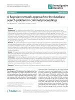
The pituitary gland and the hypothalamus are both located within the cranial region (Fig-
ure 1.1). The pituitary gland is sometimes known as the ‘master gland’ of the endocrine
Figure 1.1: Layout of the human pituitary. The hypothalamus is connected to the pituitary
through the infundibulum. Electrical and chemical signals are conducted through the infundibu-
lum to regulate the production and release of the pituitary hormones. (Picture from [1]. Copyright
c
McGraw-Hill Companies, Human Anatomy, 2005).
system, because it controls the functions of the other endocrine glands. Located at the
base of the brain, it consists of two structurally and functionally distinct parts: the anterior
1