Role of the survival proteins hsp27 and survivin in a small molecule sensitization to TRAIL mediated apoptosis 2
Bạn đang xem bản rút gọn của tài liệu. Xem và tải ngay bản đầy đủ của tài liệu tại đây (9.44 MB, 125 trang )
84
RESULTS
LY30 restores HeLa cells sensitivity to TRAIL-induced cell death.
Dose response curves for TRAIL and LY30 were established in HeLa cells by
evaluating the effect of increasing doses of TRAIL (0 to 250 ng/mL) and LY30 (0 to
25 µM), alone or in combination, on cell viability after 16h by using the crystal violet
assay (Figure 13A). HeLa cells did not exhibit a decrease in cell viability upon
TRAIL treatment even at the highest dose used (250 ng/mL). However, pre-
incubation with different doses of LY30 for 1h significantly reduced cell viability
(50% viability with 25 !M LY30 and 20 ng/mL TRAIL in combination) as compared
to both treatment alone (85% viability for 25 µM LY30 and 100% viability for 20
ng/mL), hence corroborating our earlier finding that LY30 is able to sensitize HeLa
cells to TRAIL-induced cell death [156]. Interestingly, the observed sensitization was
enhanced in a dose dependent manner as a function of TRAIL dose but not of LY30
dose. Further cell viability assays for TRAIL treatment alone were carried out over a
longer time frame (up to 72h) (Figure 13B). HeLa cells viability was not decreased
upon treatment with TRAIL for a longer period. Only HeLa cells treated with 250
ng/mL of TRAIL for 72h showed a modest effect of TRAIL on cell viability.
For subsequent experiments, the chosen doses of LY30 and TRAIL were 25
!M and 20 ng/mL respectively. At those doses, HeLa cells viability was decreased in
a time dependent manner (Figure 13C).
85
LY30 and TRAIL combined treatment decreases HeLa cells ability to form
colonies
Given the ability of LY30 and TRAIL combination to induce cell death upon
short-term treatment, we were interested in understanding the effect of LY30+TRAIL
on the long-term survival/proliferative capacity of tumor cells. This was done by
evaluating the effect of the combined drug treatment on HeLa cells colony forming
ability.
HeLa cells were exposed to LY30 for 1hr before incubation with TRAIL for
6h. An equal number of cells were then seeded onto 100mm Petri dishes and allowed
to form colonies over a period of 10 to 14 days.
After staining the cells with crystal violet, we observed a marked reduction in
the number of colonies formed for cells treated with both compounds (Figure 14A). A
significant decrease in clonogenic ability (35% of untreated cells) was observed for
cells under combinatorial treatment as compared to cells treated singly with either
TRAIL or LY30 (Figure 14B). Interestingly, TRAIL alone, and to a lesser extent
LY30 alone, induced a significant decrease of the size of the colonies (40% when
compared to control) (Figure 14C). The combined treatment further reduced the size
of the colonies. This observation explains the discrepancy between the actual number
of colonies counted after TRAIL treatment and the apparent low number of colonies
in the picture itself.
86
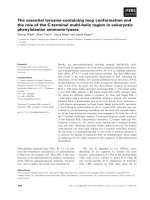
Figure 13: Effect of LY30 treatment on TRAIL-mediated apoptosis.
Hela cells exposed to TRAIL (0-250 ng/mL) for 16h with or without 1h pre-
incubation with various doses of LY30 (0-25 !M) (A), treated with TRAIL alone (0-
250 ng/mL) for 24h, 48h or 72h (B) or treated with 20ng/mL of TRAIL for different
times (4h-24h) with or without 1h pre-incubation with 25 !M of LY30 (C). The
percentage of cell survival was determined by crystal violet assay. Data shown are the
mean±S.D. of three independent experiments. NT: Non-treated; LY: treatment with
LY30; T: treatment with TRAIL; LYT: treatment with LY30 and TRAIL.
87
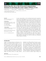
Figure 14: Effect of LY30 and TRAIL treatment on colony forming ability of
HeLa cells.
HeLa cells were exposed to 20 ng/ml TRAIL for 6h with or without pre-incubation
with 25 !M LY30. The cells were then re-seeded onto 100 mm Petri dishes and
allowed to form colonies over 10 to 14 days, followed by staining with crystal violet
(A) for colony counts (B) and colony size (C) determination. Data shown are the
mean±S.D. of three independent experiments. *p<0.01
88
LY30, alone or in combination with TRAIL, induces an early mitochondrial
membrane potential depolarization (!"
m
) and mitochondrial aggregation
It has been shown that mitochondrial outer membrane permeability (MOMP)
involves a drop in the mitochondrial transmembrane potential (!"
m
). We set out to
investigate the effect of the combined treatment of TRAIL and LY30 on !"
m
.
HeLa cells were treated with TRAIL for 1h to 4h with or without pre-
incubation with LY30 for 1h followed by !"
m
analysis with TMRE by laser scanning
cytometry. The uncoupler CiCCP was used as a positive control for depolarization
(Figure 15A).
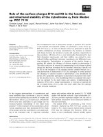
Results show that LY30, alone or in combination with TRAIL, was able to
induce a drop of the mitochondrial transmembrane potential in a time-dependent
manner (Figure 15B). In addition, an alternate analysis of the data for the 4h time
point indicated that LY30+TRAIL treatment, and to a lesser extent LY30 alone, was
inducing an early mitochondrial aggregation (Figure 16A and B) – a phenomenon
shown to occur prior MOMP [418].
89
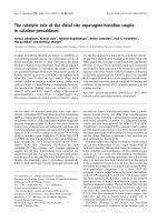
Figure 15: Effect of LY30 and TRAIL treatment on mitochondrial membrane
polarization.
HeLa cells were treated for 1h to 4h with 20 ng/mL of TRAIL with or without pre-
incubation with 25 µM LY30. Live cells were then incubated with TMRE and
Hoescht before analysis by Laser Scanning Cytometry. The uncoupler CiCCP (5 !M)
was used as a positive control for depolarization of the mitochondrial membrane (A).
Histogram representing mitochondrial membrane polarization (B). n=2.
Polarized!
Depolarized!
0%!
10%!
20%!
30%!
40%!
50%!
60%!
70%!
80%!
90%!
100%!
NT!
LY!
T!
LY+T!
CCCP!
NT!
LY!
T!
LY+T!
CCCP!
NT!
LY!
T!
LY+T!
CCCP!
NT!
LY!
T!
LY+T!
CCCP!
Depolarized!
Polarized!
Rest!
NT!
CiCCP!
A!
B!
1h! 2h! 3h! 4h!
90
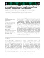
Figure 16: Effect of upon LY30 and TRAIL treatment on mitochondrial
aggregation.
(HeLa cells were treated for 4h with 20 ng/mL of TRAIL with or without pre-
incubation with 25 !M LY30. Live cells were then incubated with TMRE and
Hoescht before analysis by Laser Scanning Cytometry. Scattergram representing the
area covered by mitochondria against the ratio of the highest mitochondrial staining
intensity to the total mitochondrial staining (A). Corresponding histogram of the
mitochondrial aggregation (B). n=2.
0!
4!
8!
12!
16!
20!
NT!
LY!
T!
LY+T!
% of total number of cell!
NT! LY!
T! LYT!
A!
B!
91
LY30 and TRAIL treatment engages the mitochondrial apoptotic pathway
The release of apoptogenic factors like cytochrome c or Smac/DIABLO (cyt c
and Smac) from the mitochondria intermembrane spaces marks an important step in
the intrinsic apoptotic pathway as it engages the apoptotic cascade. Earlier results
showed that LY30 combined to TRAIL is able to induce MOMP, as evidenced by a
drop in !"
m
as well as mitochondrial aggregation, hence we set out to investigated
subsequent mitochondrial events such as the release of apoptogenic factors.
HeLa cells were treated with TRAIL for 6h, 12h or 18h with or without pre-
incubation with LY30 for 1h followed by subcellular fractionation and analysis of cyt
c and Smac release into the cytosol by Western blotting. The purity of the cytosolic
and mitochondrial fraction was confirmed by monitoring the absence of VDAC and
CuZnSOD in the cytosolic or mitochondrial fraction, respectively.
As seen in Figure 17, both cytochrome c and Smac were detected in the
cytosolic fraction after 12h upon LY30 and TRAIL treatment. At 18h, both proteins
were found in greater amount in the cytosol, a phenomenon coupled with a decrease
in the level of both proteins in the mitochondrial fraction.
These data indicate that LY30-mediated sensitization to TRAIL-induced cell
death engages the mitochondrial apoptotic pathway.
92
Figure 17: Effect of LY30 and TRAIL treatment on cytochrome c and Smac
release from the mitochondria.
Hela cells were exposed to 20 ng/ml TRAIL for 6h, 12h or 18h with or without pre-
incubation with 25 µM LY30 for 1h. Levels of cytochrome c and Smac in the
mitochondrial and cytosolic fractions were then assayed by western blotting.
93
LY30 and TRAIL combined treatment induces, and is dependent on, caspase
activation
Death receptor mediated apoptosis requires the activation of the caspase
cascade, which is responsible for the cleavage of cellular substrates to bring about the
controlled demise of cells. In order to find out whether caspase were involved in
LY30 sensitization to TRAIL-mediated cell death in our cell system, we set out to
investigate the activation of caspases in HeLa cells.
Cells were treated with TRAIL for different time (6h, 12h and 18h). The
activity of caspase-3, caspase-8 and caspase-9 was then assessed by caspase activity
assay with the respective fluorescence-conjugated caspase substrates (DEVD-AFC,
IETD-AFC and LEHD-AFC). Pre-treatment of HeLa cells with LY30 followed by
TRAIL treatment resulted in a strong amplification of caspase-3, -8 and -9 activities
as compared to untreated cells, with a peak of activity observed at 12h for all three
caspases (Figure 18A, B and C). TRAIL treatment alone was unable to induce the
activation of any of the caspases investigated and exposure of HeLa cells to LY30
resulted in minimal caspase activation (less than 2 fold increase) after 6h and 12h of
treatment. However, LY30 treatment resulted in an increased caspase-8 and -9
activity at 18h. In addition, we observed the appearance of a 17 kDa cleaved fragment
of caspase-3 at 8h following treatment with LY30 and TRAIL (Figure 18D),
indicating an activation of pro-caspase-3 via proteolytic cleavage. The caspase-3-
mediated cleavage product of one of the caspase-3 substrate, the DNA repair enzyme
PARP, was also detectable at 8h. At 20h, the 17 kDa cleaved fragment of caspase-3
can observed following LY30 treatment, alone or in combination with TRAIL.
However, unlike LY30 treatment alone, the combined treatment resulted in a marked
decrease in the level of pro-caspase-3 indicating a strong activation of caspase-3 prior
94
and up to 18h. This observation was confirmed by the complete cleavage of PARP
after 18h of the combined treatment, as opposed to the incomplete cleavage observed
for the treatment with LY30 alone.
In order to validate the involvement of caspase-mediated cell death in our
system, we investigated the effect of the pan-caspase inhibitor zVAD-fmk, as well as
the effect of specific inhibitors, on the observed caspase activation. The activation of
all three caspases resulting from the combined treatment of TRAIL and LY30 was
blocked by zVAD-fmk at 12h (Figure 18A, B and C). Moreover, specific inhibitors
for caspase-3 and -8 activity (z-DEVD-fmk and z-IETD-fmk, respectively) showed
similar results. However, the caspase-9 inhibitor (z-LEHD-fmk) was unable to block
caspase-9 acitvity as efficiently as the other inhibitors, and therefore was not used in
subsequent experiments. In addition, z-VAD-fmk, z-DEVD-fmk and z-IETD-fmk
were able to rescue HeLa cells exposed to LY30+TRAIL treatment (Figure 19).
Taken together, these results show that the combination of LY30 and TRAIL
induces a caspase-dependent apoptotic cell death in HeLa cells.
95
Figure 18: Effect of LY30 and TRAIL treatment on caspase activation.
HeLa cells were exposed to 20 ng/ml TRAIL for 6, 12 or 18h with or without pre-
treatment with 25 !M LY30 for 1h, in the presence or absence of z-VAD-fmk, z-
DEVD-fmk, z-IETD-fmk or z-LEHD-fmk (50 !M each). Whole cell lysates were
used to determine the activities of caspase-3, -8 and-9 using fluorescent-conjugated
substrates. Data shown are the mean±S.D. of three independent experiments
(A)(B)(C). HeLa cells were exposed to 20 ng/ml TRAIL for 4h, 8h or 20h with or
without pre-treatment with 25 !M LY30 for 1h Whole cell lysates were then used to
assay for caspase-3 processing by Western blotting (D). *p<0.01
96
Figure 19: Effect of caspases inhibitors on cell viability upon LY30 and TRAIL
treatment.
HeLa cells were exposed to 20 ng/ml TRAIL for 16h with or without pre-incubation
with 25 !M LY30 for 1h, in the presence or absence of z-VAD-fmk, z-DEVD-fmk, z-
IETD-fmk or z-LEHD-fmk (50 !M each). The percentage of cell survival was
determined by crystal violet assay. Data shown are the mean±S.D. of three
independent experiments. *p<0.01
0!
20!
40!
60!
80!
100!
120!
-!
zVAD!
zDEVD!
zIETD!
Cell viabilty (% of control)!
NT!
LY!
T!
LYT!
*!
*!
*!
97
LY30 and LY30+TRAIL treatment decrease Hsp27 protein level in cleared
RIPA cell lysates
Several studies have previously shown that both quercetin and LY29 (which is
derived from quercetin) were be able to regulate Hsp27 expression at the transcription
level by interfering with Hsp27 primary transcription factor Hsf-1 [294, 413-415].
Since LY30 is derived from LY29, we decided to investigate whether LY30, alone or
in combination with TRAIL, also had an effect on Hsp27 expression.
HeLa cells were treated with TRAIL for 20h with or without pre-incubation
with LY30 for 1h followed by cell lysis in RIPA buffer. Cell lysates were then cleared
(i.e. centrifuged) and the supernatants were analyzed by western blotting.
Results show that LY30 treatment, alone or in combination with TRAIL,
induced a decrease in Hsp27 protein levels in cleared RIPA lysates after 20h as
evidenced by Western blot analysis (Figure 20). However, no change could be
observed in Hsp70 or Hsp90 protein levels, suggesting that LY30 had an effect
specifically on Hsp27.
98
Figure 20: Effect of LY30 and TRAIL on Hsp27 and other Hsps expression.
HeLa cells were exposed to 20 ng/ml TRAIL for 20h with or without pre-incubation
with 25 !M LY30 for 1h. Levels of Hsp27, Hsp70 and Hsp90 in the cell lysates were
then assayed by Western blotting. Actin was used as a control for equal loading
between the wells.
99
LY30-mediated decrease in Hsp27 protein level is not due to transcriptional
regulation nor proteasomal degradation
Intrigued by this reduction in Hsp27 protein level upon LY30 treatment, we
decided to examine Hsp27 mRNA expression as well the effect of the inhibition of
proteasomal degradation.
HeLa cells were treated for 4h to 24h with TRAIL with or without pre-
incubation with LY30 followed by total RNA extraction, reverse-transcription and
PCR using primers specific for hsp27.
Interestingly, no decrease in Hsp27 total RNA could be detected at any of the
time points tested (Figure 21A), indicating that LY30-mediated decrease in Hsp27
expression is not occurring through transcriptional regulation.
Surprisingly, HeLa cells pre-treated with the proteasomal inhibitor lactacystin
prior exposure to TRAIL, alone or in combination with LY30, showed a similar
decrease in Hsp27 protein level as the one observed in cell untreated with lactacystin
(Figure 21B).
Taken together, these results indicate that the decrease in Hsp27 protein level
observed upon LY30 treatment is not due to transcriptional regulation of Hsp27 nor is
it due to an increase in Hsp27 protein degradation.
100
Figure 21: Effect of LY30 treatment on Hsp27 transcriptional regulation and
proteasomal degradation.
HeLa cells were exposed to 20 ng/ml TRAIL for 4h, 8h, 12h, 18h or 24h with or
without pre-incubation with 25 !M LY30 for 1h. Total RNA levels of Hsp27 was
then determined by PCR. Actin was used as a control for equal loading (A). HeLa
cells were exposed to 20 ng/ml TRAIL for 16h with or without pre-incubation with 25
!M LY30 for 1h in the presence or absence of 10 !M lactacystin. Hsp27 protein level
in cell lysates was then assayed by Western blotting (B).
101
LY30, alone or in combination with TRAIL, does not affect Hsp27 expression
but instead induces a long lasting translocation of Hsp27 to a nuclei-enriched
fraction
As the previous results showed, the decrease in Hsp27 protein level upon
LY30 treatment is not due to transcriptional regulation or increased protein
degradation. Interestingly, Hsp27 is known to translocate to the nucleus and/or to
form high molecular size aggregates upon certain cellular stresses. Hence, if the cell
lysis was incomplete with the RIPA buffer used, a significant fraction of total Hsp27
protein could be lost during the clearing of the samples. Similarly, high molecular size
aggregates, with little solubility, could remain in the pellet. In order to test this
hypothesis, we decided to analyze the two fractions (supernatant and pellet) obtained
after cell lysis in RIPA and clearing by centrifugation.
HeLa cells were treated with TRAIL for 6h or 16h, with or without pre-
incubation with LY30 for 1h. The cells were lysed using RIPA buffer and the lysates
were centrifuged. Both the supernatant and the pellet (resuspended in RIPA buffer)
were analyzed by Western blotting.
As previously shown, there was a marked reduction of Hsp27 protein level in
the supernatant fraction after 16h of treatment with LY30 and, to a greater extent, in
the cells treated with both drugs. This decrease in the supernatant fraction was
mirrored by an increase in Hsp27 protein level in the nuclei-enriched pellet. Such an
increase was also observed in the supernatant fraction at 6h in the LY30+TRAIL
treated cells although to a lesser extent (Figure 22A).
After showing that Hsp27 was able to translocate to the pellet fraction after
exposure to LY30, we decided to re-examine whether LY30 could have an effect on
102
Hsp27 expression. To that end, HeLa cells were treated with TRAIL for 4h, 8h or 20h
with or without pre-incubation with LY30 for 1h followed by cell lysis in RIPA
buffer. Whole cell lysates (i.e. no clearing by centrifugation) were then analyzed by
western blotting. As seen in Figure 22B, no change in Hsp27 expression at the protein
level could be observed for any of the treatments or any of the time points when
whole cell lysates were used instead of cleared lysates. This result indicates that LY30
does not have an effect on Hsp27 expression but is able to influence Hsp27
localization in the cell.
To clear out any doubt about an incomplete cell lysis being responsible for the
apparent decrease in Hsp27 protein level upon treatment with LY30, we analyzed
samples that were sonicated prior to centrifugation in order to insure a complete lysis.
As seen in Figure 22C, no more Hsp27 could be detected in the pellet fraction coming
from sonicated cells, confirming that the cell lysis was only partial.
As stated above, Hsp27 translocation could be observed after 6h of treatment
and was even more pronounced after 16h. Interestingly, HeLa cells exposed to a 1h
sub-lethal heat-shock showed a much faster nuclear translocation of Hsp27 (Figure
23) as most Hsp27 protein was detected in the nucleus after 1h. However, as soon as
the cells were allowed to recover at 37°C, Hsp27 rapidly translocated back into the
cytoplasmic compartment and could no longer be detected in the nucleus after 6h of
recovery. This indicates that LY30-induced nuclear translocation of Hsp27 is a slow
but long-lasting effect as compared to the fast and transient nuclear translocation
observed during and after a heat-shock.
Those results suggest that an incomplete cell lysis resulting in a reduction of
nuclear content in the supernatant was responsible for the apparent decrease in Hsp27
103
protein level. In addition, it indicates that Hsp27, upon LY30 treatment, translocates
to a nuclei-enriched pellet fraction.
104
Figure 22: Effect of LY30 and TRAIL treatment on Hsp27 cellular localization.
HeLa cells were treated for 6 or 16h as described earlier. Hsp27 expression in the
pellet and supernatant fractions was then assessed by Western blotting (A). HeLa cells
were treated for 4h, 8h or 20h as described earlier. Hsp27 expression in whole cell
lysates was then assessed by Western blotting (B). HeLa cells were treated for 16h as
described earlier. Hsp27 expression in the pellet and supernatant fractions of
sonicated cell lysates was then assessed by Western blotting (C).
105
Figure 23: Effect of heat-shock on Hsp27 cellular localization.
HeLa cells were heat-shocked at 44°C for 1h and allowed to recover at 37°C for up to
6h. Hsp27 levels in the nuclear and cytosolic fraction at different times before and
after the heat-shock were then assayed by Western blotting.
106
Hsp27 specifically translocates to the nucleus upon exposure to LY30
In order to ascertain that Hsp27 was translocated to the nucleus, we set out to
monitor the presence of Hsp27 in different cellular fractions obtained by using
classical cellular fractionation methods.
HeLa cells were treated with TRAIL for 16h with or without pre-incubation
with LY30 for 1h followed by either mitochondrial or nuclear subcellular
fractionation and analysis by Western blotting.
As seen in Figure 24A, Hsp27 protein level decreases in the cytosolic fraction
after treatment with LY30 but concomitantly increases in the nuclei-enriched fraction,
hence confirming previous results. Moreover, no change in Hsp27 protein level can
be observed in the mitochondrial fraction. In addition, a classical nuclear fractionation
led to similar results (Figure 24B), confirming that Hsp27 does indeed specifically
translocate to the nucleus after treatment with LY30.
107
Figure 24: Nuclear translocation of Hsp27 upon LY30 and TRAIL treatment.
Hela cells were treated for 16h with 20 ng/mL TRAIL with or without pre-incubation
with 25 !M LY30 for 1h. Hsp27 levels in the mitochondrial, cytosolic and nuclear
fraction obtained after cell fractionation (A) or specific nuclear fractionation (B) were
then assayed by Western blotting.
108
Hsp27 phosphorylation increases upon exposure to LY30
After confirming that LY30 could induce Hsp27 translocation to the nucleus,
we were interested to find out the mechanisms involved in the observed translocation.
As stated earlier, Hsp27 is known to be able to translocate to the nucleus when cells
are exposed to certain stresses. Even though the precise mechanism by which Hsp27
enters or leaves the nucleus is still unknown, it is commonly accepted that it occurs
via passive transport through nuclear pores. Therefore, the size of Hsp27, or rather
Hsp27 oligomers, has to be small enough in order to permit entry into the nucleus.
Since Hsp27 oligomerization is dependent on its phosphorylation, we first set out to
analyze the phosphorylation status of Hsp27 upon treatment with LY30 and TRAIL.
HeLa cells were treated with TRAIL, with or without pre-incubation with
LY30 for 1h, for different times followed by analysis by Western blot.
As seen in Figure 25A, a marked increase in phosphorylation of Hsp27 on
Ser-82 residue can be observed as early as 30 minutes after addition of LY30 in the
medium. Hsp27 phosphorylation reached a peak between 1h and 3h of treatment
before decreasing slightly. Interestingly, Hsp27 phosphorylation remained high up to
16h. In addition, it can be noted that TRAIL alone does not induce Hsp27
phosphorylation but is able to enhance the phosphorylation induced by LY30 to some
extent (Figure 25B).
Interestingly, in the supernatant and pellet fractions of cells treated for 16h
with TRAIL, with or without pre-incubation with LY30, a strong phosphorylation of
Hsp27 on ser82 residue could still be observed upon LY30 treatment in both
fractions, confirming the long lasting effect of LY30 on Hsp27 phosphorylation