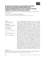
Studies on interactions between HCRSV coat protein and host proteins from kenaf
Bạn đang xem bản rút gọn của tài liệu. Xem và tải ngay bản đầy đủ của tài liệu tại đây (1.78 MB, 190 trang )
STUDIES ON INTERACTIONS BETWEEN
HCRSV COAT PROTEIN AND HOST PROTEINS IN
KENAF
Zhang Xin
(B.Sc, CAU)
A THESIS SUBMITTED
FOR THE DEGREE OF DOCTOR OF PHILOSOPHY
DEPARMENT OF BIOLOGICAL SCIENCES
NATIONAL UNIVERSITY OF SINGAPORE
2010
i
ACKNOWLEDGEMENTS
My first thank goes to my supervisor, Prof. Wong Sek Man for his excellent
instructions and constant support. I really appreciate the research study in the
molecular virology lab and the opportunity to learn under his guidance. I have also
learnt a lot from Prof. Wong about his wisdom and social experience which have
enlightened me. I really appreciate his patience and warm heart to help me to overcome
difficulties in my studies. My sincere thanks also go to my PhD committee members,
Associate Professor Yu Hao and Assistant Professor Cynthia He for their helpful
discussions and advice during the committee meetings.
Special thanks to all the members of the Plant Molecular Virology Lab: Li Weimin
for the clone of pGBKT7-CP and L10a, Cheng Ao for the cDNA clone pGST-CP, Niu
Shengniao for her technical advice. I also thank the undergraduate students in the lab
Fiona Setiawan, Sharon Kok, Tan Shihao, Lin Bitong and Xie Zhicheng. I also would
like to thank my former lab members Lim Chin Chin, Meng Chunying, Xie Juntao
and current lab members Sunil Kumar Tewary, Zhuang Linjie, Yi Wen and Gao
Ruimin for all of their help and encouragement during my PhD study in the lab.
Special appreciation goes to Wang Cheng, for his continuous care and love; special
thanks also go to Tu Haitao, Zhang Jingfeng, for their help and the precious
friendship.
ii
Sincere thanks go to Mr Chong PL and Madam Loy GL of DBS for their help with
transmission electron microscopy work. I also want to thank Ms Tong Yan for her
help in my confocal laser microscopy study.
I wish to pay special tributes to my family members for their encouragement
throughout all these years. Special thanks to my former teachers in China Agricultural
University, Associate Professor Cheng Yuqin, Professor Fan Zaifeng, Professor Li
Huaifang for their continuous encouragement and care. Finally, grateful thanks go to
the National University of Singapore for awarding me the NUS research scholarship.
iii
Table of Contents
ACKNOWLEDGEMENTS i
Table of Contents iii
LIST OF TABLES x
LIST OF FIGURES xi
ABBREVIATIONS i
SUMMARY viii
PUBLICATIONS x
CHAPTER 1 Introduction 1
1.1 Symptoms and developmental abnormalities 1
1.1.1 Virus infection affected host gene expression and proteins 2
1.1.2 Virus infection initiated plant response 4
1.1.3 Virus infection altered hormone metabolism 8
1.1.4 Virus infection triggered plant defense mechanism 9
1.2 Virus-host interaction 12
1.2.1 Virus-host interactions that coordinate replication or translation 12
1.2.2 Virus-host interactions that coordinate movement 14
1.2.3 Virus-host interaction that suppresses resistance of plant 16
iv
1.3 Protein-protein interaction study 18
1.3.1 Yeast Two-Hybrid (Y2H) Screening 19
1.3.2 Other methods to study protein-protein interaction 23
1.4 Virus-induced silencing of genes in plant 27
1.5 The functions of viral coat protein in virus infection and virus-host
interaction 28
1.6 Studies on HCRSV and HCRSV CP 30
1.6.1 HCRSV 30
1.6.2 Kenaf 33
1.6.3 Interaction studies on HCRSV CP 33
1.7 Rationales and objectives of this project 34
CHAPTER 2 General materials and methods 37
2.1 Media and buffers 37
2.2 Plant materials and inoculation 37
2.2.1 Plant materials and growth conditions 37
2.2.2 Plant inoculation 37
2.3 Molecular cloning 38
2.3.1 Polymerase chain reaction (PCR) 38
2.3.2 Purification of PCR fragments and DNA fragments from agarose gel 38
2.3.3 Ligation of DNA inserts into plasmid vectors 38
v
2.3.4 Preparation of competent E. coli 39
2.3.5 Transformation of bacteria with plasmids 39
2.3.6 Plasmid purification from E. coli 40
2.3.7 DNA sequencing 40
2.4 Y2H study 41
2.4.1 Preparation of yeast competent cells (LiAC method) 41
2.4.2 Transformation of plasmids into the yeast cells 42
2.4.3 Purification of plasmids from yeast cells 42
2.5 In vitro transcription of DNA with T7/T3 RNA polymerases 43
2.5.1 Preparation of infectious transcripts 43
2.5.2 Inoculation of in vitro transcription product onto true leaves 44
2.6 Analysis of RNA from plants 44
2.6.1 Isolation of total RNA from plants 44
2.6.2 Real-time PCR 45
2.7 Protein expression and purification 46
2.7.1 Plasmid construction and transformation 46
2.7.2 Induction of protein expression 47
2.7.3 Protein purification 47
2.7.4 Protein concentration measurement 48
2.7.5 Sodium dodecylsulfate-polyacrylamide electrophoresis (SDS-PAGE) 48
2.7.6 Staining of SDS gel by coomassie brilliant blue 49
vi
2.7.7 Immunodetection of proteins 50
2.7.8 In vitro translation of DNA with T7 RNA polymerases 50
2.7.9 Detection of the in vitro translation product 51
2.8 Protein extraction from virus-infected plants 52
2.9 Isolation and transfection of kenaf protoplasts 53
2.9.1 Isolation of kenaf protoplasts 53
2.9.2 PEG transfection of protoplasts 54
2.9.3 Confocal study of protoplasts with fluorescence 54
2.10 Reverse Transcription-PCR 54
CHAPTER 3 Screening and identification of host proteins interacting with
HCRSV CP 56
3.1 Introduction 56
3.2 Materials and methods 57
3.2.1 Construction and screening of kenaf cDNA library 57
3.2.2 Sequencing and cloning 59
3.2.3 Confirmation of the interaction 59
3.2.4 5‟ RACE PCR to amplify the complete sequence of C2 domain
containing protein 61
3.3 Results 62
3.3.1 Kenaf cDNA Library constructed 62
3.3.2 Sulfite oxidase in kenaf (HcSO) 62
vii
3.3.3 Identification of interaction between HCRSV CP and HcSO and
mapping of the interacting domains 66
3.3.4 C2 domain-containing protein in kenaf 68
3.3.5 Putative CsP 13.9 70
3.4 Discussion 72
CHAPTER 4 Plant sulfite oxidase plays important roles in the symptom
development of Hibiscus chlorotic ringspot virus in Kenaf 75
4.1 Introduction 75
4.2 Material and Methods 77
4.2.1 Plant materials and construction of plasmids 77
4.2.2 Co-localization of HCRSV CP and HcSO using BiFC 80
4.2.3 Protein expression and pull down assay of CP with HcSO 81
4.2.4 Determination of HcSO transcript level and sulfate level in mock and
HCRSV infected kenaf leaves 82
4.2.5 Biochemical assays of SO and H
2
O
2
-generating activities 83
4.2.6 Transmission electron microscopy (TEM) and immuno-EM study 83
4.2.7 Gene-silencing study of HcSO 85
4.3 Results 86
4.3.1 HcSO interacts with HCRSV CP in kenaf protoplasts 86
4.3.2 HCRSV CP bound to HcSO in vitro 89
4.3.3 HCRSV infection induces peroxisome proliferation and aggregation in
viii
kenaf cells 91
4.3.4 HCRSV infection leads to an up-regulation of HcSO gene transcript and
SO activity 93
4.3.5 HcSO was successfully silenced by TCV silencing vector 98
4.4 Discussion 103
CHAPTER 5 Hibiscus chlorotic ringspot virus coat protein interacts with plant
60S ribosomal protein RPL10A 109
5.1 Introduction 109
5.2 Materials and methods 111
5.2.1 Preparation of Hibiscus stem sap proteins 111
5.2.2 Preparation of HCRSV virions and coat protein subunits 111
5.2.3 HCRSV CP affinity chromatography 112
5.2.4 Sequence analysis of putative CP-interacting protein 112
5.2.5 Molecular cloning of L10A protein from kenaf plant 113
5.2.6 Rapid amplification of cDNA ends (RACE) and nested PCR 113
5.2.7 Yeast two-hybrid analysis 116
5.2.8 In vitro translation 116
5.2.9 Pull down assay 117
5.3 Results 118
5.3.1 Identification of RPL10A 118
5.3.2 Sequence analysis of putative L10A protein in kenaf plant 118
ix
5.3.3 Amplification of RPL10A gene using two degenerate primers generates a
band with a molecular size at around 300bp 121
5.3.4 Y2H study 129
5.3.5 Pull down assay 131
5.4 Discussion 134
CHAPTER 6 Conclusions and future work 136
6.1 Conclusion and discussion 136
6.2 Future work 138
References 140
x
LIST OF TABLES
Table 1.1 Two hybrid screening for interaction between viral proteins and host
proteins and the significance of the interaction 20
Table 1.2 Y2H method used to study interacting partners of viral proteins 22
Table 3.1 Primers for cDNA library construction and RACE PCR of C2 domain
containing protein. 60
Table 3.2 Clones with cDNA sequences matching putative proteins from NCBI
database 63
Table 4.1 Primers used in this study 79
Table 5.1 Primers used in the PCR experiment of RPL10A study 115
Table 5.2 Interaction between HCRSV CP and L10A and identification of
domains of CP that are involved in the interaction in yeast cells 130
xi
LIST OF FIGURES
Figure 1.1. Schematic representation of HCRSV genome organization 32
Figure 3.1. Amino acid sequence alignment of the SO from kenaf (HcSO) and
other plants 65
Figure 3.2. Interaction between HCRSV CP and HcSO and identification of
domains of CP that are involved in the interaction in yeast cells 67
Figure 3.3. Amino acid sequence alignment of C2 domain containing protein
from kenaf (HcSO) and similar proteins in other plants 69
Figure 3.4. Alignment of CsP13.9 with putative chaperon P13.9 [Castanea sativa],
and putative bundle sheath defective protein [Oryza sativa Japonica Group]
71
Figure 4.1. BiFC co-localization of HCRSV CP and HcSO in kenaf protoplasts. 87
Figure 4.2. Subcellular localization of HCRSV CP and HcSO in kenaf epidermal
cells (a to c) and colocalization of the two proteins to peroxisomes which were
counter-stained with a peroxisome-specific anti-SKL antibody in protoplasts
(d to i). 88
Figure 4.3. In vitro binding assay of bacterial-expressed GST-CP of HCRSV and
in vitro translated product (ivt) of HcSO 90
Figure 4.4. HCRSV infection enhanced biogenesis and aggregation of
peroxisomes in kenaf cells 92
Figure 4.5. Quantitative analysis of transcript expression of SO gene 94
xii
Figure 4.6. SO activity and H
2
O
2
-generating activity from mock-inoculated
(mock), HCRSV-infected (HCRSV) and diluted HCRSV infected (100X dil)
kenaf leaf extracts (10 µg). 95
Figure 4.7. Comparison of H
2
O
2
generating activity between mock-inoculated
(M), HCRSV (V) infected kenaf leaves and HCRSV infected kenaf leaves
under stress (SV) 13dpi. 97
Figure 4.8. Comparison of sulfate levels between mock-inoculated and
HCRSV-infected kenaf leaves 99
Figure 4.9. Study on the effect of silencing of HcSO on the symptom development
of HCRSV infection and quantitative analysis of transcript expression of
HcSO gene 102
Figure 4.10. The proposed hypothesis on the important roles of SO for the
appearance of disease symptom…………………………………………………108
Figure 5.1. Purification of CP-interacting proteins by affinity chromatography
119
Figure 5.2. Identification of p26 as a 60S ribosomal protein RPL10A 120
Figure 5.3. PCR of RPL10A gene using two degenerate primers generated a size
of around 300 bp 122
Figure 5.4. Alignment of the PCR product by degenerate primers 123
Figure 5.5. 5’ Race PCR product done with the touch down step incorporated is
in gel photo (a) and nested PCR in gel photo (b) 124
Figure 5.6. 5’ RACE PCR result showed three RPL10A sequences with a small
xiii
difference 125
Figure 5.7. The PCR product of cDNA of full length RPL10A using L10A-5’
Long Distance Primer and L10A-3’ Long Distance Primer 127
Figure 5.8. Alignment of kenaf RPL10A with 60S ribosomal protein from other
plants 128
Figure 5.9. Purification of GST-CP and GST protein through Glutathione
Sepharose 4B column chromatography 132
Figure 5.10. The in vitro translation product of RPL10A protein (arrow) from the
cloned RPL10A cDNA sequence obtained 133
i
ABBREVIATIONS
Abbreviations used for plant viruses
AMV Alfalfa mosaic virus
BMV Brome mosaic virus
CaLCuV Cabbage leaf curl virus
CaMV Cauliflower mosaic virus
CMV Cucumber mosaic virus
CNV Cucumber necrosis virus
CPMV Cowpea mosaic virus
CVYV Cucumber vein yellowing virus
HCRSV Hibiscus chlorotic ringspot virus
INSV Impatiens necrotic spot virus
MDMV Maize dwarf mosaic virus
MCMV Maize chlorotic mottle virus
PapMV Papaya mosaic virus
PNRSV Prunus necrotic ringspot virus
PVX Potato virus X
RDV Rice dwarf virus
RYMV Rice yellow mottle virus
SMV Soybean mosaic virus
SYNV Sonchus yellow net virus
ii
SuCMoV Sunflower chlorotic mottle virus
TBSV Tomato bushy stunt virus
TCV Turnip crinkle virus
TEV Tobacco etch virus
TMV Tobacco mosaic virus
ToMV Tomato mosaic virus
TuMV Turnip mosaic virus
ZYMV Zucchini yellow mosaic virus
Other abbreviations:
aa amino acid(s)
AD activating domain
Aux/IAA auxin/indole acetic acid
AOX alternative oxidase
amiRNA artificial microRNA
BD binding domain
BiFC bimolecular fluorescence complementation
bp base pair(s)
BSA bovine serum albumin
C- terminal carboxyl terminal
CP coat protein
Co-IP co-immunoprecipitation
iii
DIG digoxigenin
DCL4 DICER-like 4
dsRNAs double-stranded RNAs
DIC differential interference contrast
DMSO dimethylsulfoxide
DNA deoxyribonucleic acid
DNase deoxyribonuclease
dNTP deoxyribonucleotide triphosphate
dpi days post inoculation
dsDNA double-stranded DNA
DTT dithiothreitol
eIF(iso)4E eukaryotic initiation factor iso 4E
eEF1A eukaryotic translation elongation factor 1A
EDTA ethylenediaminetetraacetic acid
EtBr ethidium bromide
EtOH ethanol
E.coli Escherichia coli
g grams or gravitational force
G guanosine
GFP green fluorescence protein
GST glutathione S-transferase
GFP green fluorescent protein
iv
GW glycine/tryptophan
GA Gibberellic acid
GOX glycolate oxidase
HR hypersensitive response
HS heat shock
HSP heat-shock proteins
HC-Pro helper component-proteinase
h hour(s)
HRP horseradish peroxidase
IPTG Isopropyl β-D-1-thiogalactopyranoside
IRES internal ribosome entry site
ivt in vitro translated product
IVT in vitro transcription product
ITC isothermal titration calorimetry
kDa kilodaltons
kb kilobase(s)
lacZ -galactosidase gene
LB Luria-Bertani
M molar
MCS multiple cloning site(s)
mg milligram(s)
min minute(s)
v
ml milliliter(s)
mM millimole
micro
g microgram(s)
l microliter(s)
m micrometre
miRNA micro RNAs
MP movement protein
MMLV moloney murine leukemia virus
nat-siRNA natural-antisense transcript-derived
small-interfering RNAs
NbPCIP1 Nicotiana benthamiana PVX CP-interacting protein 1
NLPs nucleocapsid-like particles
nt nucleotides
nr non-redundant
ORF open reading frame
Oligo oligodeoxyribonucleotide
PAGE polyacrylamide gel electrophoresis
PABP poly A binding protein
PBS phosphate buffered saline
PCD programmed cell death
PR pathogenesis-related
vi
PSO plant sulfite oxidase
PTGS post-transcriptional gene silencing
RACE rapid amplification of cDNA ends
R genes plant resistance genes
ROS reactive oxygen species
RdRp RNA-dependent RNA polymerase
RC replication complex
RT-PCR reverse-transcription PCR
RdRp RNA-dependent RNA polymerases
RISC RNA-induced silencing complex
RNAi RNA interference
RBS ribosome-binding site(s)
RNA ribonucleic acid
RNase ribonuclease
rpm revolutions per minute
SAR systemic acquired response
sgRNA subgenomic RNA
siRNAs small interfering RNAs
SKL serine-lysine-leucine
SL stem-loop
sRNA small RNA
SDS sodium dodecyl sulfate
vii
sec second(s)
SO sulfite oxidase
TAP tandem affinity purification
tasiRNAs trans-acting small-interfering RNAs
TEM transmission electron microscopy
TGS transcriptional gene silencing
TrAPs transcriptional-activator proteins
T thymidine
TBS Tris-buffered saline
UAS upstream activating sequence
UV ultraviolet
UTR untranslated region
v/v volume per volume
VIGS virus-induced gene silencing
w/v weight per volume
X-gal 5-bromo-4-chloro-3-indolyl -D-galactopyranoside
Y2H yeast two hybrid
viii
SUMMARY
Hibiscus chlorotic ringspot virus (HCRSV) infection can cause severe chlorotic
ringspot symptoms and stunted growth on Hibiscus which is an ornamental plant
grown in many parts of the world. HCRSV coat protein (CP) is an important viral gene
product required for encapsidation and viral systemic movement. To better understand
the roles of HCRSV CP in viral infection and its interactions with host proteins, a
kenaf cDNA library was constructed and screened using a yeast two-hybrid (Y2H)
system to identify CP-interacting proteins. Using the Y2H system, several putative
proteins that interact with HCRSV coat protein were identified. These proteins include
sulfite oxidase (SO), a putative major latex-like protein, a putative chaperon P13.9, a
C2 domain-containing protein, a ricin domain-containing protein and putative
alpha-D-xylosidase like protein. The interaction of CP and SO was confirmed by in
vitro binding assay. Bimolecular fluorescence complementation (BiFC) assay was
used to colocalize the two proteins in vivo. The interaction was found to be associated
with peroxisomes by immunofluorescent labeling using anti serine-lysine-leucine
(SKL) signal peptide antibody. A second Y2Hstudy showed that both the P and S
domains of CP could interact with SO. This is probably due to the exposure of these
two domains on the outer surface of the capsid. In addition, Transmission electron
microscopy (TEM) revealed that peroxisomes were aggregated in HCRSV infected
cells and the CP was localized within the peroxisomes by immuno-gold labeling.
Biochemical assays of the total protein from kenaf leaf extracts showed that SO
ix
activity and SO-dependent H
2
O
2
-generating activity in the HCRSV-infected leaves
increased compared to the mock-inoculated kenaf plants. Thus, it is speculated that the
interaction of HCRSV CP and SO is important for the plant to up-regulate the sulfate
level and SO activity, which may be responsible for the virus accumulation or plant
defense during the virus infection.
Taken together, it is shown that HCRSV infection upregulates plant sulfite oxidase
(PSO) transcripts and increases sulfate levels in kenaf. A model is proposed on how
HCRSV is able to overcome plant resistance responses to establish infection in plants.
The interaction between ribosomal protein L10A and HCRSV CP was also further
studied (Chapter 5).
x
PUBLICATIONS
Paper:
Zhang, X. & Wong, S. M. (2009). Hibiscus chlorotic ringspot virus upregulates plant
sulfite oxidase transcripts and increases sulfate levels in kenaf (Hibiscus cannabinus
L.). J Gen Virol 90, 3042-3050.
Posters and presentations:
1. Zhang Xin, Sharon Kwok and Wong Sek Man. 2007. Screening of interactions
between HCRSV CP and proteins in kenaf using yeast-two-hybrid system. Poster
presentation for the 2nd Asian Conference on Plant Pathology, Indonesia.
2. Zhang Xin and Wong Sek Man. 2008. Screening of interactions between HCRSV
CP and proteins in kenaf using yeast-two-hybrid system. Poster presentation for the
9th International Conference of Plant pathology, Italy.
3. Zhang Xin and Wong Sek Man. 2009. Studies on interactions between HCRSV coat
protein and host proteins in kenaf. Oral presentation for the 14th Biological Sciences
Graduate Congress, Bangkok, Thailand.
1
CHAPTER 1
Introduction
Virus infection may disrupt host gene expression and physiology, causing disease
symptoms and developmental abnormalities. This may involve a virus-host interaction
which happens in the process of replication, movement, suppression of resistance. In
response to the virus infection, the plant also has its own mechanism to defense
against the viral infection. Hibiscus chlorotic ringspot virus (HCRSV) infection can
cause severe disease symptoms and stunted growth on kenaf which is an ornamental
plant grown in many parts of the world. HCRSV coat protein (CP) is an important
viral gene product required for encapsidation and viral systemic movement.
1.1 Symptoms and developmental abnormalities
Plant viruses are obligate intracellular parasites that do not have their own
translational machinery in a host cell. Therefore they do not separate themselves from
the host cells and use the host translational machinery to replicate in the cells and
synthesize viral proteins. Usually the plants infected with virus grow slower or flower
earlier than the mock inoculated plants. Virus infection may cause the display of the
disease symptoms including developmental abnormalities, chlorosis, or necrosis. The
disruption of the host processes could be one of the reasons that cause the cells to live