Investigation of new properties and applications of quadruplex DNA and development of novel oligonucleotide based topoisomerase i inhibitors
Bạn đang xem bản rút gọn của tài liệu. Xem và tải ngay bản đầy đủ của tài liệu tại đây (2.62 MB, 158 trang )
INVESTIGATION OF NEW PROPERTIES AND APPLICATIONS
OF QUADRUPLEX DNA
AND DEVELOPMENT OF NOVEL OLIGONUCLEOTIDE-
BASED TOPOISOMERASE I INHIBITORS
WANG YIFAN
NATIONAL UNIVERSITY OF SINGAPORE
2008
INVESTIGATION OF NEW PROPERTIES AND APPLICATIONS
OF QUADRUPLEX DNA
AND DEVELOPMENT OF NOVEL OLIGONUCLEOTIDE-
BASED TOPOISOMERASE I INHIBITORS
WANG YIFAN
(B.Sc., Soochow University, China)
A THESIS SUBMITTED
FOR THE DEGREE OF DOCTOR OF PHILOSOPHY
DEPARTMENT OF CHEMISTRY
NATIONAL UNIVERSITY OF SINGAPORE
2008
i
Acknowledgements
I would like to express my wholehearted gratitude to my supervisor, Associate
Professor Li Tianhu for his profound knowledge, invaluable guidance, constant
support, inspiration and encouragement throughout my graduate studies. He is not
only an extraordinary supervisor, a complete mentor, but a truly friend. The
knowledge, both scientific and otherwise, that I accumulated under his supervision,
will aid me greatly throughout my life.
I also give my sincere thanks to all the members of the Li group: Li Xinming,
Li Ming, Liu Xiaoqian, Xu Wei, Magdeline Tao Tao Ng and Chua Sock Teng, for
their cordiality and friendship. We had a great time working together.
I wish to express my deepest appreciation to my family and my boyfriend for
their love and support. Without their help, I can not complete this work.
Last but not least, my acknowledgement goes to National University of
Singapore for awarding me the research scholarship and for providing financial
support to carry out the research work reported herein.
ii
Table of Contents
Acknowledgements i
Table of Contents ii
Summary viii
List of Tables x
List of Figures xi
Chapter 1 Introduction 1
1.1. Basic Information about DNA 1
1.2. G-Quadruplex Form of DNA 3
1.2.1. Guanine Quartets
3
1.2.2. G-Quadruplexes
1.2.2.1 Discovery of G-Quadruplex DNA
1.2.2.2 Structural Polymorphism of G-Quadruplex Structures
1.2.2.2.1 Strand Stoichiometry
1.2.2.2.2 Strand Polarity Polymorphism
1.2.2.2.3 Connecting Loops
1.2.2.3 Possible Roles of G-Quadruplex in vivo
1.2.2.3.1 G-Quadruplex-Interactive Proteins
1.2.2.3.2 Telomere Protection and Elongation
1.2.2.3.3 Interaction of Small-Molecule with G-Quadruplex
4
4
4
5
5
6
7
8
9
10
1.3. i-Motif Structure of DNA 10
1.3.1. Discovery of i-Motif Form of DNA
1.3.2. Stoichiometries and Topologies of i-Motif DNA
11
11
iii
1.3.3. Possible Biological Role of i-Motif Structure of DNA
13
Chapter 2 Construction of i-Motif-Based DNA Machines 14
2.1. Background and Aims
2.1.1. Biomolecular Machines in Organisms
2.1.2. DNA-Based Artificial Molecular Machines
2.1.3. Quadruplex DNA-Based Molecular Machines
2.2. Our Strategies in Design of i-Motif-Based DNA Machines
2.3. Synthesis of Our Newly Designed i-Motif-Based DNA Machines
2.4. Operation of Our i-Motif-Based DNA Machines
2.4.1 First Half and Second Half of Operating Cycle
2.4.2 Cyclic Operation of i-Motif-Based DNA Machine
2.4.3 Calculation of Mechanical Energy Released by our i-Motif-
Based DNA Machine
2.5. Conclusions
14
14
15
18
22
26
28
28
35
36
38
Chapter 3 Search and Confirmation of G-Quadruplex-Based
Deoxyribozymes
39
3.1. Background and Aims
3.2. Confirmation of Self-Cleaving Action of a Particular G-
Quadruplex
3.3. Effect of Certain Factors on the G-Quadruplex-Based Self-
Cleavage Reaction
3.3.1 Metal Ion Dependence
3.3.2 pH Dependence
39
40
43
43
46
iv
3.3.3 DNA Concentration Dependence
3.3.4 Determination of Rate Constants of the G-Quadruplex-Based
Self-Cleavage Reactions
3.3.5 Potassium Ion Concentration Dependence
3.3.6 The formation of G-Quadruplex by Oligonucleotide 1
3.4. Conclusions
47
47
50
52
55
Chapter 4 Construction of Fluorescein-Tagged Circular G-
Quadruplexes 56
4.1. Background and Aims
4.2. Construction of Circular Oligonucleotides on the Basis of
Unimolecular G-Quadruplex
4.2.1 Design and Synthesis of Circular Oligonucleotide on the Basis
of Unimolecular G-Quadruplex
4.2.2 Confirmation of Circular Nature of Our Ligation Product
4.2.3 Conformation Dependence of the Circularization Reactions
4.2.4 Loop-Size Dependence of Our Circularization Reactions
4.2.5 Alkali-Ion Dependence of Our Circularization Course
4.2.6 pH Dependence of the Designed Ligation Reactions
4.2.7 Potassium Ion-Concentration Dependence of Our Ligation
Reaction
4.2.8 Verification of Formation of G-Quadruplex by Newly
Synthesized Circular Oligonucleotides
4.3. Construction of Fluorescein-Tagged Circular Oligonucleotides
4.3.1 Design and Synthesis of Fluorescein-Tagged Circular
56
58
58
63
65
67
69
70
70
72
74
v
Oligonucleotides
4.3.2 Structural Verification of Fluorescein-Tagged Circular
Oligonucleotides
4.3.3 Fluorescence Measurement of Fluorescein-Tagged Circular G-
Quadruplex
4.4. Conclusions
74
76
78
79
Chapter 5 Development of New Oligonucleotides-Based Topoisomerase
I Inhibitors
81
5.1. Background and Aims
5.1.1 DNA Topoisomerases
5.1.2 Mode of Action of DNA Topoisomerase I
5.1.3 Topoisomerase I Inhibitors
5.2. Construction of C3-Spacer-Containing Circular Oligonucleotides
as Topoisomerase I Inhibitors
5.2.1 General Design Strategy
5.2.2 Synthesis and Characterization of the C3-Spacer-Containing
Circular Oligonucleotides
5.2.3 Inhibitory Effect of the C3-Spacer-Containing Circular
Oligonucleotides against Topoisomerase I Inhibitors
5.2.4 Confirmation of the Existence of Topo I-DNA Covalent
Conjugate
5.2.5 Examination of Resistance of Oligonucleotide 1 against Repair
Enzyme
5.2.6 Position Dependence of C3-Spacer Modification on the
81
83
83
85
86
86
88
90
92
94
vi
Inhibitory Efficiency of Topoisoemrase I
5.3. Gap-Containing Unimolecular Oligonucleotides as Topoisomerase I
Inhibitors
5.3.1 Design of Gap-Containing Oligonucleotides as
Topoisomerase I Inhibitors
5.3.2 Examination of Inhibitory Effect of Gap-Containing
Oligonucleotides as Topoisomerase I Inhibitors
5.4.Conclusions
95
98
99
100
107
Chapter 6 Materials And Methods 108
6.1. Materials
6.1.1. Oligonucleotides
6.1.2. Enzymes
6.1.3. PBR 322 DNA
6.1.4. Buffer
6.2. Methodology
6.2.1. 5’ End Labeling of DNA (T4 Polynucleotide Kinase Method)
6.2.2. Polyacrylamide Gel Electrophoresis (PAGE)
6.2.3. DNA Purification (Desalting)
6.2.4. Preparation of N-Cyanoimidazole
6.2.5. Chemical Ligation Reactions of Unimolecular G-Quadruplex
using N-Cyanoimidazole
6.2.6. Self-Cleavage Reactions of Oligonucleotide 1
6.2.7. Fluoresence Measurement
6.2.8. Thermal Stability Analysis of Oligonucleotides by UV
108
108
108
114
115
116
116
117
118
119
119
120
120
vii
Spectroscopy
6.2.9. CD Measurement
6.2.10. Empirical Estimation of Duplex Melting Temperature
6.2.11. General Procure for Exonuclease VII Hydrolysis
6.2.12. Partial Hydrolysis of the Identified Circular Product by
DNase I
120
120
121
122
122
References 123
List of Publications 140
viii
Summary
Some sequences of DNA that possess certain guanine or cytosine-riched
stretches are capable of associating into two types of four-stranded DNA structures,
namely G-quadruplex and i-motif respectively. It has been suggested in the past that
some of these quadruplex structures could exist in some biologically important
regions of DNA such as at the end of chromosomes and in the regulatory regions of
oncogenes. In addition, due to their distinctive structural characteristics, quadruplex
structures of DNA have been widely used as building blocks in various
nanotechnological applications, such as G-quadruplex nanodevices and i-motif
nanoswitches. With the aim of exploring new properties and applications of
quadruplex DNA during my graduate studies, we have (1) constructed i-motif DNA-
based molecular devices that are operable through variations of their surrounding pH
values; (2) developed certain fluorescence-tagged circular G-quadruplexes to be used
as molecular probes; and (3) investigated the factors that affect the G-quadruplex that
could undergo self-cleavage reactions. Finally, we have designed and synthesized
certain dumbbell-shaped oligonucleotides and further examined their inhibitory
effects on the activities of human topoisomerase I.
In Chapter 2, design and synthesis of a novel quadruplex DNA machine is
presented that was capable of converting chemical energy into elastic potential
energy. As a consequence of this energy converting process, Watson-Crick hydrogen
bonding interaction between two complementary 11-mer oligonucleotides was forced
to break down, leading to a free energy change of 12.46 kcal mol
-1
.
In Chapter 3, self-cleavage reaction of a guanine-riched oligonucleotide was
thoroughly studied during our investigation. Subsequent examinations on certain
factors that affect self-cleavage reactions of G-quadruplexes are described, such as
ix
variation of metal ions, pH values and concentration of DNA. In addition, kinetic
analysis of self-cleavage of G-quadruplex was also carried out. It is our hope that the
results reported in this chapter could be helpful for searching for new G-quadruplex
structures that could perform self-cleavage reactions.
In Chapter 4, our studies of synthesis and characterization of unimolecularly
circular G-quadruplex on the template basis of G-quadruplex through chemical
ligations of guanine-riched oligonucleotides are described. Loop-size effect of ligation
reaction, conformation dependence of circularization course, effects of alkali ions and
pH values as well as concentration of potassium ions on the circularization reactions
were investigated during our studies. The potential application of the obtained
unimolecularly circular G-quadruplex in certain biological processes is also presented
in this chapter.
In Chapter 5, design and synthesis of a series of dumbbell-shaped circular
oligonucleotides containing internal C3-spacers are presented. Our studies
demonstrated that this C3-spacer-containing oligonucleotide displays an IC
50
value of
33 nM in its inhibition on the activity of human topoisomerase I, which is much
efficient than those of camptothecins (anticancer drugs currently in clinical use).
x
List of Tables
Table
No.
Page
No.
2-1 Calculations of the free energy changed during the formation of
duplex structure from its single-stranded form.
37
4-1 Sequences of oligonucleotides used in the current study.
73
5-1 Inhibitory efficiency (IC
50
) of some C3-spacer-containing
oligonucleotides on the activity of human Topo I.
96
5-2 Sequences and C3-spacer modifications of oligonucleotides prepared
during this study.
96
xi
List of Figures
Figure
No.
Page
No.
1-1 Structures of four types of nitrogenous bases
2
1-2 Base Pairing in DNA Double Helix
2
1-3 Structures of Guanine Quartets
3
1-4 G-quadruplex structures formed from one, two or four strands
5
1-5 Stoichiometries of G-Quadruplex structures
5
1-6 Different strand polarity arrangements of G-quadruplexes
6
1-7 Strand connectivity alternatives for bimolecular guanine tetrad
structures
6
1-8 Strand connectivity alternatives for unimolecular guanine tetrad
structures
7
1-9 Possible biological roles of G-quadruplexes
9
1-10 Illustration of C·C
+
interaction in i-motif structure of DNA
11
1-11 i-motif structures with (a) four, (b) two and (c) one strand(s)
12
2-1 The F
0
F
1
-ATPase molecular motor
15
2-2 DNA-based twisting molecular switch
16
2-3 DNA tweezers
17
2-4 DNA walkers
18
2-5 A quadruplex-duplex exchange nanomachine
19
2-6 A switchable aptamer device
20
2-7 A Proton-Fuelled DNA Nanomachine
22
2-8 Illustration of our designed DNA-based molecular machine
23
2-9 Schematic representation of our strategy for designing a new energy-
converting DNA machine capable of breaking down Watson-Crick
interaction.
25
xii
2-10 Polyacrylamide gel electrophoretic analysis of oligonucleotides as
components of the artificial DNA machines designed in our study.
28
2-11 Structure of Bodipy 493/503 modification on 5’ end of oligonucleotide
29
2-12 Fluorescence Spectroscopic analysis of formation and disintegration of
duplex structure associated with the artificial devices designed in the
current studies.
30
2-13 Analysis of dissociation and formation of duplex structure correlated
with the artificial machines using fluorescence spectroscopy.
32
2-14 Confirmation of presence of duplex structure between Sequence 1 and
Sequence 2 (State 1 in Figure 2-2C) at pH > 6.2.
34
2-15 Examination of operability of artificial DNA machines using
fluorescence spectroscopy.
35
2-16 UV melting curve of the 11 base pairs duplex entity.
37
3-1 Schematic representation of a self-cleavage process of G-quadruplex
DNA in this study.
40
3-2 Diagrammatic illustration of a possible self-cleaving reaction at one of
the two phosphodiester bonds between A
14
and A
15
of Oligonucleotide
1.
41
3-3 Polyacrylamide gel electrophoretic analysis of self-cleavage of DNA.
42
3-4 Polyacrylamide gel electrophoretic analysis of self-cleavage of DNA
visualized by SYBER GREEN staining.
43
3-5 PAGE analysis of self-cleavage of Oligonucleotide 1 in the presence of
20 mM alkaline metal ions (Li
+
, Na
+
, K
+
, Rb
+
and Cs
+
).
44
3-6 PAGE analysis of self-cleavage of Oligonucleotide 1 in the presence of
1 mM transition metal ions (Zn
2+
, Pb
2+
, Ni
2+
, Co
2+
and Mn
2+
).
45
3-7 PAGE analysis of self-cleavage of Oligonucleotide 1 in the presence of
20 mM alkaline earth metal ions (Mg
2+
, Ca
2+
, Sr
2+
and Ba
2+
).
45
3-8 pH dependent of self-cleavage of Oligonucleotide 1 vary from 5.0 to
9.0.
47
3-9 PAGE analyses of self-cleavage of Oligonucleotide 1 in different DNA
concentrations vary from 1 nM to 100 nM.
47
3-10 Time dependence of self-cleavage reaction of Oligonucleotide 1.
48
3-11 Determination of observed rate constants of Oligonucleotide 1 in its
xiii
self-cleavage reactions.
49
3-12 Time dependence of self-cleavage reaction of Oligonucleotide 1 in the
absence of magnesium ions.
50
3-13 Effect of potassium ion concentration on the self-cleavage reaction of
Oligonucleotide 1.
51
3-14 PAGE analysis of self-cleavage of Oligonucleotide 1 in the presence of
80 mM alkaline metal ions (Li
+
, Na
+
, K
+
, Rb
+
and Cs
+
).
51
3-15 CD spectroscopic analysis of Oligonucleotide 1 in the presence of K
+
.
52
3-16 Comparison CD studies of Oligonucleotide 1 in the presence of
different alkaline metal ions (Li
+
, Na
+
, K
+
and Rb
+
).
52
3-17 Comparison CD studies of Oligonucleotide 1 in the presence of
different alkaline earth metal ions (Mg
2+
, Ca
2+
, Sr
2+
and Ba
2+
).
53
3-18 Comparison CD studies of Oligonucleotide 1 in the presence of
different transition metal ions (Zn
2+
, Pb
2+
, Ni
2+
, and Mn
2+
).
54
4-1 Schematic representation of G-quadruplex formed unimolecularly (a),
bimolecularly (b) and through the association of four strands of
oligonucleotides (c).
57
4-2 Diagrammatic illustration of our strategy for constructing
unimolecularly circular G-quadruplex through chemical ligation.
59
4-3 Possible folding patterns of certain fluorescence-tagged circular G-
quadruplexes.
60
4-4 Illustration of different loop geometries possessed by unimolecular G-
quadruplexes.
60
4-5 Construction of unimolecularly circular oligonucleotides on the
template basis of G-quadruplex and time course of the ligation reaction
62
4-6 Hydrolysis of the identified circular products by exonuclease.
64
4-7 Partial hydrolysis of the identified circular products by DNAse I.
64
4-8 Effect of mismatched sequences on the circularization reaction.
66
4-9 Effect of recessive sequences on the circularization reaction.
67
4-10 Effect of loop size on the circularization reaction.
68
4-11 Effect of alkali ions on the circularization reaction.
69
xiv
4-12 pH dependency of the circularization reaction.
70
4-13 Effect of potassium-ion concentration on the circularization reaction.
71
4-14 CD spectra of circular oligonucleotides of
<GGTTTTGGGGTTTTGGGGTTTTGGGGTTTTGG> (20 µM) in 10
mM Tris·HCl buffer (pH 7.0)
73
4-15 Schematic representation of our synthetic route toward fluorecein-
labeled circular G-quadruplex.
75
4-16 Electrophoretic analysis of fluorecein-labeled circular G-quadruplex.
76
4-17 Hydrolysis of fluorecein-labeled circular products by exonuclease VII.
77
4-18 Partial hydrolysis of the fluorecein-labeled circular products by DNAse
I.
78
4-19 Fluorescence emission spectra of fluorescein-labeled circular G-
quadruplex (a) and non-fluorescein-labeled linear oligonucleotide,
sequence 2 (b).
79
5-1 Superhelical tension generated by DNA unwinding and resolved by
DNA topoisomerases.
82
5-2 Type I and Type II DNA topoisomerases.
83
5-3 Mode of action of DNA topoisomerase I.
84
5-4 Chemical structures of Camptothecin, Topotecan and irinotecan.
86
5-5 Schematic representation of a C3-spacer-containing dumbbell-shaped
oligonucleotide designed in our studies.
87
5-6 Diagrammatic illustration of anticipated inhibitory mechanisms of a
C3-spacer-containing oligonucleotide (Oligonucleotide 1) on the
activities of human topoisomerase I in our studies.
88
5-7 Illustration of the ligation reaction of Oligonucleotide 1
89
5-8 Polyacrylamide gel electrophoretic analysis of formation of
Oligonucleotide 1.
89
5-9 Polyacrylamide gel electrophoretic analysis of circularity of
Oligonucleotide 1 in its backbone.
90
5-10 Agarose gel electrophoretic analysis of inhibitory effect of
Oligonucleotide 1 (b) and Oligonucleotide 2 (c) on the activities of
human topoisomerase I.
92
xv
5-11 Correlations between concentration of oligonucleotide 1 and percent
inhibition on topoisomerase I activity.
92
5-12 Denaturing polyacrylamide gel electrophoretic confirmation of
formation of Topo I-Oligonucleotide 1 covalent conjugates.
93
5-13 Polyacrylamide gel electrophoretic analysis of hydrolytic products of
Oligonucleotide 1, 2 and 3 generated by T7 endonuclease I.
95
5-14 Sequences of oligonucleotides used in the study of Topoisomerase I
Inhibitors.
99
5-15 Illustration of possible mechanism for gap-containing oligonucleotides
as Topoisomerase I Inhibitors.
100
5-16 Agarose gel electrophoretic analysis of inhibitory effect of Duplex 3 on
human topoisomerase I.
101
5-17 Correlations between percent inhibition on topoisomerase I activity and
concentration of Duplex 3.
102
5-18 Agarose gel electrophoretic analysis of inhibitory effect of Duplex 2 on
human topoisomerase I.
103
5-19 Correlations between percent inhibition on topoisomerase I activity and
concentration of Duplex 2.
103
5-20 Agarose gel electrophoretic analysis of inhibitory effect of Duplex 1 on
human topoisomerase I.
104
5-21 Correlations between percent inhibition on topoisomerase I activity and
concentration of Duplex 1.
105
5-22 Agarose gel electrophoretic analysis of inhibitory effect of Duplex 3 on
human topoisomerase I without preincubation.
106
5-23 Correlations between percent inhibition on topoisomerase I activity and
concentration of Duplex 3 without preincubation.
106
1
Chapter 1
Introduction
1.1 Basic Information about DNA
Deoxyribonucleic acid (DNA) is a type of biomacromolecule that contains
genetic information used for the functioning of living organisms.
1
The major role of
DNA in vivo is its long-term storage of genetic information. From the perspective of
chemistry, DNA is a long polymer built up on simple units called nucleotides, linked
together through a backbone made of sugars and phosphate groups.
1, 2
A single strand
form of DNA is a long chain composed of different nucleotides. Each nucleotide
consists of a sugar, a phosphate and a nitrogenous base. There are four different types
of bases in DNA (Figure 1-1), and each base is usually abbreviated by the first letter
of its name: Adenine (A), Thymine (T), Guanine (G) and Cytosine(C). Two strands of
nucleotides usually wrap around each other, which are twisted together into a long
helix; like a ladder twisted about its long axis (Figure 1-2).
2
The backbone of sugar-
phosphate linkages forms the uprights of the twisted ladder. The rungs of the ladder
are made up of base pairs, which are almost always found connected to each other.
Each twist of the ladder contains approximately 10 rungs, which is 0.34 nm apart. In a
complete helix, A always lines up with T and G goes with C. In these combinations,
the different bases fit together perfectly like a lock and key, which is termed with
“Watson-Crick base pairing” (Figure 1-2).
2
2
Adenine - A
Cytosine - C
Guanine - G
Thymine - T
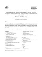
Figure 1-1. Structures of four types of nitrogenous bases
Sugar phosphate backbone
Cytosine
Adenine
Guanine
Thymine
Base pairs
Nitrogenous base
Figure 1-2. Base Pairing in DNA Double Helix
3
1.2 G-Quadruplex Form of DNA
1.2.1 Guanine Quartets
DNA commonly exists in the form of duplex structure in which two self-
complementary strands are held together by Watson–Crick base pairs. Besides this
form of duplex DNA, certain guanine-riched DNA sequences can form four-stranded
structures, namely G-quadruplexes.
3-6
The basic building block of G-quadruplex is the
guanine quartets (also known as guanine tetrads) composed of four guanine bases
arrayed in a square planar configuration, in which each base is both the donor and
acceptor of two hydrogen bonds with its neighbors (Figure 1-3). More precisely, the
guanine quartet arises from the association of four guanines into a cyclic Hoogsteen
hydrogen bonding arrangement that involves N1, N7, O6 and N2 of each guanine
base.
7-10
Positively charged metal ions can be sandwiched between the quartets. Their
presence in the central cavity of the quadruplex helps maintain the stability of the
tetraplex structure.
3
In addition, the G-quartet could form a particularly effective
stacking unit when placed next to each other, resulting in a strong attraction that
contributes substantially to the stability of the overall structure.
11-19
N
N
N
N
O
N
H
H
H
H
N
N
N
N
O
N
N
N
N
N
O
N
N
N
N
N
O
N
H
H
H
H
H
H
H
H
H
H
H
H
M
+
/M
2+
Figure 1-3. Structures of Guanine Quartets
4
1.2.2 G-quadruplexes
In certain guanine-riched strands, two or more G-quartets can stack upon each
other to form four-stranded structures with a guanine tetrad core. These structures are
known as G-quadruplexes.
3
The term G-quadruplex refers to any four-stranded DNA
structure containing guanine quartets without reference to strand connectivity.
3-5
G-quadruplexes exhibit an unusual dependence on specific metal ions, usually
K
+
and occasionally Na
+
,
20
which results in very tight metal binding via inner sphere
coordination. The cavity between G-quartets is well suited to coordinating the right
size of cations because the two planes of quartets are lined by eight carboxyl O6
atoms from guanine. It was reported that a wide variety of cations are capable of
occupying the central cavity of quadruplex structures, including monovalent ions such
as NH
4
+
and Tl
+
and divalent cations such as Sr
2+
, Ba
2+
, and Pb
2+
.
21
1.2.2.1 Discovery of G-quadruplex DNA
It was known since early 19
th
century that guanosine and its derivatives could
form viscous gels in water.
22
Until 1962, David R. Davies et. al.
23
proposed on the
basis of X-ray diffraction data that four guanine bases form a planar structure through
Hoogsteen hydrogen bonding interaction.
22
Subsequent NMR studies of these gels
further suggested that cations such as Na
+
and K
+
could coordinate to the O6 atoms of
each guanine base and strongly influence the specific type of structure adopted by the
gels.
24
1.2.2.2 Structural Polymorphism of G-quadruplex Structures
One of the most intriguing aspects of G-quadruplex is their extensive
polymorphism which arises from variation of strand stoichiometry, strand polarity and
connecting loop.
11-15
Quadruplexes typically contain 1, 2, or 4 nucleic acid strands,
5
giving rise to unimolecular, bimolecular or four-stranded structures and display a
wide variety of topologies (Figure 1-4). These tetraplex structures can exist in
different isomeric forms caused by different strand polarities of adjacent backbones.
Certain guanine-riched sequences can, for example, orient themselves in all parallel,
three parallel and one anti-parallel, adjacent parallel or alternating anti-parallel.
10
Some of the polymorphisms are discussed in the following sections.
b
c
a
G
G
G
G
G
G
G
G
G
G
G
G
G
G
G
G
G
G
G
G
G
G
G
G
G
G
G
G
G
G
G
G
G
G
G
G
G
G
G
G
G
G
G
G
d
G
G
G
G
G
G
G
G
G
G
G
G
Figure 1-4. G-quadruplex structures formed from one, two or four strands
1.2.2.2.1 Strand Stoichiometry
G-quadruplexes could be formed by association of one (Figure 1-5A), two
(Figure 1-5B), or four strands (Figure 1-5C) of oligonucleotides. The structural
assemblies of unimolecular, biomolecular and tetramolecular G-quadruplexes could
display different physical and chemical properties.
Figure 1-5. Stoichiometries of G-Quadruplex structures
1.2.2.2.2 Strand Polarity Polymorphism
The additional structural characteristic of G-quadruplex is the relative
arrangement of adjacent backbones, which could have different polarities. The four
6
strands of oligonucleotides in a G-quadruplex can be all parallel (Figure 1-6A), three
parallel and one anti-parallel (Figure 1-6B), adjacent parallel (Figure 1-6C), or
alternating anti-parallel (Figure 1-6D). Many guanine-riched oligonucleotides have
been determined either with NMR
26
or crystallography
27
, which displayed different
strand polarities as shown in Figure 1-6.
Figure 1-6. Different strand polarity arrangements of G-quadruplexes
1.2.2.2.3 Connecting Loops
The loops that connect guanine quartets participating in the formation of
unimolecular or bimolecular G-quadruplexes can run in different ways. The two
strands involved in bimolecular G-quadruplexes can have loops that connect guanine
tracts either diagonally or edgewise.
25
Figure 1-7. Strand connectivity alternatives for bimolecular guanine tetrad structures
Diagonal loops are expected to protrude on opposite ends of the guanine tetrad
core (Figure 1-7A). When the two loops connect guanine tracts edgewise, they can be
either on the same or on opposite sides of the tetrad core. Loops on the same side of
the core can be either parallel (Figure 1-7B) or anti-parallel (Figure 1-7C). When the
7
two loops protrude on opposite sides of the core, they can run in two different
directions (Figure 1-7D and 1-7E).
For unimolecular G-quadruplexes, structural isomers of G-quadruplex caused
by loop-connecting fashion are fewer. In order to avoid the clash of two diagonal
loops on the same side, the three loops can join either in the order adjacent-adjacent-
adjacent (Figure 1-8A) or adjacent- diagonal-adjacent (Figure 1-8B). On the other
hand, there are some examples of parallel strands connecting via loops running on the
outside of the guanine tetrad core (Figure 1-8C), which indicates that the spectra of
unimolecular structures may be more complex than prospected here.
28
Figure 1-8. Strand connectivity alternatives for unimolecular guanine tetrad
structures
1.2.2.3 Possible Roles of G-quadruplex in vivo
Little attention was paid to the phenomenon of guanine tetrads for more than
20 years since it was elucidated in 1962 by David R. Davies. Until 1980s, emerging
interest in G-quadruplex structure was stimulated by several implications of its
existence in various biologically important genomic regions such as telomeres.
29, 30
For example, these structures were suggested to participate in telomere regulations.
In addition, it is believed that G-quadruplex is responsible for the switch
recombination to bring different constant regions next to variable regions during the
differentiation of B lymphocytes.
31
8
In addition, telomeres are the specialized ends of linear chromosomes
comprising tandemly-repeated short DNA sequences.
32
Various proteins are involved
in regulating the structure and function of human telomeres, including telomerase and
some telomere-interacting proteins such as Pot1, TRF1 and TRF2. It is well known
that telomeres are essential for genome integrity and appear to play an important role
in cellular aging and cancer. In almost all organisms, the telomeric DNA sequence has
a G-rich 3’ overhang, such as “TTAGGG” in vertebrates or “TTGGGG” in ciliate
Tetrahymena. The length of the sequences can range from a dozens to thousands of
such repeats. Generally, the last few hundred based of G-rich strand in telomeres is
thought to be in single-stranded form.
32
Besides present at the ends of telomeres,
guanine-rich sequences are found in a number of important DNA regions, such as in
the immunoglobulin switch regions and gene promoter region of c-myc and other
oncogenes.
36
Moreover, several G-quadruplex-binding proteins have been identified
over the past 10 years.
32-35
It consequently becomes apparent that G-quadruplex could
play certain significant roles in various types of biological processes.
1.2.2.3.1 G-quadruplex-Interactive Proteins
Many proteins, mostly from ciliates and yeast, have been found to bind to G-
quadruplex structures.
32-40
Among these, yeast RAP1 protein
34
and beta-subunit of
Oxytricha telomere binding protein
37
are the most interesting ones because they not
only bind to G-quadruplex but also facilitate the formation of these structures. In
addition, four helicases, the Simian Virus (SV) 40 large T-antigen,
41
Bloom’s
syndrome helicase (BLM) from yeast, and Werner syndrome helicase from humans
42
have been found to unwind G-quadruplex DNA. Another enzyme that could interact
with quadruplex structures is human DNA topoisomerase I (Topo I).
43
Arimondo et.
al. demonstrated that Topo I can bind to both linear, four-stranded quadruplexes and