Development of sphingosine kinase (SPHK) inhibitors and the role of sphingolipids in adult stem cell proliferation and differentiation 2
Bạn đang xem bản rút gọn của tài liệu. Xem và tải ngay bản đầy đủ của tài liệu tại đây (395.93 KB, 32 trang )
Chapter2 Development and Evaluation of Human SPHK Inhibitors
32
CHAPTER 2 DEVELOPMENT AND EVALUATION OF HUMAN
SPHK INHIBITORS
As discussed in Chapter 1, SPHK and its product S1P, play important role in many
cellular processes, such as in the regulation of intracellular calcium signals, in
angiogenesis and control of cell adhesion molecule expression, and chemotaxis. More
particularly, in immune cells, it was shown that the SPHK/S1P pathway may promote
inflammation by triggering the release of proinflammatory mediators (Taha et al., 2006,
Melendez, 2008). SPHK and S1P are very tightly involved in several pathological
processes, indicating that the SPHK/S1P pathway represents an interesting target for the
development of novel therapeutics. In particular, compounds having the ability to
modulate the levels of S1P would have a high potential for the treatment of diseases
wherein S1P is believed to be involved, such as cardiovascular diseases-including
atherosclerosis, thrombosis and dyslipidemia, diabetes including type I and type II, stroke,
autoimmune and inflammatory diseases such as multiple sclerosis, psoriasis and
inflammatory arthritis, allergic diseases such as asthma and dermatitis, T helper-1 related
diseases, chronic obstructive pulmonary disease, cancer and neurodegenerative disorders
(Kuokkanen et al., 1997; Nair et al., 1977; Enlund et al., 1999; Hong et al., 1999; Xia et
al. 2000).
Another important potential of SPHK inhibitors is that, they may be capable to promote
the stem cells differentiation, therefore, shortening the incubation duration for stem cells
differentiating into pure subpopulation(s), as discussed in Chapter 1.
Unfortunately, there are no specific inhibitors for SPHK commercially available yet.
DMS is currently the most widely used inhibitor of SPHK, but it has been shown to be
Chapter2 Development and Evaluation of Human SPHK Inhibitors
33
not specific to SPHK. It was found to inhibit not only two isozymes of human SPHKs
(SPHK1 & SPHK2), but other kinases such as PKC (Igarashi and Hakomori 1989;
Igarashi et al., 1989). Some attempts to produce inhibitors of SPHK have been carried
out yielding promising compounds (French et al., 2003, Kim et al., 2005), but they are
lack of proper validation.
In this study, novel inhibitors were designed and synthesized based on the natural
substrate of SPHK (D-erythro-Sphingosine). These compounds are aimed to be more
specific inhibitors for SPHK, and even isotype-specific (SPHK1 vs. SPHK2). With the
newly developed inhibitors, a better understanding of the role of SPHK in the process of
stem cells differentiation will be studied as well. In this chapter, only the compounds
synthesis and evaluation are discussed. Compounds function on stem cells differentiation
will be addressed in Chapter 3.
The inhibitors of human SPHK used in this study were designed as the analogues of the
natural substrate, D-erythro-sphingosine, by modifying several functional chemical
groups of it. The synthetic compounds were then evaluated for their efficiency and
specificity to inhibit human exogenous and endogenous SPHK; the compounds were also
tested for their potential cytotoxicity effects, and counter screened against other kinases
including DAGK and PKCα. The materials and methods are addressed below, followed
by the results and discussion.
2.1 MATERIALS AND METHODS
Unless otherwise stated, all chemicals were purchased from Sigma-Aldrich, Singapore.
2.1.1 Synthesis of Human SPHK Inhibitors
Chapter2 Development and Evaluation of Human SPHK Inhibitors
34
2.1.1.1 Compounds Designed As Analogues of Sphingosine
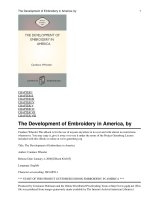
Earlier studies have shown that D-erythro-sphingosine (Figure 2.1a) is a natural substrate
for SPHKs. The design of the compounds was based on investigations of: 1) changing the
fatty acid carbon chain length; 2) converting the existing double bond to either a single or
triple bond, or 3) changing the hydroxyl group to other functionalities (Figure 2.1b).
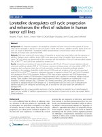
Scheme 1 summarises the experimental design, including compounds structures and key
procedures to synthesize them. During the first round testing, only six compounds were
finally synthesized and evaluated.
It should be noticed that Scheme 1 is the synthesis flow for the whole project of
developing SPHK inhibitors, which has been filed as a patent, while this thesis only
covered six analogues described here. However, in order to keep the story as a whole one,
Scheme 1 is still used here to describe the flow.
HO C
13
H
27
OH
NH
2
HO C
13
H
27
OH
NH
2
D-erythro-sphingosine
(a)
The functional groups which would be modified
(b)
(1)
(2)
(3)
Figure 2.1 (a) D-erythro-sphingosine. (b) Positions of modifications on D-erythro-
sphingosine
Chapter2 Development and Evaluation of Human SPHK Inhibitors
35
HO
OH
NHBoc
ONBoc
C
O
H
ONBoc
CR
H
HO
ONBoc
CR
OH
H
2a
2b
R
HO
NHBoc
R
OH
3a
3b
HO
OH
NH
2
R
HO
NH
2
R
OH
4a 4b
ONBoc
CR
O
5
ONBoc
C
O
6
R
HO
R
O
NHBoc
7
HO
NHBoc
R
8
O
+
Amberlyst 15, MeOH
Phenol, TMS-Cl, CH
2
Cl
2
R
BuLi, THF
-40/-15
o
C
MnO
2
CH
2
Cl
2
10% Pd/C, H
2
,
Ph
2
S, MeOH
HO
NH
2
R
9
O
1
HO
OH
NHBoc
R
3a or 3b
MnO
2
CH
2
Cl
2
(a)
(b)
2a1: R = C
4
H
9
2a2: R = C
8
H
17
2a3: R = C
13
H
27
2b1: R = C
4
H
9
5a: R = C
4
H
9
5b: R = C
8
H
17
5c: R = C
13
H
27
8a: R = C
4
H
9
8b: R = C
8
H
17
8c: R = C
13
H
27
Scheme 1: Synthesis of Analogues of Sphingosine
2.1.1.2 Chemical Experimental Procedures for the Synthesis of Compounds
2.1.1.2.1 Starting Materials and Apparatus
All chemical reagents and solvents were obtained from Sigma Aldrich, Merck, Lancaster,
or Fluka, and were used without further purification. Analytical thin layer
chromatography (TLC) was carried out on pre-coated silica plates (Merck silica gel 60,
F254) and visualized with UV light or stained with phosphomolybdic acid (PMA) stain.
Chapter2 Development and Evaluation of Human SPHK Inhibitors
36
Flash column chromatography was performed with silica (Merck, 70-230 mesh).
1
H
NMR and
13
C NMR spectra were measured on a Bruker ACF 300 or AMX 500 Fourier
Transform spectrometer. Chemical shifts were reported in parts per million (δ), relative to
the internal standard of tetramethylsilane (TMS). The signals observed were described as
follows: s (singlet), d (doublet), t (triplet), m (multiplet). The number of protons (n) for a
given resonance was indicated as nH. Mass spectra were performed on a Finnigan/MAT
LCQ mass spectrometer under electron spray ionization (ESI). Optical rotations were
determined with a JASCO DCP-1000 digital polarimeter and were the average of at least
10 measurements. The purity of all synthesized compounds was >95% as estimated by
1
H
NMR analysis.
As described in the Scheme 1 above, starting from S-(-)-1,1-dimethylethyl-4-formyl-2,2-
dimethyloxazolidine-3-carboxylate, also known as Garner’s aldehyde 1, and using the
synthetic route shown in Scheme 1, six analogues of sphingosine were synthesized.
In the first step, acetylides of various chain lengths, obtained from the treatment of
alkynes with BuLi, were coupled diastereoselectively with 1 (Scheme 1) giving both the
erythro-isomer 2a and the threo-isomer 2b (Garner et al., 1996). Subsequent ring opening
of 2a using Amberlyst 15 yielded 3a, in high yields (Herold et al., 1988).
2.1.1.2.2 Synthesis Procedure for the Six Compounds as described in the Scheme 1
Synthesis of compounds 2a ((S)-tert-Butyl-4-((R)-1-hydroxy-3-alkyl-2-ynyl)-2,2-
dimethyloxazolidine- 3-carboxylate), and 2b ((S)-tert-Butyl-4-((S)-1-hydroxy-3-alkyl-2-
ynyl)-2,2-dimethyloxazolidine-3-carboxylate).
Garner’s aldehyde 1 (0.432 ml, 2 mmol) in tetrahydrofuran (12 ml) was added, via a
cannula, to a solution of the respective alkyne (2.72 mmol) and BuLi (1.45 ml, 2.32
Chapter2 Development and Evaluation of Human SPHK Inhibitors
37
mmol) in tetrahydrofuran (25 ml), at -23 ºC. The reaction mixture was stirred under
nitrogen for 4 hours at -23 ºC. When TLC indicated the complete consumption of the
starting materials, the reaction mixture was quenched with distilled water (10ml) and
extracted with ethyl acetate. The combined organic layer was washed with ammonium
chloride, dried over magnesium sulfate, filtered, concentrated and purified by column
chromatography (ethyl acetate: hexane = 1:8) to yield 2a and 2b.
Synthesis of Compound 3a (tert-Butyl-(2S,3R)-1,3-dihydroxyalk-4-yn-2-ylcarbamate).
Compound 2a (0.2534 mmol) was dissolved in anhydrous methanol (15 ml). Amberlyst
15 resin (1.2 wt. eq) was added and the reaction mixture was allowed to shake for 48
hours. After which, the reaction mixture was filtered through Celite and evaporated in
vacuo, and the residue was purified by filtration through silica gel with Hexane/ethyl
acetate 1:1 to give 3a.
2.1.2 Evaluation of the Synthetic Human SPHK Inhibitors
2.1.2.1 Compound Function on Exogenous SPHK Activity
After the six analogues of sphingosine were synthesized, the inhibitory function of them
on SPHK activity was first investigated by an in vitro SPHK assay (addressed in Section
2.1.2.1.5 SPHK Assay) using cell lysate which predominantly contained SPHK1 or
SPHK2 protein. CHO cells were transfected to over-express SPHK1 or SPHK2, to study
the function of the synthetic compounds on the specific inhibitory potential against
SPHK1 or SPHK2 activity.
Chapter2 Development and Evaluation of Human SPHK Inhibitors
38
2.1.2.1.1 CHO cells culture
CHO cells were cultured in Dulbeco’s modified Eagle’s medium (DMEM),
supplemented with 10% heat-inactivated FBS (GIBCO), 1% 2mM L-glutamine,
10mg/mL streptomycin and 10U/mL penicillin. The cells were cultured in an incubator at
37°C, 5% CO
2
in a humidified environment.
2.1.2.1.2 Over-expression of SPHK1 and SPHK2 in CHO Cells
A plasmid containing the SPHK1-cDNA fused to an enhanced green fluorescence protein
cDNA (EGFP-SPHK1) has previously been developed in the laboratory (Melendez et al.,
2000). The plasmid details are shown in Figure 2.2. The construct was made using the
restrictive sites for the enzymes NheI and EcoRI, and inserting the SPHK1 cDNA.
Figure 2.2 Map of EGFP-SPHK1 plasmid.
The SPHK2 clone was purchased from iDNA Technology (Open Biosystems), with
SPHK2 cDNA was inserted into a pCMV-SPORT6 vector.
Both plasmids were amplified in DH5α (E.coli) system and purified using QIAfilter Midi
Cartridges (QIAGEN).
Chapter2 Development and Evaluation of Human SPHK Inhibitors
39
Over-expression of SPHK1 and SPHK2 was achieved by transfecting the SPHK1 or
SPHK2 plasmids into CHO cells using Lipofectamine
TM
2000 (Invitrogen), following the
instructions provided by the manufacturer. Briefly, CHO cells were transfected with 24μg
plasmid/DNA using Lipofectamine
TM
2000 (Invitrogen) as a carrier, following the
instructions provided. The cells had been plated in 100mm tissue culture dishes.
Transfection efficiency for SPHK1 was detected by fluorescence microscope (by
detecting green fluorescence) and SPHK activity assay. For SPHK2, since there is no
reporter system (like GFP) in the plasmid, the transfection efficiency was measured by
SPHK activity assay comparing transfected to un-transfected cell lysates. The SPHK
activity assay utilizes P
32
-γ-ATP to generate a radio-labeled product (S1P), the gold-
standard method to measure and quantify SPHK activity in vitro. More details about this
assay will be addressed below.
2.1.2.1.3 Cell lysis
CHO cells were collected and re-suspended in suitable amount of SPHK buffer
(described below). Cells were lysed by several cycles of freeze-thawing and total cell
lysates were obtained from the supernatant after centrifugation. Protein concentration in
the cell lysates was measured using the Bradford assay.
2.1.2.1.4 Protein Quantification by Bradford Assay
Bradford assay was used to quantify proteins in cell lysates. It is a rapid and accurate
method commonly used to determine the total protein concentration of a sample. The
assay is based on the notion that the absorbance maximum for an acidic solution, of
Coomassie Brilliant Blue G-250, shifts from 465 nm to 595 nm when it binds to proteins.
Both hydrophobic and ionic interactions stabilize the anionic form of the dye, causing a
Chapter2 Development and Evaluation of Human SPHK Inhibitors
40
visible color change. Within the linear range of the assay (~5-25μg/mL), the more protein
present, the more the Coomassie Blue binds changing the absorbance of the sample.
After getting cell lysates from the CHO cells, over-expressing SPHK1 or SPHK,
compounds were tested for their inhibitory function on these two proteins using the
SPHK assay (described below).
2.1.2.1.5 SPHK Assay
2.1.2.1.5.1 Buffers, Solutions and Substrate Preparation
D-erythro-sphingosine was prepared in ethanol at 50mM in a screw-capped glass tube
and store at -70°C.
Bovine serum albumin (BSA) (tissue culture grade) was prepared in PBS to generate a
final concentration of 4mg/ml.
20mM ATP was freshly prepared in 200mM MgCl
2
solution.
γ[
32
P]ATP (10mCi/ml) Redivue
TM
was purchased from GE Healthcare Bio-Sciences and
the final working concentration was 2μCi/sample.
SPHK buffer: 20mM Tris-HCl (pH7.4), containing 20% glycerol, 1mM mercaptoethanol,
1mM EDTA, 1mM sodium orthovanadate (SOV), 1mM PMSF, 1mM Aprotinin, 1mM
Leupeptin and 1mM Pepstatin A. The buffer was stored at 4℃ and the protease inhibitors
(Aprotinin, Leupeptin, Pepstatin A, PMSF and SOV) were freshly added each time
before using.
Substrate preparation: 1mM D-erythro-sphingosine was prepared by mixing 20μl
sphingosine (50mM) with 1ml BSA (4mg/ml). This solution was routinely vortexed (1-2
min) before using it, to generate sphingosine-BSA complexes. These complexes ensure
Chapter2 Development and Evaluation of Human SPHK Inhibitors
41
enough contact area for sphingosine to react with SPHK as sphingosine alone is not
readily phosphorylated by SPHKs.
Radio-labeled ATP/Mg
2+
mixture was fleshly prepared, before each experiment, by
mixing 10mCi/ml [
32
P]-γ-ATP with unlabeled ATP-MgCl
2
(5mM ATP “cold”)
followed by vortex, to generate a mixture of “hot” and “cold” ATP of 2μCi/sample.
2.1.2.1.5.2 Reaction Procedures
80μg of cell lysate, over-expressing SPHK1 or SPHK2, was placed into conical glass
tubes and the tubes were placed on ice, 10μl of sphingosine-BSA complex was added,
with or without the synthesized compounds. Compounds, as well as DMS (used as a
control), were tested at five different concentrations: 10μM, 25μM, 50μM, 75μM, and
100μM. The reaction mixture was supplemented with SPHK buffer to a final volume of
190μl, finally 10μl of ATP mixture was added and the tubes were then vortexed.
Reactions were started by placing the samples at 37°C, and reactions were terminated
after 30 minutes. To terminate the reactions, 20μl of HCl (1N) was added. To extract the
lipids, 0.8ml of chloroform: methanol: HCl (100:200:1, v/v) mixture was added to the
samples, mixed and left to stand at room temperature for 5-10 minutes. The organic and
aqueous phases were separated by adding 240μl of chloroform and 240 μl of 2N KCl.
The mixture was then vortexed, and rested at room temperature for 5-10 min, followed by
a 5-10 min centrifugation at 400g. 50μl of samples from the organic phase, which contain
radio-labeled S1P, were spotted onto a TLC plate (silica G50, Watman), using a positive
displacement pipette. After the sample spots were completely dry, the TLC plate was
placed into a TLC chamber containing the resolution solvent: 1-butanol:methanol:acetic
acid:water (80:20:10:20, v/v). The organic solvent mixture helps to run the lipid
Chapter2 Development and Evaluation of Human SPHK Inhibitors
42
components in the samples, which are then separated in the TLC plate. The plate was run
in the chamber until the solvent front reached 2cm from the top of the plate, then
removed from the chamber and air dried in a fume hood. Radioactive signal on the TLC
plate was captured by exposing the plate to a Typhoon
TM
scanner and the intensity of the
radioactive signals were visualized and quantified using a Typhoon
TM
phosphor-imager.
2.1.2.2 Compounds Function on Endogenous Human SPHK1 Activity
After detecting compound function on over-expressed SPHK1 and SPHK2 containing
lysates, their inhibitory function on endogenous SPHK activity was investigated, this
assay also helps to evaluate the membrane penetration ability of the compounds.
In this study, U937 cells, differentiated into macrophages, were used, as the lab has
previously shown that the anaphylatoxin C5a stimulates SPHK1 in these cells, without
stimulating SPHK2.
2.1.2.2.1 Human Histiocytic Lymphoma U-937 Cells culture
The U-937 cell line was purchased from ATCC (Rockville, MD). It is a human leukemic
pro-monocyte lymphoma cell line. The cells were cultured in RPMI 1640 supplemented
with 10% FBS (GIBCO, Invitrogen Singapore), 2mM glutamine, 10U/ml penicillin and
10mg/ml streptomycin at 37C, 5% carbon dioxide in a humidified atmosphere.
The cells were differentiated into a more macrophages adding 1mM of dbcAMP to the
culture media and continuing the culture for further 48 hours.
2.1.2.2.2 Compounds Function on endogenous SPHK1 activity
Differentiated U937 cells were pretreated with 10μM of each of the synthesized
compounds, or with 10μM of DMS, for 30 minutes prior to stimulation. Cells were then
stimulated with 5nM of C5a and warmed to 37°C for different the times indicated in the
Chapter2 Development and Evaluation of Human SPHK Inhibitors
43
figures. Stimulation was stopped by adding v/v of ice-cold 1x PBS. The cell samples
were collected (by centrifugation) and lysed (Section 2.1.2.1.3 Cell lysis), and enzyme
activity was detected by SPHK assay stated above in Section 2.1.2.1.5 (SPHK assay).
2.1.2.3 Compounds Specificity Testing
As was stated in the beginning of this chapter, one of the motivations for synthesizing
new inhibitors for SPHK was the present lack of specific inhibitors. Therefore, the newly
synthetic compounds were tested not only on SPHK, but on some other enzymes like
DAGK and PKC for their specificity.
The most widely used SPHK inhibitor DMS, also inhibits DAGK and some PKC
isoforms, suggesting that any novel SPHK inhibitors may possibly also inhibit DAGK
and PKC. Therefore bacterial DAGK and human PKCα were chosen as counter-screens
for compounds specificity testing.
2.1.2.3.1 DAGK Assay
DAGK assay was established and described by Bollag and Griner et al. (1998). In this
assay, DAGK is incubated with its substrate (diacylglycerol), and a mixture of “cold” and
radio-labeled ATP, the radio-labeled lipid product generated reflects the amount of
DAGK activity. It is similar to the SPHK assay which was described above.
Initially, 15μl of 60mU/ml DAGK and different amounts of DAG was mixed with ATP,
including radio-labeled ATP (2μCi/sample), for optimizing the amount of DAG that
should be used. The radio-labeled ATP (2μCi/sample) used was a mixture of 10mCi/ml
radio-labeled ATP and 5mM non-radio-labeled ATP. The optimized DAG concentration
was determined by the lowest amount tested that generated detectable clear signals.
Chapter2 Development and Evaluation of Human SPHK Inhibitors
44
After the amount of DAG was optimized, the DAGK assay was carried out as a counter
screening for all of the synthetic compounds. Compounds, as well as DMS as a control,
were tested at five concentrations: 10μM, 25μM, 50μM, 75μM, and 100μM. The lipids
were separated using the TLC method as for the SPHK assay, and the results were
visualized and quantified using Typhoon
TM
phosphor-imager.
2.1.2.3.2 PKC Assay
Another counter screening in this study was to evaluate the compounds on PKC activity.
PepTag
®
assay for non-radioactive detection of PKC (Promega) was used. This PKC
assay is a non-radioactive assay, which utilizes fluorescent peptide substrates that are
highly specific for PKCs. The method is very simple: the active PKC phosphorylates its
specific substrate, and thus the peptide net charge was altered from +1 to -1.
Electrophoresis is then used to separate the phosphorylated and non-phosphorylated
peptides, as the change in the net charge will change the migratory properties of the
peptide during electrophoresis.
In this study, recombinant human PKC alpha was used. Our compounds, as well as DMS
as a control, were tested at five different concentrations: 10μM, 25μM, 50μM, 75μM,
and 100μM in this assay. The assay was carried out following the protocol manufacturer
provided. Briefly, 5μl of PKC reaction 5xBuffer, 5μl of PepTag C1 peptide (0.4μg/μl),
and sonicated PKC activator 5X Solution were mixed with deionized water to make up a
final volume of 25μl, to prepare a reaction solution mixture. Initially, the reaction
mixture was incubated for 2 minutes at 30°C. 5ng human PKC alpha was immediately
added into the pre-warmed reaction mixture and incubated at 30°C for 30 minutes. The
incubation was terminated by placing the reaction tubes in a 95°C heat block for 10
Chapter2 Development and Evaluation of Human SPHK Inhibitors
45
minutes. 1μl of 80% glycerol was then added and mixed to ensure the samples remaining
in the wells when running electrophoresis, and then the samples were loaded on a 0.8%
agarose gel. After which phosphorylated and non-phosphorylated peptides were separated
by standard electrophoresis, corresponding bands can be excised for quantification by
spectrophotometry, densitometry or spectrofluorometry.
2.1.2.4 Compounds Cytotoxicity Testing
25μM of compounds and DMS were tested for their cytotoxicity in U937 cells.
U937 cell were incubated with 25μM of compounds or DMS for 24 hours and 48 hours
in 96-well plates (5,000 or 10,000 cells/well). The ratio of dead cells versus total cells
was measured by cell counting using trypan blue.
2.1.3 Statistical Analysis
Results are expressed as mean ± SD. Significance between mean values was determined
by Student’s t-test. Samples were analyzed by two-sample equal variance, and two-tailed
distribution, with a value of P< 0.05 considered significant.
2.2 RESULTS
2.2.1 Synthesis of D-erythro-sphingosine Analogues
Six analogues of sphingosine were finally synthesized for the first round testing, and their
analytical data are listed below. The six compounds structures and yields are summarized
in table 2.1.
(S)-tert-Butyl-4-((R)-1-hydroxyhept-2-ynyl)-2,2-dimethyloxazolidine-3-carboxylate
(2a1). [α]
25
= -44.28° (c = 99.5 x 10
-3
g/ml, CH
2
Cl
2
);
1
H NMR (500 MHz, C
6
D
6
) δ4.71
Chapter2 Development and Evaluation of Human SPHK Inhibitors
46
(m, 1H), 4.18 (m, 1H), 3.84 (m, 2H), 2.10 (t, 2H), 1.75 (s, 3H), 1.53 (s, 3H), 1.46 (s, 9H),
1.41 (m, 4H), 0.87 (t, 3H);
13
C NMR (500 MHz, C
6
D
6
) δ 154.8, 95.6, 86.6, 81.3, 80.1,
64.8, 63.8, 63.1, 30.4, 28.9, 27.8, 27.7, 25.6, 25.3, 21.6, 18.2, 13.2; HRMS calculated for
C
17
H
29
O
4
N
+ Na: 334.1994, found 334.1989. Yield: 76.4%.
(S)-tert-Butyl-4-((R)-1-hydroxyundec-2-ynyl)-2,2-dimethyloxazolidine-3-carboxylate
(2a2). [α]
25
= -44.62° (c = 43.5 x 10
-3
g/ml, CH
2
Cl
2
);
1
H NMR (300 MHz, C
6
D
6
) δ4.75
(m, 1H), 4.10 (m, 1H), 3.78 (m, 2H), 2.04 (t, 2H), 1.68 (s, 3H), 1.44 (s, 3H), 1.37 (s, 9H),
1.20 (m, 12H), 0.90 (t, 3H);
13
C NMR (300 MHz, C
6
D
6
) δ154.8, 95.6, 86.6, 81.3, 80.1,
65.9, 65.5, 64.8, 64.2, 63.9, 62.9, 32.8, 30.2, 30.1, 29.8, 29.6, 29.0, 26.7, 23.6, 19.7, 14.9;
HRMS calculated for C
21
H
37
O
4
N + Na: 390.2615, found 390.2621. Yield: 12.46%.
(S)-tert-Butyl-4-((S)-1-hydroxyhept-2-ynyl)-2,2-dimethyloxazolidine-3-carboxylate
(2b1). [α]
25
= -49.16° (c = 84.0 x 10
-3
g/ml, CH
2
Cl
2
);
1
H NMR (300 MHz, C
6
D
6
) δ4.85
(m, 1H), 4.08 (m, 1H), 3.79 (m, 2H), 2.01 (t, 2H), 1.56 (s, 3H), 1.40 (s, 3H), 1.36 (s, 9H),
1.32 (m, 4H), 0.76 (t, 3H);
13
C NMR (300 MHz, C
6
D
6
) δ 154.8, 95.6, 86.6, 81.3, 80.1,
65.9, 64.7, 64.2, 31.6, 29.1, 28.9, 28.8, 26.7, 26.4, 22.8, 19.3, 14.3; HRMS calculated for
C
17
H
29
O
4
N + Na: 334.1994, found 334.1987. Yield: 15%.
tert-Butyl-(2S,3R)-1,3-dihydroxynon-4-yn-2-ylcarbamate (3a1). [α]
25
= -13.55° (c =
1.100 x 10
-3
g/ml, DMSO);
1
H NMR (300 MHz, CDCl
3
) δ4.59 (m, 1H), 4.06 (m, 1H),
3.74 (m, 2H), 2.21 (t, 2H), 1.44 (s, 9H), 1.24 (m, 4H), 0.89 (t, 3H);
13
C NMR (300 MHz,
CDCl
3
) δ 156.2, 87.9, 80.0, 64.4, 62.6, 55.7, 30.4, 28.3, 21.8, 18.3, 13.5; HRMS
calculated for C
14
H
25
O
4
N + Na: 294.1676, found 294.1681. Yield: 64.44%.
tert-Butyl-(2S,3R)-1,3-dihydroxytridec-4-yn-2-ylcarbamate (3a2). [α]
25
= -20.57° (c =
1.060 x 10
-3
g/ml, DMSO);
1
H NMR (300 MHz, CDCl
3
) δ4.57 (m, 1H), 4.08 (m, 1H),
Chapter2 Development and Evaluation of Human SPHK Inhibitors
47
3.72 (m, 2H), 2.18 (t, 2H), 1.43 (s, 9H), 1.24 (m, 12H), 0.85 (t, 3H);
13
C NMR (300 MHz,
CDCl
3
) δ 156.2, 87.9, 80.0, 64.3, 62.6, 55.8, 31.7, 29.1, 29.0, 28.8, 28.4, 28.2, 22.5, 18.6,
14.0; HRMS calculated for C
18
H
33
O
4
N + Na: 350.2302, found 350.2314. Yield: 72.98%.
tert-Butyl-(2S,3R)-1,3-dihydroxyoctadec-4-yn-2-ylcarbamate (3a3). [α]
25
= -63.31° (c
= 0.420 x 10
-3
g/ml, DMSO);
1
H NMR (300 MHz, CDCl
3
) δ4.58 (m, 1H), 4.05 (m, 1H),
3.70 (m, 2H), 2.19 (t, 2H), 1.44 (s, 9H), 1.24 (m, 22H), 0.86 (t, 3H);
13
C NMR (300 MHz,
CDCl
3
) δ 156.2, 88.0, 80.0, 64.4, 63.6, 62.6, 55.8, 31.8, 29.6, 29.3, 29.0, 28.8, 28.5, 28.3,
22.6, 18.6, 14.0; HRMS calculated for C
23
H
43
O
4
N + Na: 420.3084, found 420.3088.
Yield: 63.88%.
In fact, compound 3a3 was synthesized from compound 2a3 (followed the procedure
stated in the Section 2.1.1.2.2 Synthesis procedure for the six compounds). The reason
why compound 2a3 was not in the final list and not being tested further, is mainly
because the yield of compound 2a3 is really low (~7%). Therefore, obtaining compound
3a3 requied a large amount of the starting material-Garner’s aldyhide, which is very
expensive. Comparing the the structures of compounds 2a3 and 3a3, it is deduced that
compound 3a3, which does not contain a (O-C-Nboc) ring, is more like to sphingosine, in
terms of the structure similarity. Therefore, all compound 2a3 obtained was converted
into 3a3 in the first round testing, and no extra 2a3 was left that could be used in the
further screening.
The structure and analytical data of 2a3 is shown below:
ONBoc
CC
13
H
27
H
HO
Chapter2 Development and Evaluation of Human SPHK Inhibitors
48
(S)-tert-Butyl-4-((R)-1-hydroxyhexadec-2-ynyl)-2,2-dimethyloxazolidine-3-
carboxylate (2a3): [α]
25
= -40.09° (c = 26.0 x 10
-3
g/ml, CH
2
Cl
2
);
1
H NMR (500 MHz,
C
6
D
6
) δ4.60 (m, 1H), 4.09 (m, 1H), 3.79 (m, 2H), 2.06 (t, 2H), 1.68 (s, 3H), 1.44 (s, 3H),
1.37 (s, 9H), 1.33 (m, 22H), 0.92 (t, 3H);
13
C NMR (300 MHz, C
6
D
6
) δ 154.8, 94.6, 85.6,
80.2, 79.7, 79.2, 65.0, 64.2, 63.8, 63.2, 62.8, 61.9, 32.0, 29.8, 29.6, 29.4, 29.2, 28.9, 28.7,
28.0, 25.8, 25.4, 23.2, 22.7, 18.7, 14.0; HRMS calculated for C
26
H
47
O
4
N + Na: 460.3397,
found 460.3406. Yield: 7%.
Table 2.1
Compounds 2a1(compound1=CP1) 2b1 (compound2=CP2) 2a2 (compound3=CP3)
Structures
Product Light yellow liquid Light yellow liquid Colorless liquid
Yield 76.4% 15% 12.46%
Compounds 3a1(compound4=CP4) 3a2 (compound5=CP5) 3a3 (compound6=CP6)
Structures
Product Light yellow liquid Light yellow liquid White solid
Yield 64.44% 72.98% 63.88%
Structures of six obtained compounds (analogues of sphingosine)
2.2.2 Compounds Inhibitory Function on SPHK Activity in vitro
2.2.2.1 Over-expression of SPHK1 and SPHK2
2.2.2.1.1 EGFP-SPHK1 Plasmid Verification
The EGFP-SPHK1 was constructed in the laboratory, and was amplified and purified
followed by 1% of agarose-gel electrophoresis verification (Figure 2.3). The primers for
ONBoc
CC
4
H
9
H
HO
ONBoc
CC
4
H
9
OH
H
ONBoc
CC
8
H
17
H
HO
HO
OH
NHBoc
C
4
H
9
HO
OH
NHBoc
C
8
H
17
HO
OH
NHBoc
C
13
H
27
Chapter2 Development and Evaluation of Human SPHK Inhibitors
49
the PCR reaction were designed to amplify a 200bp fragment of the SPHK1 cDNA. Lane
3 demonstrated the correct size of PCR products using the plasmid as the template.
Figure 2.3 Amplified EGFP-SPHK1 verification. (From left to right) Lane1: 100bp
DNA ladder; Lane2: control (without DNA template); Lane3: plasmid amplified; Lane4:
stock plasmid
2.2.2.1.2 Over-expression of SPHK1 and SPHK2
As it was described in Section 2.1.2.1 Compound Function on Exogenous SPHK Activity,
EGFP-SPHK1 transfection efficiency was detected by fluorescence microscopy and
SPHK assay.
Figure 2.4 shows the same visual field of the EGFP-SPHK1 transfected CHO cells under
normal and fluorescence microscope. It can be clearly seen that the transfection was very
successful and the over-expression was shown to yield a substantial increase in SPHK
activity, 18-fold for SPHK1 and 5-fold for SPHK2, in transfected cell lysates. The results
are shown in Figure 2.4C. These transfections allow us to differentiate between SPHK1
or SPHK2 activities when testing for compound selectivity.
Chapter2 Development and Evaluation of Human SPHK Inhibitors
50
Figure 2.4 Transfection efficiency of SPHK1 and SPHK2. CHO cells transfected
with the EGFP-SPHK1 shown under normal microscope (A) or fluorescence microscope
(B). Results shown are representative of three independent experiments. Figure C, SPHK
activity from CHO cell-lysates that had been transfected with EGFP-SPHK1 and SPHK2,
and compared to un-transfected-control cell lysate. Results are the average U+U the SD of
triplicates samples from three independent experiments
2.2.2.2 Compounds Functions on SPHK1 and SPHK2 Activity in vitro
Transfected CHO cell lysates were used in the validation. The most widely used SPHK
inhibitor – DMS – was used as a control when compounds were tested in both SPHK1
and SPHK2 assays.
Figure 2.5A and Figure 2.5B show the six compounds and DMS functions on cell lysates
that over-express SPHK1 or SPHK2, respectively.
DMS showed a clear dose-dependent character in inhibiting both exogenous SPHK1 and
SPHK2 activities (Figure 2.5A and 2.5B). In both tests, 100μM DMS was shown to
Transfection efficiency
0%
300%
600%
900%
1200%
1500%
1800%
2100%
untransF EGFP-SPHK1-
transF
SPHK2-transF
SPHK activity by SPHK assa
y
C
A
B
Chapter2 Development and Evaluation of Human SPHK Inhibitors
51
inhibit half amount of SPHK1 or SPHK2 activity, indicating that DMS is not a specific
inhibitor for either SPHK1 or SPHK2.
Figure 2.5 Inhibitory functions of DMS and compounds on SPHK1 and SPHK2
activity. A, Inhibition of SPHK1 activity by various concentrations of compounds and
DMS is shown. B, Inhibition of SPHK2 activity by various concentrations of compounds
and DMS is shown. Results are the average U+U the SD of triplicates samples from three
independent experiments. (n=3±SD, *P<0.05, vs. EGFP-SPHK1 transF in A, or vs.
SPHK2-transF in B;
#P<0.05. Student’s t-test).
It is very clear that none of the six compounds showed to inhibit SPHK2 activity (Figure
2.5B). However, all of them showed to inhibit SPHK1 to different degrees (Figure 2.5A).
A
B
*
*
#
*
*
*
*
*
*
*
Chapter2 Development and Evaluation of Human SPHK Inhibitors
52
Among all six compounds, CP3 showed the best inhibition at 100μM as it inhibited
around 57% of the SPHK1 activity, which was better than that of DMS at the same
concentration (~ 49%). CP4 and CP5 showed good inhibition as well, as they inhibited
around 34% and 32% SPHK1 activity, respectively.
From these experiments one may conclude that all six compounds are more specific to
inhibit human SPHK1 than SPHK2, at least in these in vitro tests, on cell lysates that
contained high amounts of the SPHKs.
2.2.3 Compounds Inhibitory Function on Endogenous SPHK1 Activity
Our laboratory (Ibrahim et al. 2004; Melendez et al. 2004) has previously shown that the
anaphylatoxin C5a, a physiologically-relevant pro-inflammatory factor, triggered SPHK1
(but not SPHK2) activity in neutrophil and macrophages. Therefore, C5a triggering
endogenous SPHK1 was used as a model to investigate the inhibitory role of the synthetic
compounds, and DMS, on receptor-coupled activation of endogenous SPHK1.
Briefly, U937 cells differentiated macrophage-like cells were pretreated with 10μM
inhibitors (compounds or DMS), followed by stimulation with C5a for different time.
All compounds and DMS showed inhibitory function on the C5a-medited endogenous
SPHK1 activity (Figure 2.6). Among all compounds tested, CP3 and CP6 demonstrated
the best inhibitory effect, which was comparable or even better than DMS. Moreover,
these two compounds, as well as DMS, were shown to inhibit basal level of SPHK
activity in these cells.
Chapter2 Development and Evaluation of Human SPHK Inhibitors
53
Figure 2.6 Inhibitory functions of compounds and DMS on endogenous SPHK1
activity triggered by C5a in U937 cell-differentiated macrophage-like cells. Cells were
pretreated with different inhibitors for 30 minutes, followed by C5a stimulation for 0, 2, 5,
10, 15, or 30 minutes. SPHK activity in the cells without any compounds/DMS or C5a
stimulation was set as 100%. Results are the average U+U the SD of triplicates samples from
three independent experiments.
2.2.4 Counter Screening Assays for Compound Specificity
2.2.4.1 Role of the Compounds on DAGK Activity
2.2.4.1.1 Optimization of the amount of DAG used in DAGK Assay
The amount of DAG used in the assay was optimized first.
Five different concentrations of DAG (16nmol, 32nmol, 160nmol, 800nmol, and
4000nmol) were tested and the radio-labeled product signals are shown in Figure 2.7.
Results suggest that under all five concentrations tested, radio-labeled product signals
were detectable and the signals increased when the amount of DAG increased.
70
90
110
130
150
0 5 10 15 20 25 30
C5a
C5a +DMS
C5a + CP1
C5a + CP2
C5a + CP3
C5a + CP4
C5a + CP5
C5a + CP6
Time
SPHK1 activity % over basal
Chapter2 Development and Evaluation of Human SPHK Inhibitors
54
In order to have the best sensitive control for inhibition, the smallest amount of DAG
(16nmol), required to obtain a DAGK activity signal, was selected and used in the DAGK
assay.
Figure 2.7 Optimization of DAG used in DAGK assay. Five different concentrations
of DAG (16nmol, 32nmol, 160nmol, 800nmol, and 4000nmol) were tested and samples
were kept in triplicates for each concentration used. The bands shown are from the
DAGK assay. Triplicate samples were run for each concentration.
2.2.4.1.2 Compounds Function on DAGK Activity
Compounds and DMS potential inhibitory effects on DAGK are shown in Figure 2.8.
It is apparent that DMS could inhibit DAGK activity in a dose-dependent manner. At
100μM, DMS inhibited around half of DAGK activity. However, none of six compounds
tested showed any inhibitory effect on DAGK activity, at any of the five concentrations
(10μM, 25μM, 50μM, 75μM, and 100μM) tested.
Chapter2 Development and Evaluation of Human SPHK Inhibitors
55
Figure 2.8 Effects of DMS and compounds on DAGK activity. DMS and six
compounds (CP1-CP6) were tested on their potential inhibitory effects on DAGK activity
at 10μM, 25μM, 50μM, 75μM, and 100μM. DAGK activity in untreated controls (ctrl)
was set as 100%. The DAGK activity in samples pretreated with the compounds and
DMS were compared with the control. Results are the average U+U the SD of triplicates
samples from three independent experiments. *P<0.05 vs. ctrl without DMS/CPs.
2.2.4.2 Compounds Function on PKC Activity
DMS and two PKC inhibitors (100nM Bis and RO-31-8220) were used first on human
PKC alpha activity to set the controls. Five different concentrations (10μM, 25μM, 50μM,
75μM, and 100μM) of DMS were tested. The PKC inhibitors Bisindolilamide (Bis) and
RO-31-8220 were tested at 100nM as positive controls. Results from DMS, Bis, and RO-
31-8220 are shown in Figure 2.9.
*
*
Chapter2 Development and Evaluation of Human SPHK Inhibitors
56
DMS, Bis and RO-31-8220 function on PKC activity
0.00%
20.00%
40.00%
60.00%
80.00%
100.00%
120.00%
ctrl 10uM
DMS
25uM
DMS
50uM
DMS
75uM
DMS
100uM
DMS
100nM
Bis
100nM
RO-31-
8220
PKC activity
Figure 2.9 DMS and two PKC inhibitors (Bis and RO-31-8220) functions on human
PKC alpha activity. PKC activity without any inhibitors (ctrl) was set as 100%. The PKC
activity in samples pretreated with the inhibitors were compared with the control. Results
are the average
U+U the SD of triplicates samples from three independent experiments.
(*P≈0.03, vs. ctrl)
The six compounds were then tested, on the PKC activity assay, at five different
concentrations (10μM, 25μM, 50μM, 75μM, and 100μM), and the results are shown in
Figure 2.10. This figure shows that none of six compounds inhibited human PKC alpha
activity, perhaps only a very marginal inhibition may be suggested at the highest doses
tested (100μM), but the statistics analysis showed it was not significant (*P>0.05).
*
*
*