Analysis of p13k independent survival pathways in the prostate cancer cell line LN cap 1
Bạn đang xem bản rút gọn của tài liệu. Xem và tải ngay bản đầy đủ của tài liệu tại đây (1.9 MB, 140 trang )
i
ANALYSIS OF PI3K-INDEPENDENT SURVIVAL
PATHWAYS IN THE PROSTATE CANCER CELL LINE
LNCaP
OLIVIA CHAO SU PING
B.
App
.Sc. (Hons.)
A THESIS SUBMITTED FOR THE DEGREE OF
DOCTOR OF PHILOSOPHY
DEPARTMENT OF MICROBIOLOGY
NATIONAL UNIVERSITY OF SINGAPORE
2007
ii
Acknowledgements
I wish to express my deepest gratitude to my mentor and supervisor, Associate
Professor Marie-Véronique Clément, Department of Biochemistry, for introducing me
to the field of apoptosis and generously sharing her vast knowledge on the subject.
The work in this thesis would never have been completed without her constant
guidance, everlasting optimism and unending support in all matters, academic and
otherwise. Furthermore I want to thank her for her exceptional patience and faith in
my abilities to accomplish the work involved. I also wish to thank my co-supervisor
Associate Professor Ren Ee Chee, Department of Microbiology, for opening the door
to the field of scientific research and his support through the years. I am also indebted
to Professor Shazib Pervaiz and his lab in Department of Physiology for their helpful
collaboration in knowledge and expertise.
My warmest thanks to my lab colleagues and friends for making the
sometimes long days in the lab more bearable. I want to thank them for being my
teachers, my extra pair of hands when two weren’t enough, my ‘cheerleaders’ when
the going gets tough and most of all for being my friends.
Finally, my deepest gratitude to my family for their support and encouragement
throughout the years, and a special thanks to Seng, who has stood by me through the
good and bad times. I would not have been able to do this without his unfailing love
and faith in me.
iii
Contents
Acknowledgements ii
Contents iii
Summary vi
List of Figures viii
List of Tables x
Abbreviations xi
CHAPTER 1: INTRODUCTION 1
1.1 APOPTOSIS 1
1.1.1 Overview of Apoptosis 1
1.1.2 Molecular mechanisms of apoptosis: Caspases as the central
executioner of Apoptosis 2
Activation of caspases. 4
Extrinsic and Intrinsic Apoptotic Pathway. 5
Substrates of caspases 7
1.2 BCL-2 FAMILY 8
1.2.1 Role of Mitochondria in Apoptosis 8
1.2.2 Bcl-2 family proteins 10
The Multidomain Pro-survival proteins 12
The Multidomain Pro-apoptotic proteins 13
The BH3-only Pro-apoptotic proteins 15
1.2.3 Interactions Among Bcl-2 family members. 17
1.3 DEFECTS IN APOPTOSIS AND CANCER. 18
1.3.1 Mutations that Confer Apoptosis Resistance. 19
1.4 ANTI-APOPTOTIC MECHANISMS: GROWTH FACTOR
SIGNALING. 22
1.4.1 Growth Factor Signaling. 22
1.4.2 Aberrant Growth Factors Signaling in Cancer Cells 24
1.4.3 Growth Factors-Regulated Survival Signaling Pathways: PI3K-
Akt Pathway. 27
Aberrations of PI3K-Akt Signaling in Cancer. 27
PI3K-Akt Signaling. 28
Akt-mediated Survival Signaling 29
1.4.4 Growth Factors-Regulated Survival Signaling Pathways: Ras-Raf-
MEK-ERK Pathway 32
Aberrations of Ras-Raf-MEK-ERK Signaling in Cancer 32
Ras-Raf-MEK-ERK signaling 33
p90 Ribosomal S6 Kinase (RSK) 38
Ras-Raf-MEK-ERK-mediated Survival Signaling 40
iv
1.5 ANTI-APOPTOTIC MECHANISMS: REDOX REGULATION OF
CELL SIGNALING. 45
1.5.1 Sources of Reactive Oxygen Species (ROS) and Redox Balance. 45
1.5.2 ROS in Cell Signaling. 48
1.5.3 ROS in Tumorigenesis. 54
1.5.4 The Pro-Survival Role of Superoxide anion in Cancer 55
1.6 PROSTATE CANCER. 59
1.6.1 Prostate Cancer Development and Progression. 59
1.6.2 Survival Signals in Prostate Cancer Development and Progression. 63
1.6.3 Growth Factor Signaling and Survival in Prostate Cancer 65
1.7 AIM OF STUDY. 66
CHAPTER 2: MATERIALS AND METHODS 68
2.1 MATERIALS 68
2.1.1 Chemicals 68
2.1.2 Antibodies 69
2.1.3 Plasmids used in the study 70
2.1.4 Cell lines and cell culture 71
2.2 METHODS 71
2.2.1 Treatment of Cells 71
2.2.2 Cell Viability Assay (Crystal Violet Assay). 72
2.2.3 DNA Fragmentation Assay 73
2.2.4 Caspase Activity Assay 74
2.2.5 Transient Transfection 75
2.2.6 β-galactosidase Survival Assay 76
2.2.7 SDS-PAGE and Western Immunoblotting 77
2.2.8 Intracellular Superoxide Measurement. 78
2.2.9 RNA Interference (RNAi) Assay. 79
2.2.10 Subcellular Fractionation. 79
2.2.11 In vitro Akt Kinase Assay. 81
2.2.12 Immunofluorescence Assay for Bax Activation. 82
2.2.13 Statistical Analysis. 83
CHAPTER 3: RESULTS 84
3.1 GROWTH-FACTOR REGULATION OF CELL SURVIVAL. 84
3.1.1 PI3K-Akt pathway is the major survival pathway in serum-
deprived LNCaP cells 84
3.1.2 Serum and Epidermal Growth Factor activate an alternative
survival mechanism that is PI3K-independent 93
3.1.3 EGF but not serum-mediated survival is MEK-dependent. 99
3.1.4 EGF inhibits LY-induced apoptosis via inactivation of Bad. 106
3.1.5 Serum-mediated inhibition of LY-induced cell death is
independent of Bad inactivation 114
3.1.6 LY-mediated apoptosis is Bad-dependent 116
3.1.7 RSK1 is the Bad kinase activated by EGF in LNCaP cells. 121
v
3.1.8 Role of ErbB receptors in EGF- and serum-mediated survival of
LNCaP cells 130
3.1.9 Bax is required for LY-mediated apoptosis in LNCaP cells 138
3.1.10 Serum promotes cell survival in LY-treated LNCaP cells via
inhibition of Bad and Bax translocation 141
3.2 REACTIVE OXYGEN SPECIES REGULATION OF CELL
SURVIVAL. 147
3.2.1 Intracellular superoxide anions modulate serum’s inhibition of
LY-induced cell death. 147
3.2.2 Intracellular superoxide and activation of MEK-ERK-RSK
pathway. 153
3.2.3 Bad phosphorylation is regulated by intracellular O
2
·
−
level. 158
CHAPTER 4: DISCUSSION 164
4.1 EGF AND SERUM ACTIVATE PI3K-AKT-INDEPENDENT
SURVIVAL PATHWAY(S) IN LNCAP CELLS. 165
4.2 EGF-MEDIATED SURVIVAL IS DEPENDENT ON MEK-ERK
ACTIVATION IN LNCAP CELLS. 166
4.3 EGF-MEDIATED SURVIVAL REQUIRES EGFR’S TYROSINE
KINASE ACTIVITY. 167
4.4 PHOSPHORYLATION AND INACTIVATION OF BAD IS AN
IMPORTANT MECHANISM OF GROWTH FACTOR MEDIATED
SURVIVAL IN PROSTATE CANCER CELLS. 169
4.5 BAD EXPRESSION REGULATES CANCER CELLS
SENSITIVITY TO APOPTOTIC TRIGGERS. 172
4.6 SERUM-MEDIATED SURVIVAL IS INDEPENDENT OF MEK-
ERK- AND PI3K-AKT-INDEPENDENT PATHWAY IN LNCAP
CELLS. 174
4.7 CROSSTALK BETWEEN PI3K-AKT PATHWAY AND MEK-ERK
PATHWAY IN LNCAP CELLS. 175
4.8 PRESENCE OF SERUM INDUCES A NON-CONDUCIVE
ENVIRONMENT FOR TRANSLOCATION OF BAD AND BAX TO
THE MITOCHONDRIA. 177
4.9 INCREASED LEVEL OF SUPEROXIDE ANION PROMOTES
LNCAP CELLS SURVIVAL. 179
4.10 CONCLUSION. 182
References 184
Publications and Presentations 245
vi
Summary
Understanding the mechanisms behind tumor cells ability to evade cell death
when confronted with multiple apoptotic signals during the course of cancer
development and progression as well as during treatment with anti-cancer drugs, is of
key importance towards development of efficient targeted therapy for different types
of cancer. In prostate cancer (PCa), one of the most common mutations found is
inactivation mutation of PTEN (phosphatase and tensin homologue deleted on
chromosome 10), resulting in constitutive activation of PI3K-Akt signaling,
recognized as a major survival pathway in PCa cells. However, there is increasing
evidence supporting the existence of PI3K-Akt-independent survival pathways in PCa.
Deregulation of growth factor signaling is often observed during the course of PCa,
and is proposed to gain importance as the tumor progresses towards androgen-
independence. In this study, we provide evidence for the role of growth factors- and
serum-mediated activation of PI3K-Akt-independent survival signaling in PCa. Using
LNCaP prostate cancer cell line which harbors a PTEN frameshift mutation, we
showed that EGF activated the MEK-ERK signaling pathway to promote LNCaP
survival independently of PI3K-signaling. Inhibition of apoptosis by EGF was
mediated mainly through EGF’s ability to phosphorylate the pro-apoptotic BH3-only
Bcl-2 protein, Bad, at Ser75, which has been shown to sequester the protein in the
cytosol, preventing Bad from antagonizing pro-survival Bcl-2 functions. Moreover we
demonstrated that RSK1 as the kinase activated downstream of MEK-ERK signaling
responsible for phosphorylating Bad. Using siRNA strategy, we demonstrated that
silencing Bad inhibited apoptosis similar to the level afforded by EGF, whereas
silencing of Bax, a multidomain pro-apoptotic Bcl-2 protein, completely inhibited
vii
apoptosis, supporting the role of Bad as an “enabler” and Bax as an “effector” of
apoptosis in LNCaP cells.
Moreover, we show that serum-mediated survival, unlike EGF, was
independent of MEK-ERK signaling. Although serum also phosphorylated Bad on
Ser75, it was sensitive to inhibition of PI3K signaling and not MEK signaling,
implying that serum phosphorylation of Bad was not the mechanism behind serum-
mediated survival under those conditions. We proceeded to demonstrate that serum
inhibited translocation of both Bad and Bax to the mitochondria in a PI3K-
independent manner, which likely accounts for serum-mediated survival. Additionally
we show that while EGF transmits it survival signals through EGFR tyrosine kinase
activation (not ErbB2), serum-mediated survival signaling did not require EGFR or
ErbB2 tyrosine activity.
Previous studies in our lab demonstrated the role of increased O
2
·
−
in
inhibition of apoptosis by diverse apoptotic triggers in tumor cells. While others have
shown growth factors and serum increase production of O
2
·
−
via NADPH oxidase, we
found that serum did not induce significant increase in O
2
·
−
levels in LNCaP cells.
However, when LNCaP’s steady-state level of O
2
·
−
was decreased using an inhibitor
of NADPH oxidase, DPI, serum-mediated survival was abrogated, while increasing
O
2
·
−
levels using DDC an inhibitor of superoxide dismutase, protected the cells.
Interestingly, we also show that phosphorylation of Bad and ERK1/2 was sensitive to
regulation by O
2
·
−
levels. However, further studies are required to elucidate the
molecular targets of O
2
·
−
in promoting survival as well as their regulation of Bad and
ERK1/2.
viii
List of Figures
Figure I: Bcl-2 family members 11
Figure II: Schematic diagram of Growth Factor-mediated survival signaling via
PI3K-Akt pathway. 30
Figure III: Domain structure and regulatory phosphorylations sites of RSK1. 39
Figure IV: Schematic diagram of Growth Factor-mediated survival signaling via
Ras-Raf-MEK-ERK pathway. 44
Figure V: Production of ROS in cells. 48
Figure 1: PI3K-Akt pathway is constitutively activated in LNCaP in the absence of
growth factors. 86
Figure 2: Restoring a functional PTEN sensitizes LNCaP cells to cell death in the
absence of growth factors. 88
Figure 3: LY294002, a specific inhibitor of PI3K, sensitizes LNCaP cells to cell death
in the absence of growth factor 91
Figure 4: LY-mediated cell death is caspase-3- and caspase-9-dependent but not
caspase-8 92
Figure 5: Serum and EGF increased viability of LY-treated LNCaP cells 94
Figure 6: Serum and EGF decrease LY-mediated DNA fragmentation. 95
Figure 7: Serum and EGF decrease LY-mediated caspase-3 and caspase-9 activation.
96
Figure 8: Serum and EGF does not activate PI3K-Akt pathway in the presence of
LY294002. 97
Figure 9: EGF induces robust and sustained ERK phosphorylation in LNCaP cells.100
Figure 10: EGF- and serum induced-ERK phosphorylation in LNCaP cells is MEK-
dependent. 102
Figure 11: EGF but not serum-mediated decrease in DNA fragmentation is inhibited
by U0126, a specific inhibitor of MEK1/2. 104
Figure 12: EGF but not serum-mediated decrease in caspase-3 activation is inhibited
by U0126 105
Figure 13: Serum and EGF does not alter Bcl-2 or Bcl-xL protein expression 108
Figure 14: Serum induces phosphorylation of endogenous Bad at Ser75. 109
Figure 15: LY294002 induces total dephosphorylation of Bad 110
Figure 16: EGF activation of the MEK-ERK pathway leads to strong phosphorylation
of Bad 112
Figure 17: Ser75 is the major phosphorylation site of Bad by EGF 113
ix
Figure 18: Serum-mediated phosphorylation of Bad is dependent on PI3K but not
MEK activity 115
Figure 19: Bad is required for LY-mediated cell death execution in LNCaP cells. 117
Figure 20: EGF prevents LY-mediated translocation of Bad to the mitochondria 119
Figure 21: RSK is strongly phosphorylated by EGF via MEK-dependent pathway. 122
Figure 22: RSK1 not RSK2 is the major Bad kinase activated by EGF in LNCaP cells.
124
Figure 23: Silencing RSK1 not RSK 2 attenuates EGF-mediated inhibition of
apoptosis. 125
Figure 24: Silencing RSK1 or RSK2 does not attenuate serum-mediated inhibition of
apoptosis. 126
Figure 25: Transfection of dominant-negative RSK1 decreases phosphorylation of
Bad by EGF 128
Figure 26: Transfection of dominant-negative RSK1 attenuates EGF-mediated
inhibition of apoptosis 129
Figure 27: Dose-response of LNCaP cell viability and caspase-3 activation to ErbB
receptor kinase inhibitors 133
Figure 28: Effects of AG1478 and AG879 on EGFR and ErbB2 phosphorylation by
EGF and serum. 134
Figure 29: Inhibition of EGFR and ErbB2 activity do not prevent serum-mediated
inhibition of LY-induced death in LNCaP cells. 137
Figure 30: Bax is required for induction of apoptosis in LY-treated LNCaP cells. 140
Figure 31: LY-induced initial Bax translocation to the mitochondria is inhibited by
serum but not EGF 143
Figure 32: LY-induced initial Bax activation is inhibited by serum but EGF 145
Figure 33: DPI decrease O
2
·
−
level and abrogates serum’s protection in LY-induced
apoptosis. 149
Figure 34: DDC increase intracellular O
2
·
−
concentration in LNCaP cells. 151
Figure 35: DDC-mediated increase intracellular O
2
·
−
reverts sensitization to apoptosis
induced by DPI. 152
Figure 36: Activation of MEK-ERK pathway is regulated by intracellular superoxide.
156
Figure 37: DDC increases activation of MEK-ERK-RSK signaling cascade in a dose-
dependent manner. 157
Figure 38: DPI induces Bad dephosphorylation. 159
Figure 39: DPI-induced caspase-3 activation in LY-treated LNCaP cells requires Bad.
160
Figure 40: DDC induces Bad phosphorylation 160
Figure 41: PMA-induced O
2
·
−
production induces Bad phosphorylation and cell
survival 163
x
List of Tables
Table 1: Properties of the members of the caspase family 3
xi
Abbreviations
AFC 7-amino-4-trifluoromethyl coumarin
ATP adenosine triphosphate
Bad Bcl-2-antagonist of cell death
Bax Bcl-2-associated X protein
BCECF-AM 2’,7’-bis-(2-carboxyethyl)-5-(and-6-)-carboxyfluorescein-
acetoxymethyl
Bcl-2 B-cell CLL/lymphoma 2
Bcl-xL Bcl-x long form
BH Bcl-2 homology
Bid BH3-interacting domain agonist
Bim Bcl-2 interacting mediator of cell death
BSA Bovine serum albumin
CTKD C-terminal kinase domain
DDC Diethyldithiocarbamate
DMSO Dimethyl sulfoxide
DPI Diphenylene iodonium
DTT Dithiothreitol
EDTA Ethylenediamine tetraacetic acid
EGF Epidermal growth factor
EGFR Epidermal growth factor receptor
EGTA Ethyleneglycol tetraacetic acid
ERK Extracellular signal-regulated kinase
FBS Fetal bovine serum
FITC Fluorescein isothiocyanate
GDP Guanosine diphosphate
Glu Glutamic acid
GTP Guanosine triphosphate
H
2
O
2
Hydrogen peroxide
HA Hemagglutinin
HBSS Hank’s balanced salt solution
HEPES N-(2-hydroxylethyl)piperazine-N’-(2-ethanesulfonic acid)
IAP Inhibitor of apoptosis protein
IMS Intermembrane space
LNCaP Lymph node metastasis of prostate cancer
LY LY294002
MAPK Mitogen-activated protein kinase
Mcl-1 Myeloid cell leukemia 1
MEK Mitogen/extracellular-signal regulated kinase kinase
MOM Mitochondria outer membrane
MOMP Mitochondria outer membrane permeabilization
NADPH Nicotinamide adenine dinucleotide phosphate (reduced)
NF-κB nuclear factor immunoglobulin κ chain enhancer-B cell
Nox NADPH oxidase
NP-40 Nonidet P-40
NTKD N-terminal kinase domain
O
2
·¯ Superoxide anion
PBS Phosphate-buffered saline
xii
PCa Prostate cancer
PCD Programmed cell death
PH Pleckstrin homology
PI Propidium iodide
PI3K Phosphatidylinositol 3-kinase
PIP2 Phosphoinositide(4,5)P2
PIP3 Phosphoinositide(3,4,5)P3
PIPES Piperazine-N,N′-bis(2-ethanesulfonic acid)
PMA Phorbol 12-myristate 13-acetate
PMSF Phenylmethylsulfonylfluoride
Pro Proline
PT Permeability transition
PTEN Phosphatase and Tensin homolog deleted from chromosome 10
PTP Permeability transition pore
RFU Relative fluorescence unit
RLU Relative luminescence unit
ROS Reactive oxygen species
RSK p90 ribosomal S6 kinase
RTK Receptor tyrosine kinase
SDS Sodium dodecylsulfate
SDS-PAGE SDS-protein agarose gel electrophoresis
Ser Serine
SH2 Src Homology 2
siRNA Small interfering ribonucleic acid
SOD Superoxide dismutase
TBS Tris-buffered saline
Thr Threonine
Tyr Tyrosine
Xaa Unspecified amino acid
1
CHAPTER 1: INTRODUCTION
1.1 APOPTOSIS
1.1.1 Overview of Apoptosis
For more than a century the role of programmed cell death (PCD) in the
physiological processes of multicellular organisms have been observed and studied.
PCD is vital for the morphogenesis of embryonic tissues and later for maintenance of
homeostasis in adult organs and tissue, which is a balance of cell death versus cell
proliferation (Meier et al. 2000). It is also involved in the generation of the immune
system and removal of damaged or infected cells, halting the propagation of diseased
cells (Shibata et al. 1994; Green and Martin 1995). This systematic self-destruction
involves activation of a cascade of events that result in controlled degradation of
cellular constituents with minimal impact on neighboring cells. Eventually, the term
“apoptosis” was coined by Kerr et al. to describe the morphological processes
observed in controlled cell suicide (Kerr et al. 1972).
Apoptosis can be distinguished from other forms of cell death by a
characteristic pattern of morphological and molecular changes. Some of the key
morphological features include cell shrinkage, chromatin condensation and
margination to the nuclear periphery, plasma membrane blebbing and fragmentation
of the cell into multiple, compact, membrane-bound ‘apoptotic bodies’ (Kerr et al.
1972; Skalka et al. 1976; Arends et al. 1990; Eastman et al. 1994). During the early
stages there appears to be preservation of structure of most cellular organelles with
the exception of the mitochondria (Vander Heiden et al. 1997). On the molecular level,
there is internucleosomal DNA cleavage (Wyllie 1980) and exposure of
phosphatidylserine on the outer leaflet of plasma membrane as a signal for
2
phagocytosis (Fadok and Henson 1998). Techniques based on detecting some of these
changes are now standard tools for demonstrating apoptosis.
In contrast to the tightly regulated apoptotic cell death which is an active
process requiring energy, cell death by necrosis is an accidental passive process in
response to gross injury. The early events of necrosis features swelling of the cell and
organelles followed by rupture of the plasma membrane with little DNA degradation.
Unlike apoptosis, where apoptotic bodies are engulfed by macrophages and rapidly
removed, in necrosis, cellular contents are released into the cell’s environment
triggering an inflammatory response and damaging surrounding cells (Fiers et al.
1999). It should be mentioned that not all of these morphological criteria in the
strictest sense are seen in all cell types during cell death. While apoptosis is possibly
the most commonly occurring PCD, many forms of ‘apoptosis-like’ and ‘necrosis-
like’ PCD have been reported over the years (Leist and Jaattela 2001). Many of these
other forms of PCDs are less well-defined but undoubtedly they hold biological
significance and likely clinical significance in treatment of disease.
1.1.2 Molecular mechanisms of apoptosis: Caspases as the central executioner
of Apoptosis.
Of greater interest is that all the typical signs of apoptosis are the consequence
of a complex biochemical cascade of events. Once apoptosis is triggered, regardless
of the origin of the death signal, a common cell death machinery is eventually
activated where a family of cysteine proteases called caspases takes center stage
(Nicholson and Thornberry 1997). Caspases are evolutionarily conserved and can be
found in mammals, all the way down to insects and nematodes (Yuan et al. 1993;
White et al. 1994; Nicholson and Thornberry 1997). In fact it was the identification of
3
a set of genes dedicated to regulation of apoptosis in the nematode Caenorhabditis
elegans (Ellis and Horvitz 1986), followed by discovery of their counterparts in
humans (Yuan et al. 1993; Hengartner and Horvitz 1994) that launched the field of
apoptosis. Studies on C. elegans provided important insights into how the core
apoptotic machinery executes cell death. Two genes identified, ced-3 and ced-4, were
found to be absolutely necessary for apoptosis to occur. ced-3 was found to encode
for a protein similar to the mammalian interleukin 1β converting enzyme (ICE) (Yuan
et al. 1993). Soon more members of the CED-3/ICE family of proteases were
identified, and up to date there are at least 14 caspases identified in mammals (see
Table 1), with half of them having roles in apoptosis. The term ‘caspase’ was later
adopted for all members of this family (Alnemri et al. 1996).
m= murine b= bovine
Table 1: Properties of the members of the caspase family
(Adapted from Vermeulen et al. 2005 Ann Hematol 84:627-639 and Philchenkov et al.
2004 Exp Oncol 26,2:82-97).
Caspase Other names Tetrapeptide
preferance
Prodomain
size
Prodomain
motifs
Function
Caspase-2 ICH-1/mNedd2 DEHD/VDVAD Long CARD Apoptosis
initiator
Caspase-8 MACH/FLICE/
Mch5
LETD/IETD Long DED Apoptosis
initiator
Caspase-9 ICE-LAP6/Mch6 LEHD Long CARD Apoptosis
initiator
Caspase-10 Mch4/FLICE2 IEAD Long DED Apoptosis
initiator
Caspase-3 CPP32/Apopain/
Yama
DMQD/DEVD Short Apoptosis
effector
Caspase-6 Mch2 VEID/VEHD Short Apoptosis
effector
Caspase-7 Mch3/ICE-
LAP3/CMH-1
DEVD Short Apoptosis
effector
Caspase-1 ICE WEHD/YEVD Long CARD Inflammation
Caspase-4 TX/ICH-
2/ICE
REL
-II
LEVD/(W/L)EHD Long CARD Inflammation
Caspase-5 TY/ICE
REL
-III (W/L)EHD Long CARD Inflammation
Caspase-11
m
ICH-3 (I/L/V/P)EHD Long CARD Inflammation
Caspase-12
m
Unknown Long CARD Inflammation
Caspase-13
b
ERICE Unknown Long CARD Inflammation
Caspase-14 MICE Unknown Short Differentiation
4
Activation of caspases.
All known caspases possess a cysteine residue at their active-sites which is
crucial for their catalytic activity; and they cleave their substrate after an aspartic acid
residue (Asp-X sites) within a tetrapeptide recognition motif (see Table 1), hence the
name ‘caspase’ (cysteine aspartate-specific proteases) (Thornberry and Lazebnik
1998). The recognition motifs differ significantly among the caspases and in part
confer substrate specificity. It also explains their ability to perform diverse biological
functions. Caspases-1, -4, -5, -11 and -12 seems to be mainly involved in the
regulation of inflammatory response and little in apoptosis execution (Vermeulen et
al. 2005). In contrast, gene-knockout studies have shown that caspase-3, -8, -9, -2, -6,
-7 and -10 have been shown to play an important role in apoptosis signaling and
execution (Kuida et al. 1996; Kuida et al. 1998; Thornberry and Lazebnik 1998;
Varfolomeev et al. 1998).
Like most proteases, caspases are synthesized in the cell as inactive zymogens
called procaspase, which consists of a prodomain at the N-terminal, followed by a
large (~20 kDa), p20 and small (~10 kDa), p10 subunit. They can be activated rapidly
by proteolytic cleavage of the region between the p20 and p10 domains and also
removal of prodomain, to form the mature and active caspase which is usually a
heterotetramer consisting of two p20/p10 heterodimers (Earnshaw et al. 1999). The
cleavage sites on the procaspases are themselves Asp-X sites, indicating that caspases
are capable of being activated by autoproteolysis. Indeed caspases involved in
apoptosis can be divided into two groups: the ‘initiator’ caspases which includes
caspase-2, -8, -9 and -10 with long prodomains (more than 90 amino acid residues);
and the ‘effector’ caspases, including caspase-3, -6, and -7 with short prodomains (20-
30 amino acid residues). The long prodomain in initiator caspases contains
5
structurally related motifs; the DED (death effector domain) in caspase-8 and -10, and
the CARD (caspase recruitment domain) in caspase-2 and -9. During apoptosis,
interactions between DED or CARD motifs on the initiator caspase prodomain with
similar motifs on adaptor proteins lead to activation of the procaspase. Activated
initiator caspases then proteolytically cleaves and activates downstream effector
caspases (caspase-3, -6, -7) which proceed to execute the apoptosis program by
cleaving vital cellular proteins. This ‘caspase signaling cascade’, beginning with the
activation of initiator caspases, not only integrates upstream apoptotic signals but also
allows for amplification of caspase activity (Thornberry et al. 1997; Hirata et al. 1998;
Slee et al. 1999).
Extrinsic and Intrinsic Apoptotic Pathway.
Two major pathways of caspase activation have been characterized, namely
the extrinsic pathway and the intrinsic pathway. Apoptosis induced by aggregation of
cell surface death receptors (extrinsic pathway) like the CD95/Fas, tumor necrosis
factor receptor-1 (TNFR1) and TRAIL receptors DR4 and DR5, is initiated mainly by
caspase-8. In the case of CD95/Fas receptor, upon binding of the CD95/Fas ligand,
the death receptor oligomerizes and recruits an adaptor protein, the Fas-associated
protein with death domain (FADD) via interaction of the death domain (DD) located
on both FADD and the cytoplasmic tail of the death receptor. FADD also contain
another important domain, the death effector domain (DED), through which it recruits
procaspase-8 via homologous interaction with another DED in procaspase-8’s
prodomain region (Boldin et al. 1996). Together they form the death-inducing
signaling complex (DISC) which serves as a platform to bring procaspase-8 in close
proximity of each other (Scaffidi et al. 1998). This induced proximity results in
6
procaspase-8 dimerization and activation due to their low intrinsic proteolytic activity
(Muzio et al. 1998; Boatright et al. 2003). Active caspase-8 in turn cleaves effector
caspases like caspase-3, activating the downstream caspase signaling cascade
(Stennicke et al. 1998). Alternatively, cellular stress or DNA damage caused by
various stimuli including cytotoxic agents, UV irradiation, oxidative stress, growth
factor withdrawal and aberrant oncogene expression (Fearnhead et al. 1998; Soengas
et al. 1999; Kaufmann and Earnshaw 2000; Wang 2001) mediate caspase activation
via the intrinsic pathway. When the cell senses these cellular stress or damage, it
activates caspases from within the cell to eliminate itself. The mitochondria play a
central role in the integration and propagation of the intrinsic apoptotic signals leading
to caspase activation. In most cases, these cellular stress eventually lead to
mitochondrial dysfunction like loss of mitochondrial membrane potential and changes
in mitochondrial membrane permeability which causes the release of pro-apoptotic
factors such as cytochrome c from the mitochondria intermembrane space into the
cytoplasm (Bernardi et al. 1999; Loeffler and Kroemer 2000). Once in the cytoplasm,
cytochrome c activates Apaf-1 (apoptosis protease–activating factor 1), the C. elegans
death gene ced-4 homologue. In the presence of dATP/ATP, Apaf-1 undergoes
conformational change and forms a heptameric complex allowing for the recruitment
of procaspase-9 via interaction of their respective caspase recruitment domain
(CARD) (Li et al. 1997; Srinivasula et al. 1998; Acehan et al. 2002). This
mulitcomponent complex of approximately 700 -1400 kDa, called the apoptosome
(Cain et al. 2002), provides a high concentration and proper protein conformation
suitable for activation of procaspase-9. Activated caspase-9 goes on to cleave and
activate effector caspase like caspase-3 and caspase-7.
7
Substrates of caspases.
Once activated, effector caspases cleaves specific proteins to begin the
degradation phase that gives rise to the typical apoptotic morphology. Caspases
contribute to disassembly of the cellular structure for example by cleaving nuclear
lamins leading to nuclear shrinking and chromatin condensation (Orth et al. 1996;
Rao et al. 1996). Caspases also cleave several proteins involved in cytoskeleton
organization, including gelsolin (Kothakota et al. 1997), p21-activated kinase 2
(PAK2) (Rudel and Bokoch 1997) and focal adhesion kinase (FAK) (Wen et al.
1997), leading to membrane blebbing and changes in cell shape. Cleavage of the
inhibitor of caspase-activated DNAse (CAD), iCAD, by caspase, frees the active
endonuclease to translocate to the nucleus and degrade nuclear DNA, producing the
characteristic internucleosomal DNA fragmentation observed in apoptosis (Sakahira
et al. 1998). Caspase activation also inactivates or deregulates proteins involved in
DNA repair like poly-(ADP-ribose) polymerase (PARP) (Duriez and Shah 1997).
Other proteins involved in cell cycle regulation, transcription and cell signaling have
also been reported (Vermeulen et al. 2005).
8
1.2 BCL-2 FAMILY
1.2.1 Role of Mitochondria in Apoptosis.
The mitochondria play an essential role in apoptotic cell death in mammalian
cells. Besides mediating apoptosis in the intrinsic pathway, it also amplifies the
extrinsic apoptotic pathways in certain cell types, making it the point of convergence
for both pathways. When the mitochondrion senses the proper apoptotic signals, it
undergoes several structural and morphological changes that culminate in the release
of apoptogenic factors like cytochrome c from the mitochondrial intermembrane
space into the cytosol to trigger caspase activation (Martinou and Green 2001;
Zamzami and Kroemer 2001).
The mitochondrial event that appears vital to ensure cell death is the
permeabilization of the mitochondrial outer membrane (MOMP) (Kroemer 2002)
which precedes the release of the apoptogenic factors. The precise mechanisms of
MOMP are still much debated on; however two prominent models have been
proposed. The first model for MOMP involves the mitochondrial permeability
transition which refers to an abrupt transition in permeability of the inner
mitochondrial membrane to solutes up to 1500 Da, through formation of the
permeability transition pore (PTP) (Haworth and Hunter 1979; Hunter and Haworth
1979a; Hunter and Haworth 1979b). The PTP multiprotein complex is believed to
comprise of the voltage-dependent anion channel (VDAC) in the outer membrane, the
soluble matrix protein cyclophilin D (CyD), and the adenine nucleotide translocase
(ANT) in the inner membrane, forming a “megachannel” that spans the contact sites
between the inner and outer mitochondrial membranes (Zoratti and Szabo 1995;
Crompton 1999; Kuwana and Newmeyer 2003). Certain pro-apoptotic stimuli such as
increased Ca
2+
levels or oxidative stress induces the opening of PTP, allowing influx
9
of water and ions into the mitochondria matrix, causing the loss of mitochondrial
membrane potential (∆Ψm), uncoupling of oxidative phosphorylation and matrix
swelling. This leads to mechanical disruption of the mitochondrial outer membrane
and subsequent release of apoptogenic factors into the cytosol (Green and Reed 1998;
Kuwana and Newmeyer 2003). However, there is increasing evidence questioning the
general view that permeability transition is fundamental for MOMP. Some of the
classic features of permeability transition such as mitochondria matrix swelling do not
always occur in apoptosis (Jurgensmeier et al. 1998; De Giorgi et al. 2002), moreover,
in some cases, cytochrome c release and caspase activation occurs before any
detectable loss of ∆Ψm (Bossy-Wetzel et al. 1998; von Ahsen et al. 2000; Waterhouse
et al. 2001), implying that permeability transition is not absolutely necessary for
caspase activation and apoptosis to occur.
The second model of MOMP is highly dependent on the Bcl-2 family proteins.
The pro-apoptotic members of the Bcl-2 family, Bax and Bak, have been shown to be
essential for apoptosis since cells doubly-deficient in these two proteins do not
undergo MOMP and are resistant to cytochrome c release induced by multiple
apoptotic stimuli (Wei et al. 2001; Zong et al. 2001; Degli Esposti and Dive 2003). It
is proposed that the BH3-only members of the Bcl-2 family relay apoptotic signals
from various sources to activate Bax/Bak, thus inducing their homo-oligomerization
to form pores in the mitochondrial outer membrane large enough for the release of
apoptogenic factors. The hypothesis was borne from the observation that Bcl-2 family
protein share structural similarities with the pore-forming domains of bacterial toxins
(Muchmore et al. 1996; Sattler et al. 1997; Suzuki et al. 2000). Consistent with this,
are the findings that Bax oligomers are capable of pore formation in artificial
membranes (Antonsson et al. 1997; Antonsson et al. 2000; Saito et al. 2000). More
10
recently, Kuwana et al. demonstrated using vesicles reconstituted from isolated
mitochondrial membrane or defined liposomes, that activated or oligomerized Bax
alone is capable of forming openings in these membranes that can allow passing of
large molecules (up to 2 MDa), without the need for other mitochondrial proteins
(Kuwana et al. 2002).
However it is possible that Bcl-2 family proteins regulate MOMP by
interacting with the proteins involved in permeability transition. Interaction between
Bax and ANT (Marzo et al. 1998) and also VDAC (Shimizu et al. 1999; Adachi et al.
2004) to mediate apoptosis have been reported. Moreover mitochondria isolated from
Bcl-2 transfected cells have been shown to be resistant to mitochondria permeability
transition (Susin et al. 1996). It is certainly conceivable that more than one
mechanism may collaborate simultaneously or sequentially in permeabilizing the
mitochondrial outer membrane. While the mechanism remains controversial,
nevertheless, Bcl-2 family proteins seem to play a prominent role in regulation of
release of apoptogenic factors from mitochondria and consequently apoptosis
regardless of the mechanism involved.
1.2.2 Bcl-2 family proteins.
The Bcl-2 family proteins are key players in the initiation of the apoptosis
machinery as they regulate the release of apoptogenic factors from the mitochondria.
Presently, there are at least 20 members in the Bcl-2 family of proteins in mammalian
cells. All members of the Bcl-2 family contain one to four regions of high amino acid
sequence homology to the Bcl-2 protein, known as the Bcl-2 homology (BH) domains
(BH1-4) (see Figure I). They can be divided into two main groups – the anti-apoptotic
or pro-survival members comprising of Bcl-2, Bcl-xL (Boise et al. 1993), Bcl-w
11
(Gibson et al. 1996), Mcl-1 (Kozopas et al. 1993) and A1 (Choi et al. 1995), which
has three to four BH domains; and the pro-apoptotic members, which can be further
divided into 2 subgroups. The first pro-apoptotic subgroup, usually referred to as the
multidomain pro-apoptotic members, comprise of Bax, Bak and Bok. They are
structurally similar to Bcl-2 and possess BH1, BH2 and BH3 domains. The second
subgroup named the BH3-only members as it bears only the BH3 domain includes
Bad, Bid, Bim, Bik, Noxa, Puma, Bmf and Hrk (Bouillet and Strasser 2002). The BH
domains are important for the Bcl-2 family proteins’ function as well as for
heterodimerization among different members of the family (Yin et al. 1994; Cheng et
al. 1996a; Adams and Cory 1998; Gross et al. 1999). Additionally, most members
also feature a hydrophobic transmembrane (TM) domain at the C-terminal, which
most likely enables membrane localization (Nguyen et al. 1993). Proteins in the
different groups are capable of forming either homo-oligomers or hetero-oligomers
with one another. Upon apoptotic stimulation, the pro-survival and pro-apoptotic
members of the Bcl-2 family interact with each other to determine the cell’s fate.
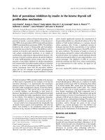
Figure I: Bcl-2 family members. Members of the Bcl-2 family are divided into three
groups- the pro-survival Bcl-2 group, and the pro-apoptotic Bax-like group and BH3-
only group. They are characterized by the existence of one to four of the conserved
Bcl-2 homology domains (BH1-BH4). Most of the members also possess a
hydrophobic region (TM) for membrane localization at the C-terminal.
BH4 BH3 TM BH1 BH2
BH3
BH3 TM
BH3 BH1 BH2 TM
Bcl-2,
Bcl-xL, A1,
Bcl-w
Bid
Pro
-
survival:
Pro-apoptotic:
BH3-only
group
Bcl-2 group
Bax-like group
Bax, Bak,
Bok
Bim, Bik,
Bad, Bmf,
Hrk, Noxa,
Puma
12
The Multidomain Pro-survival proteins.
The first member of this family, Bcl-2, is a proto-oncogene that was identified
at the chromosomal breakpoint between chromosomes 14 and 18, t(14;18) in human
follicular B-cell lymphomas (Tsujimoto et al. 1984a; Bakhshi et al. 1985; Tsujimoto
et al. 1985; Cleary et al. 1986; Tsujimoto and Croce 1986). Unlike other oncogenes at
that time, Bcl-2 expression did not promote tumorigenesis by inducing cell
proliferation. Instead its primary mode of action seemed to be the prevention of cell
death when challenged with various cytotoxic stimuli (Vaux et al. 1988; McDonnell
et al. 1989). In fact, these findings pushed the field of apoptosis center stage in cancer
research as it became clear that tumorigenesis is not merely due to excessive cell
proliferation but also impairment of apoptosis.
Members of the pro-survival group have been found localized mostly on the
membranes of cellular organelles like the mitochondrial membrane, the endoplasmic
reticulum (ER) and the nuclear envelope (Krajewski et al. 1993; de Jong et al. 1994).
Membrane targeting is most likely enabled by a highly hydrophobic transmembrane
(TM) region located at the carboxy-terminal, anchoring the protein to the membrane
on the cytoplasmic face (Nguyen et al. 1993; Kaufmann et al. 2003). Some members
of this group like Bcl-xL and Bcl-w, are found both in the cytosol and mitochondria
membrane. They reportedly associate tightly with the mitochondrial membrane upon
receiving apoptotic signal which triggers a conformational change (Hsu et al. 1997b;
Wilson-Annan et al. 2003). This localization of the pro-survival Bcl-2 family
members to mitochondria membrane following an apoptotic signal is consistence with
their main function which appears to be protecting the integrity of the outer
mitochondrial membrane thus preventing the release of cytochrome c and caspase
activation (Green and Reed 1998; Gross et al. 1999). The ability of pro-survival Bcl-2
13
members to heterodimerize with pro-apoptotic members appears to be important for
their anti-apoptotic properties (Yin et al. 1994; Sedlak et al. 1995). Crystallography
studies of some pro-survival Bcl-2 members have revealed similar core three-
dimensional structure among them (Muchmore et al. 1996; Petros et al. 2001; Petros
et al. 2004) that is important for their heterodimerization with other members of Bcl-2
family. Notably, the hydrophobic groove formed by residues from BH1, BH2 and
BH3 domain has been shown to be capable of binding an exposed BH3 α-helix of
pro-apoptotic Bcl-2 family members (Sattler et al. 1997). Heterodimerization of pro-
survival Bcl-2 members with pro-apoptotic members neutralizes them and prevents
their aggregation at the mitochondrial membrane; however the exact mechanism is
still contentious.
The Multidomain Pro-apoptotic proteins.
The multidomain pro-apoptotic members of Bcl-2 family, also sometimes
referred to as the “Bax-like” pro-apoptotic proteins, consists currently of Bax, Bak
and Bok. While Bax and Bak are widely distributed (Krajewski et al. 1994; Krajewski
et al. 1996); the less-known Bok expression is more limited to reproductive tissues
(Hsu et al. 1997a). Knockout studies in mice revealed that while Bax and Bak show
partial functional redundancy in many cell types, they are absolutely required for
inducing apoptosis triggered by various death stimuli via the mitochondrial pathway
(Lindsten et al. 2000; Wei et al. 2001; Zong et al. 2001). In healthy cells, Bax resides
in the cytosol as soluble monomeric protein while Bak is found on the outer
membrane of the mitochondria and endoplasmic reticulum in an inactive state (Wei et
al. 2000). Upon receiving apoptotic signals, Bax undergoes a conformational change
and translocates to the mitochondria where it forms homo-oligomers and inserts into