Roles of TACC related protein, mia1p in MTOCs and microtubule dynamics in schizosaccharomyces pombe 2
Bạn đang xem bản rút gọn của tài liệu. Xem và tải ngay bản đầy đủ của tài liệu tại đây (287.27 KB, 32 trang )
1
Chapter I Introduction
1.1 A general introduction to microtubules
1.1.1 Properties of microtubules
Microtubules mediate a wide variety of cellular functions. Chromosome
segregation requires assembly and function of the microtubule-based mitotic spindle (for
review, see Musacchio and Salmon, 2007); Establishment and maintenance of proper cell
morphology relies on microtubules to deposit cell growth components to precise sites (for
review, see Chang and Peter, 2003). Properties of microtubules allow them to
successfully perform these tasks. They consist of 13 protofilaments which associate
laterally to form hollow, cylindrical structures with ~25nm diameter. Each protofilament
is composed of α/β tubulin heterodimers that are longitudinally linked. However
microtubules are not static structures but are highly dynamic, undergoing repeated
transitions between growth and shrinkage, a phenomenon called dynamic instability (for
review, see Dammermann et al., 2003). In an in vitro situation, during polymerizing,
growing state, α/β-tubulin subunits are added to both ends of the microtubule with
different rates: the slow growing end terminated by α-tubulin is defined as “minus end”,
while the fast growing end capped by β-tubulin is termed “plus end” (for review, see
Dammermann et al., 2003). As tubulin heterodimers bind to the growing ends of
microtubules, β-tubulin guanosine triphosphate (GTP) within the heterodimer
subsequently hydrolyzes to guanosine diphosphate (GDP), leaving GDP bound form of
tubulin subunits in the lattice. Hydrolysis of GTP causes conformation changes in the
tubulin subunits but the resulting curved protofilaments are stabilized by the lateral bonds
within the microtubule wall, especially in the vicinity of the stable cap of GTP-containing
2
subunits at microtubule plus end. During shrinking state, the GTP cap is lost from the
plus end of microtubule and the unstable curved protofiliments peel back from the
microtubule wall. Eventually, the protofilaments disassemble into free tubulin
heterodimers (for text book, see Molecular Biology of the cell, Third Edition). The
dynamic instability of microtubules is shown on the following cartoon (for review, see
Wiese and Zheng, 2006).
However in an in vivo environment lacking a relatively high tubulin
concentration, microtubule nucleation requires another member of the tubulin family, γ-
tubulin. γ-tubulin is found in a large protein complex called γ-tubulin ring complex (γ-
TuRC) that is present both at the centrosome and non-centrosomal microtubule
3
nucleation sites to promote assembly of microtubules (Raynaud-Messina and Merdes,
2007)
1.1.2 Centrosome
The centrosome is thought to nucleate the majority of microtubules in many
animal cells. In the fluorescence microscopy analyses, the microtubules are seen in
greatest density around the nucleus and radiate out into the cell periphery during
interphase. This ordered microtubule network is organized by one key player—the
centrosome. Centrosome consists of a pair of centrioles surrounded by an amorphous
cloud of pericentriolar material (Bornens, 2002). Functions of centrioles within the
centrosome are still poorly understood, although centrioles are thought to serve as the
organizer of the pericentriolar material based on the observation of pericentriolar material
dispersion upon microinjection of antibody to disassemble the centrioles (Bobinnec et al.,
1998). Nevertheless, there is now little doubt that γ-tubulin ring complexes (γ-TuRCs),
identified within the pericentriolar material, are responsible for nucleating microtubules
(Bobinnec et al., 1998; Wiese and Zheng, 2006). The detailed function of γ-TuRC will be
discussed later in this thesis.
1.1.3 Non-centrosomal microtubule
Non-centrosomal microtubules generated by centrosome-independent
mechanisms are also found in neurons and epithelial cells as well as skeletal muscle cells
(Bartolini and Gundersen, 2006).
4
How the non-centrosomal microtubules are formed is poorly understood. Recent
data from studies of non-centrosomal microtubules propose that there are several general
mechanisms that cells might employ to nucleate non-centrosomal microtubules. Firstly,
existing microtubules may be released from the centrosome probably through action of
some severing proteins such as Katanin (for review, see Bartolini and Gundersen, 2006;
McNally and Vale, 1993). Secondly, microtubules are nucleated from the free γ-TuRCs
present in the cytoplasmic form (for review, see Bartolini and Gundersen, 2006). Thirdly,
microtubules can be nucleated from distinct non-centrosomal sites such as trans- Golgi
network in human cells (Efimov et al., 2007). Forth, existing microtubules could break
into two parts to generate free microtubule under mechanical stress (for review, see
Bartolini and Gundersen, 2006).
1.1.4 Schizosaccharomyces pombe (S. pombe) as a model for studying MT dynamics
The unicellular model organism, fission yeast Schizosaccharomyces pombe (S.
pombe) is an attractive model system to investigate various aspects of microtubule
dynamics. Straightforward genetic analyses and a fully sequenced and annotated genome
(Wood et al., 2002) are useful in dissecting molecular mechanisms of microtubule
organization. A relatively large cell size allows detailed dynamic observation of
cytoskeletal filaments and cellular components tagged with fluorescent proteins.
Importantly, fission yeast cells exhibit simple but distinct types of microtubule arrays
depending on the cell cycle stage.
So far four fission yeast tubulin genes have been identified: γ tubulin (gtb1/tug1),
two α tubulin (nda2, atb2) and a β tubulin (nda3) (Yanagida, 1987). Among these, α-
5
tubulin and β-tubulin are components of microtubules, while γ-tubulin, identified within
the microtubule organizing centers, acts as a microtubule nucleating template (for review,
see Hagan, 1998).
1.2 MTOCs and microtubule cytoskeleton in Schizosaccharomyces pombe (S. pombe)
The vegetative cell cycle of S. pombe consists of interphase (including G1, S and
G2 phases) and mitosis. To perform their functions at different cell stage, microtubules
are organized into complex arrays by MTOCs. In S. pombe cells, other than the spindle
pole body (SPB) which is equivalent to the mammalian centrosome, two other transient,
cell-cycle-regulated nucleation sites (defined here as non-centrosomal MTOCs) are
known: the interphase MTOCs (iMTOCs) and the equatorial MTOC (eMTOC) (Hagan
and Petersen, 2000; Tran et al., 2001).
1.2.1 Microtubule Nucleating sites—MTOCs in S. pombe
1.2.1.1 Spindle pole body (SPB)
The fission yeast spindle pole bodies (SPBs) are functionally analogous to
centrosomes and undergo a duplication and separation cycle, correlated with the cell
division cycle. Like the centrosome of vertebrate cells, the SPB of S. pombe spends most
of interphase in the cytoplasm, immediately next to the nuclear envelope (NE) (Ding et
al., 1997; Uzawa et al., 2004). Currently, it is still a controversial issue about the
duplicating time of SPBs: Some researchers think that it occurs in the late G2 phase as
shown in the following cartoon (from Ding et al., 1997). Another opinion is that SPBs are
duplicated at the G1/S boundary and matured in the G2 phase (Uzawa et al., 2004).
6
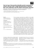
Unlike other vertebrate cells undergoing open mitosis in which the NE disassembles in
early prophase, fission yeast undergoes “closed mitosis”: The NE remains intact
throughout the cell cycle. To ensure the access of microtubules to the hereditary material,
a portion of the NE underlying the SPB pair breaks down to form an opening called
“fenestra” once the cell enters mitosis. Then the duplicated SPBs settle into fenestra to
nucleate intranuclear microtubules. During the elongation of the spindle, SPBs are always
localized at the leading edge of the NE. As anaphase proceeds, the nuclear fenestrae
close, and the SPBs are extruded back into the cytoplasm to nucleate interphase
microtubules. The summary of dynamics of the NE and the SPBs through out the cell
cycle is shown in the following cartoon (Ding et al., 1997).
7
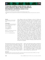
Diagram summarizing dynamics of the NE and the SPBs throughout the cell cycle.
The SPB (shaded ellipse with line) is associated with an appendage (small solid ellipse)
and lies close to the NE. Late in G2 phase, the SPB duplicates and matures, daughter
SPBs are connected by a bridge derived from the appendage. Upon mitotic entry, the NE
invaginates, perforates to form fenestra and the SPB settle into it. The two halves of the
structure separate as the spindle forms, such that each SPB occupies its own fenestra. At
the end of mitosis, the fenestrae close and extrude the SPB back into the cytoplasm for
the next interphase.
8
1.2.1.2 Non-centrosomal MTOCs
Compared to the SPBs which exist throughout the cell cycle, the iMTOCs and the
eMTOC are assembled at different cell cycle stages: The eMTOC localizes to the cell
center to nucleate the post anaphase array (PAA) at the end of mitosis (Venkatram et al.,
2005). On the other hand, the iMTOCs are only present in interphase cells and
disassemble once the cells enter mitosis. So far, all components found in the eMTOC are
also present in the iMTOCs. It is believed that disassembly of the eMTOC is synchronous
with the establishment of the iMTOCs (Zimmerman et al., 2004a).
1.2.1.2.1 Interphase microtubule organizing centers (iMTOCs)
The definition of iMTOCs is still largely controversial to date. Some researchers
refer to iMTOCs as “satellites” of γ-tubulin complex proteins (Janson et al., 2005). In my
opinion, the iMTOCs are the sites on the NE where interphase microtubules are
nucleated.
It is hard to observe the iMTOCs structures in live cells at steady state due to low
fluorescence intensity and distribution of γ-TuRC components along microtubule
bundles. However, after drug-induced microtubule depolymerization, γ-TuRC
components aggregate at microtubule nucleation sites and the iMTOCs can be observed
as several dots around the NE by tagging the components of γ-TuRC with green
fluorescent protein (GFP). Repolymerization of microtubules will be initiated from such
dots by washing out the depolymerizing agent, demonstrating the microtubule-nucleating
capability of the iMTOCs.
9
It is known that the establishment of the iMTOCs is linked to the disassembly of
the eMTOC (Zimmerman et al., 2004a), but the precise origin of the iMTOCs remains
elusive.
1.2.1.2.2 Equatorial microtubule organizing center (eMTOC)
At the end of anaphase, the mitotic spindle breaks down and microtubules
originate from the middle of the cell to create the PAA (Hagan and Hyams, 1988). The
PAA is nucleated from a distinct MTOC called the eMTOC. In fluorescence microscope
analyses, the eMTOC appear as a ring structure co-localizing with the actomyosin ring in
the middle of the dividing cell.
Assembly of the eMTOC requires the activity of a GTPase signaling cascade
known as the septation initiation network (SIN) that regulates the onset of cytokenesis
(Heitz et al., 2001). In addition, integrity of the eMTOC relies on the integrity of the F-
actin but not the microtubules: the dispersal of the eMTOC in the absence of the
actomyosin ring indicates the essential role of actomyosin ring in eMTOC formation
(Heitz et al., 2001). Recent studies found that the eMTOC breaks down into small pieces
defined as satellites that can move along the microtubules (Zimmerman et al., 2004a;
Sawin and Tran, 2006).
During the eMTOC-breakdown process, the DnaJ domain protein, Rsp1p,
functions in disassembly of the eMTOC, and possibly in assembly of the iMTOCs,
probably through interacting with the cytoplasmic hsp70 protein, Ssa1p (Zimmerman et
al., 2004a). It’s believed that Rsp1p may stimulate the ATPase activity of Ssa1p, which
then exerts an ATP-dependent conformational change on its substrate, causing the
10
protein-protein interaction to weaken and resulting in the breakdown of the eMTOC
(Zimmerman et al., 2004a). The establishment of the iMTOCs is believed to proceed
synchronously with the disassembly of the eMTOC since γ-TuRC components were
found to move from eMTOC to the iMTOCs as satellites when the eMTOC ring
constricts (Zimmerman et al., 2004a). Failure to disassemble the eMTOC in the rsp1-1
mutant leads to a defect in the organization of interphase cytoplasmic microtubules
(Zimmerman et al., 2004a).
1.2.1.2.3 γ-TuRC satellites
In addition to the MTOCs mentioned above, when fused with GFP, the γ-TuRC
components are also seen as weakly fluorescent dots traveling along the interphase
microtubules in a bi-directional manner (Zimmerman et al., 2004a). Such satellites are
capable of nucleating microtubules on the preexisted microtubule in interphase cells
(Janson et al., 2007).
1.2.2 Components of MTOCs
1.2.2.1 The γ-TuRC
Microtubule polymerization at the MTOCs requires the action of a large,
conserved multisubunit protein complex—γ-TuRC. Although there are two different
forms of the γ-tubulin complex present in higher eukaryotes, centrosomal γ-TuRC and
cytosolic γ-tubulin small complex (γ-TuSC), it seems that only the γ-TuRC possesses
microtubule nucleating capacity on its own in vitro [(Oegema et al., 1999)
11
In addition to γ-tubulin, identified as the key component of the MTOCs, two other
highly conserved proteins are found to be present in the γ-TuRC in all eukaryotes:
Spc97p and Spc98p in the budding yeast (Saccharomyces cerevisiae) or Gcp2p and
Gcp3p in human (Knop and Schiebel, 1997; Zimmerman et al., 2004b). Other
components of γ-TuRC vary in different species.
In fission yeast, the components of γ-TuRC include Tug1/ Gtb1 (γ-tubulin) (Horio
et al., 1991) together with Alp4p and Alp6p which are homologs of Spc97p and Spc98p
respectively. But unlike the S. cerevisiae γ-tubulin complex, additional members of the γ-
TuRC, Alp16p and Gfh1p, have been identified in S. pombe. Database searches revealed
that they share sequence similarity with regions of hGCP6 and hGCP4, respectively
(Fujita et al., 2002; Venkatram et al., 2004). These γ-TuRC components localize to the
SPBs both in interphase and mitotic cells and also on the iMTOCs and eMTOC (Vardy
and Toda, 2000; Fujita et al., 2002; Venkatram et al., 2004).
Among γ-TuRC components, the γ-tubulin, Alp4p and Alp6p are required for cell
viability and are essential for various aspects of microtubule functions. Since cells
lacking such components die due to defects in formation of mitotic bipolar spindles
(Tange et al., 2004), their functions in interphase microtubule organization are largely
founded on phenotypes of temperature-sensitive mutants (Vardy and Toda, 2000).
Another two γ-TuRC members—Alp16p and Ghf1p are not essential for cell viability.
They are required for the formation of normal interphase microtubules but have little
effect on the function of the mitotic spindle (Fujita et al., 2002; Venkatram et al., 2004).
This suggests that different members of the γ-TuRC play specific roles in microtubule
organization. However, defects in any of the γ-TuRC components typically exhibit
12
similar interphase phenotypes: reduced number of microtubule bundles; the microtubules
do not stop growing when their plus ends touch the cell tips, instead, they continue to
grow and produce longer microtubules which curve around the cell tips (Fujita et al.,
2002; Samejima et al., 2005).
How do γ-TuRC components in the minus ends of microtubules contribute to
microtubule dynamic is yet unclear. It is possible that in addition to the factors such as
Mal3p or Tip1p that directly binding to microtubule plus ends to prevent pre-matured
catastrophe at cell cortex until microtubules touch cell tips (Busch and Brunner, 2004),
other factors that regulating microtubule plus-end dynamic need to be loaded at the
microtubule minus ends during nucleation: One sample is kinesin Teap2p that is loaded
onto the microtubules in close proximity to the nucleus and then travels using its intrinsic
motor activity primarily at the tips of polymerizing microtubules (Browning et al., 2003;
Sawin and Tran, 2006). Also, the transforming acidic coiled-coil (TACC)-related protein,
Mia1p (or Alp7p), localizes at the SPB to recruit Alp14p (homologue of XMAP215p) to
stabilize microtubules (Sato et al., 2004). Alternatively, such changes could be due to the
disturbed balance between plus ends of microtubules and microtubule stabilizing proteins
and free tubulin subunits. Further investigations of γ-TuRC components should provide
new insights to this issue.
1.2.2.2 Mto1p and Mto2p
Recently, two other novel proteins have been identified as γ-TuRC components in
fission yeast—Mto1p (also known as Mod20p and Mbo1p) and Mto2p. These two
proteins physically interact with each other and can co-immunoprecipitate with γ-tubulin.
13
They are non-essential genes and neither of them are required for assembly of mitotic
spindle (Samejima et al., 2005). However, interphase microtubules are longer and curved
around the cell tips in both mto1Δ and mto2Δ cells, a typical phenotype of other γ-TuRC
mutants. The defects in nucleating cytoplasmic microtubules from non-spindle pole body
MTOCs—iMTOCs suggest that Mto1p and Mto2p function by recruiting the γ-tubulin
complex to non-spindle pole body MTOCs for microtubule nucleation (Sawin et al.,
2004). By database searching, several proteins (such as D. melanogaster centrosomin)
sharing similar sequence to Mto1p have been found in filamentous fungi and higher
eukaryotes suggesting that a general mechanism for the organization of noncentrosomal
MTOCs is conserved in eukaryotic cells (Sawin et al., 2004; Venkatram et al., 2005).
1.2.3 Organization of microtubule bundles
To organize an ordered microtubule network, a wide array of components is
required: Firstly, microtubule nucleators, MTOCs, need to maintain the right amount of
microtubules and localize them at precise sites. Secondly, crosslinkers are required to
bundle microtubules. Thirdly, motor proteins mediate microtubule sliding within the
microtubule bundles to facilitate the maintenance of microtubule polarity (Carazo-Salas
and Nurse, 2007).
In interphase S. pombe cells, in addition to microtubules nucleated by the SPBs
and the iMTOCs around the NE, new microtubules are also generated from the satellites
present on the preexisting microtubules (Janson et al., 2007). These microtubules are
transported to the overlap region around the NE where they are stabilized. This process
functions to maintain the overall antiparallel microtubule arrangement (Sawin and Tran,
14
2006). Recent work in microtubule dynamics found that at least two proteins are involved
in this process. One of them is the minus-end-directed protein—Klp2p.
1.2.3.1 Minus-end-directed motor protein—Klp2p
Klp2p is a kinesin of the KAR3 subfamily in fission yeast. In mitotic cells, it
localizes to kinetochores to regulate elongation of the anaphase spindle and to control
appropriate disassembly of the spindle at the completion of mitosis (Troxell, 2001).
During interphase, Klp2p localizes as numerous dots which accumulate only at plus ends
of both existing mother microtubules and newly nucleated daughter microtubules but is
absent from the minus ends of individual microtubules, suggesting that sliding forces
between overlapping microtubules are generated only at microtubule plus ends (Janson et
al., 2007), thus the plus end of a newly nucleated microtubule serves as a cargo of Klp2p
to be delivered to the minus end of mother microtubule.
In the absence of Klp2p, sliding of newly nucleated microtubules on pre-existing
microtubules is compromised, leading to defects in focusing of microtubules at the
overlapping regions (Carazo-Salas et al., 2005). This suggests that Klp2p functions as a
microtubule slider to transport newly nucleated microtubules to the overlapping region,
thus contributing to maintenance of the antiparallel linear microtubule arrays in
interphase cells.
1.2.3.2 Bundling protein—Ase1p
Another protein which functions in organizing antiparallel microtubule
arrangement is Ase1p. The yeast Ase1p belongs to the conserved ASE1/PRC1/MAP65
15
family of microtubule bundling proteins found at the mitotic spindle midzone (Chan et
al., 1999). Ase1p functions in both interphase and mitosis in S. pombe cells. During
mitosis, it localizes to mitotic spindle midzone where the plus ends of microtubule
overlap to stabilize bipolar spindle and prevents it from collapsing (Loiodice et al., 2005).
In interphase cells, Ase1p localizes at the regions where minus ends of microtubules
overlap, including regions around the MTOCs at the NE and overlapping regions of
mother-daughter microtubules when daughter microtubule is nucleated by the γ-TuRC
satellites on the mother microtubules. In ase1Δ mutants, interphase microtubules are
disorganized and fail to form overlapping antiparallel microtubule bundles (Loiodice et
al., 2005), suggesting that the role of Ase1p in S. pombe cells is to act as a microtubule
bundler.
1.2.3.3 Model of maintaining ordered microtubules
MTOCs concentrate microtubule nucleation, attachment and bundling factors
which coordinate each other in effectively organizing interphase microtubules into
ordered anti-parallel linear arrays. Recent studies in MTOCs discover more and more
components of the γ-TuRC. Their functions in organizing microtubules have been
characterized, providing more clues and leading to form a model of organizing the
microtubule bundles (Carazo-Salas and Nurse, 2007). Data published so far supported a
model in which interplay between bundling proteins—Ase1p and the minus-end directed
motor protein—Klp2p are sufficient to generate bipolar antiparallel microtubule array
(Sawin and Tran, 2006).
16
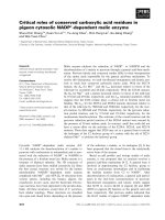
According to this model, once the γ-TuRC satellite has nucleated a new daughter
microtubule on a pre-existing mother microtubule, Ase1p and Klp2p will ensure that
mother and daughter microtubules are further organized into functional patterns. The
inherent ability of Ase1p allows it to bind only at microtubule antiparallel regions but
makes it absent from other regions on microtubule arrays. By forming oligomers, Ase1p
is capable of bringing two or more microtubule binding domains together, thus to bundle
and stabilize the antiparallel arrangement of daughter-mother microtubules (Janson et al.,
2007). Bundling activity of Ase1p is dynamic in order to allow microtubules remodeling
and sliding. Additionally, Klp2p is recruited to the growing plus-end tip of the daughter
microtubule, pulling it to move toward the cell center along mother microtubules. The
pulling forces generated by Klp2p effectively slide the daughter and mother microtubules
relative to each other (Sawin and Tran, 2006). However sliding forces are negatively
affected by the Ase1p bundling. Thus, as daughter microtubule continues to grow, more
Ase1p molecules are recruited to the increasing daughter-mother microtubule
overlapping region, causing sliding to attenuate. No further sliding occurs when Klp2p
reaches the end of the mother microtubule. In this way, microtubules are organized into
an ordered and functional array with minus-ends bundled together at the cell center and
plus-ends facing toward the cell tips. Model of maintaining ordered microtubules (from
Sawin and Tran, 2006) is shown in the following cartoon.
17
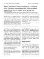
Model of interphase microtubules bundling in fission yeast. Model includes the
following steps: (1) The γ-TuRC satellites were recruited to the lattice of mother MTs to
nucleate daughter MTs. (2) Ase1p bundles and stabilizes the antiparallel arrangement of
daughter-mother MTs. (3) Klp2p is recruited to the plus-end of the daughter MT, where it
pulls the daughter MT toward the minus end of the mother MTs.
18
1.2.4 Arrangement of microtubule cytoskeleton in S. pombe
Since MTOCs alter their microtubule-nucleating activity in a cell-cycle specific
manner, the architecture of microtubule network is regulated dynamically, allowing it to
perform specific functions at different stages of the cell cycle.
1.2.4.1 Interphase microtubule cytoskeleton
1.2.4.1.1 Architecture and dynamics of interphase microtubule cytoskeleton
During interphase, 4-5 bundles of microtubules run along the long axis of a
cylindrical cell. One of them is attached to the SPB and the rest to the multiple iMTOCs
at the nuclear envelope. Thus the minus ends of microtubules are anchored / organized at
the MTOCs while the plus ends extend to the cell tips (Drummond and Cross, 2000; Tran
et al., 2001). Such an anti-parallel arrangement can be detected in the cells with α-tubulin
tagged with GFP under the fluorescence microscope: higher intensity of fluorescence
appears in the cell center where minus ends of microtubules overlap around the nuclear
envelope.
Interphase microtubules are highly dynamic. Generally, microtubules are
nucleated from the MTOCs on the nuclear envelope and continue to grow until they hit
the cell tip, pausing there for a while (Brunner and Nurse, 2000), then undergo
depolymerization. The transition from polymerization to depolymerization is defined as
“catastrophe”. Like in higher eukaryotic cell, such interphase dynamic microtubule arrays
play an important role in establishment and maintenance of cell polarity and overall
intracellular organization.
19
1.2.4.1.2. Functions of interphase microtubule cytoskeleton
1.2.4.1.2.1 Maintaining cell morphology
Fission yeast cells grow longitudinally by restricting the growth at two opposite
cell tips (Nurse, 1994). To maintain its rod-shape morphology, fission yeast has
developed a mechanism to position polarity factors at precise location. The role of a
dynamic microtubule array is to provide tracks for transporting those polarity factors to
the sites where they are needed (Chang and Peter, 2003). Polarity of microtubules is
required in this process: microtubule plus ends need to be facing the cell tips to ensure
polarized growth at the cell tips by depositing the kelch-repeat protein, Tea1p, at the cell
tips. There, Tea1p functions in polarity establishment by recruiting a complex, formin-
binding protein—Bud6p and the formin, For3p, to assemble actin cables (Feierbach et al.,
2004). Cells with abnormal microtubules often face difficulties in delivering the polarity
factors to the precise sites, causing misplaced growth and producing bent, branched, or T-
shaped cells (Beinhauer et al., 1997; Brunner and Nurse, 2000).
A model for establishment of cell polarity based on current knowledge has been
generated: the cell end marker, Tea1p, plays a critical role in maintaining cell
morphology by binding to the plus ends of growing interphase microtubules in a Tip1p
and Tea2p-dependent manner (Chang and Peter, 2003). When the growing microtubules
reach the cell tips and dwell there for a while, the microtubule plus end associated
proteins such as Tea1p are released to the cell tips (Behrens and Nurse, 2002). On the
other hand, a complex at the cell tips is waiting to dock the released Tea1p. One
candidate of this complex is a novel membrane-anchoring protein—Mod5p. This protein
contains a prenylation site (CaaX motif) responsible for its plasma membrane localization
20
(Snaith and Sawin, 2003). Coincident with microtubule shrinkage, Tea1p is released from
microtubules and stabilized at the plasma membrane in a Mod5p-dependnent manner.
Once in the cell tip, Tea1p functions to regulate cell polarity by recruiting other polarity
factors (Feierbach et al., 2004). This model for microtubule regulation of cell polarity in
fission yeast is shown in the following cartoon (Chang and Peter, 2003).
21
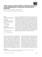
Role of microtubules in regulating cell polarity in fission yeast. a, Tea1p binds to the
growing plus end of MTs (green) in a Tip1p and Tea2p dependent manner. b, Tea1p are
released to the cell tip when MT has shrunk back. c, Tea1p recruits a large protein
complex of cell polarity factors to the cell tips. d, The complex includes For3p and
Bud6p, which function to assemble actin cables (red) at the cell tip.
22
1.2.4.1.2.2 Controlling central localization of nucleus
The interphase microtubules are also involved in positioning the nucleus at the
cell center, which in turn provides a critical cue for depositing the components of the cell
division plane later mitosis (Tran et al., 2001). Since minus ends of interphase
microtubules are attached to the nuclear envelope, transient forces are transduced back to
the nucleus where the minus ends of microtubules are anchored, hence pushing the
nucleus to move away from the cell tips (Tran et al., 2001). Owing notably to the
symmetrical longitudinally arrangement of microtubules in S. pombe cells, a balance of
the pushing forces is achieved by the polymerizing microtubules, centering the nucleus
between the two cell tips. Mutants with only one or two microtubule bundles often
exhibit misplaced a nucleus due to the reduced ability in maintaining the balance of the
pushing forces (as showed in my own data). As a consequence, the cell division plane in
these mutants is often misplaced (Sato et al., 2004; Zimmerman and Chang, 2005).
1.2.4.2 Mitotic microtubule cytoskeleton
1.2.4.2.1 Architecture of mitotic microtubule cytoskeleton
The interphase microtubules together with large iMTOCs are disassembled once
cells enter mitosis. The duplicated SPBs separate and nucleate microtubules to form
mitotic spindle, which is composed of three kinds of microtubules: kinetochore
microtubules that emanate from SPBs to capture kinetochores, polar microtubules from
opposite SPBs that overlap each other in the cell center, and astral microtubules that
extend outward from the SPBs to the cell periphery (Ding et al., 1993).
23
To ensure faithful segregation of chromosomes, the kinetochore microtubules
capture the kinetochores and eventually move the chromosomes to the metaphase plate
(McCully and Robinow, 1971; Uzawa and Yanagida, 1992). Subsequently they shorten
and pull the separated chromosomes to the poles. The polar microtubules then extend and
slide apart from each other at the anti-parallel region to separate the genomes (for text
book, see the molecular biology of Schizosaccharomyces pombe; Uzawa and Yanagida,
1992). At the end of anaphase, the mitotic spindle disassembles and the eMTOC appears
at the medial cell division site to nucleate microtubules that form the PAA (Hagan, 1998).
Microtubules are symmetrical at both side of the eMTOC with their plus ends reaching
toward cell tips in two radial arrays. Eventually, as the eMTOC constricts, the nucleation
sites of the PAA fuse together, causing microtubules to divide into two parts to each
daughter cell (Zimmerman et al., 2004a).
1.2.4.2.2 Functions of mitotic microtubule cytoskeleton
The function of mitotic spindle is to ensure efficient and high-fidelity
chromosome segregation, which requires the concerted effort with the spindle assembly
checkpoint machinery (Zhou et al., 2002). Before the transit from metaphase to anaphase,
chromosomes need to be lined up in the center of the cell with both sister kinetochores
attached by the microtubules from the two opposite SPBs (Zhou et al., 2002). As the
spindle elongates, DNA masses are equally separated into two daughter cells. If the
microtubules within the spindle fail to attach to the kinetochores, a component of the
spindle check-point, Mad2p, will localize to the unattached kinetochore to prevent
downstream protein separase from cleaving cohesin links between sister chromotins, thus
24
arresting cells in metaphase (He et al., 1997). Therefore, the order and the timing of
dynamic changes in chromosomal morphology and behavior must be tightly controlled to
coincide with the cytoskeletal changes, ensuring the faithful distribution of genome in
both daughter cells.
Changes in microtubule distribution and MTOCs during cell cycle progression in
S. pombe are summarized in the following cartoon.
25
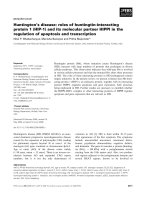
Model of MTs and MTOCs organization throughout the cell cycle in fission yeast.
During interphase, 4-5 MT bundles ran along the long axis of cells. One of them is
nucleated by the SPB, the rest are nucleated from the iMTOCs. Upon mitotic entry, the
interphase MT bundles disassemble and two SPBs (one of them is duplicated in
interphase) separate to form the mitotic spindle, then spindle elongates. At the end of
mitosis, the eMTOC appear in the middle of cell to nucleate the PAA. As the eMTOC
constricts with the actomyosin ring, it breakdown to small pieces called satellites that can
move along the MTs. Subsequently, cell divides into two daughter cells.
*
*
*
*
*
*
*
*
*
*
iMTO
SPB
eMTOC
satellite