Cancer cell migration in 3d collagen gel matrix system
Bạn đang xem bản rút gọn của tài liệu. Xem và tải ngay bản đầy đủ của tài liệu tại đây (4.26 MB, 160 trang )
CANCER CELL MIGRATION IN 3D COLLAGEN
GEL MATRIX SYSTEM
SUN WEI
(B.Eng. Tsinghua Univ, Beijing)
(MSc, NUS)
A THESIS SUBMITTED
FOR THE DEGREE OF DOCTOR OF PHILOSOPHY
NATIONAL UNIVERSITY OF SINGAPORE
2010
i
Acknowledgements
My sincere thanks must first go to my supervisor, Professor Lim Chwee Teck. He is not only
my academic advisor, but also a role model for us – a young generation of engineers who are
determined to pursue a career out of the conventional engineering domain, by solving long
standing questions in life sciences and medical sciences with engineering approaches or by
adding to the human’s understanding of lives and diseases from new perspectives. Although he
never advocated with words, his achievements greatly inspired me when I made up my mind to
switch my career, otherwise I will probably be designing an energy system for a building right
at this moment. I believe he as a good example will attract more and more young people with
engineering background to contribute to improving the health of mankind. He first directed me
to this topic – studying cancer cell migration in 3D – in Feb 2007. Through guiding me to read
relevant literatures, introducing me to the group leading the field, Professor Lim opened the
door to a great new world for me and brought me the compass. He is very supportive while I
went through the period of protocol optimization, through generously funding the reagents,
consumables, microscope and accessories and also investing in a very expensive software
package for image analysis. I had never ever worried about purchasing anything for the
experiments throughout the 4 years, all thanks to his great support. As the research progressed,
he went through the difficulties, unforeseeable adversaries together with me, keeping me
encouraged all the time. To solve technical problems, we had many discussions within the
group and also with scientists of similar interests, who offered constructive advice. To my
surprise, he is not only making his best effort, but also engaging his friends to make their best
effort to help my research. When I was confused about choosing the direction, he helped me to
set realistic goals. His insight and detailed guidance ensured that the research is always on the
right track and the project is making progresses.
ii
All in all, without Professor Lim as my supervisor, I will not be able to take this endeavour to
explore cancer metastasis, not able to find the right paths, to proceed and complete the project,
never mention presenting this thesis to all.
I am also deeply indebted to my co-supervisor Professor Raj Rajagopalan. Among the
committee who interviewed me for admission into NGS, it is his support on me that made
everything possible. Ever since joining his research group in the second year of my study, he
treated me same as the graduate students that he guides as main supervisor. On the project
requiring collaboration between Nicholas and me, he invested tremendous time and energy to
guide us. His foresight of frontier science, his wisdom, enthusiasm and intelligence guided my
research like a lighthouse. Whenever I had difficulties in anything during the study, I would
like to turn to him for advice because I know he is kind and ready to help me. He always
outlined the tasks and dissected them into detailed steps which were in turn explained in clarity
with his experience. As a result, I often came into his office confused, with fear over the
challenges, and left his office with confidence, courage, and sometimes a big smile – thanks to
his humour. He really cares about the development of his graduate students and gives us career
guide from time to time. I benefitted from Prof Raj’s supervision a great deal and really feel that
he is a great academic father to me. All in all, I feel lucky to have good supervisors in the years
working towards a PhD.
Besides, I am very grateful to all the past and current lab officers: Ms Tan Phay Shing, Eunice,
Mr Hairul Nizam Bin Ramli and Ms Charlene Wang (Prof Seeram Ramakrishna’s Laboratory)
for their good maintenance to the laboratory and their timely help throughout the course of my
PhD study.
Since I became a member of NanoBiomechanics Lab four years ago, I often feel lucky to have
a wonderful lab environment. In this friendly and conducive lab, I am able to conduct
experiments smoothly, to have my bench work time more enjoyable, my everyday life filled
with fun. My gratitude goes to everyone of my lab friends, for being kind colleagues to work
iii
with, for offering me help whenever requested, sharing your knowledge and experience to me
without reservation, encouraging me in many ways and sharing countless cheerful occasions
with me.
I appreciate being a student of NUS Graduate School for Integrative Sciences & Engineering
(NGS), the privileged graduate school in Singapore, for a few reasons. NGS offers me not only
generous funding, but also a great opportunity to build up the background and carry out
interdisciplinary research, because we are empowered by the innovative curriculum setting and
the encouraging environment in NGS. The community of NGS did take care of each of us
postgraduate students with attention. I also benefitted from support of the NGS office, for
which I can never forget the kind assistance and guidance from Ms. Ivy Wee, Ms. Irene Chuan
and Ms Neo Cheng Bee and many others, during the course of my stay in NGS.
Last, but most importantly, I would also like to thank my family for their love and
understanding throughout. My husband, parents and parents-in-law all supported my study
whole-heartedly. Their encouragement and caring kept me accompanied all the time despite
that I was thousands of miles away. Without my family standing behind, it would not be
possible for me to start and complete the PhD study.
iv
Table of Contents
Acknowledgements i
Table of Contents iv
Summary vii
List of Tables viii
List of Figures ix
List of Symbols xvi
Chapter 1: Introduction 1
1.1. The significance of cancer metastasis research 1
1.1.1.
The urgency of cancer research 1
1.1.2.
Cancer metastasis research 2
1.2.
Quantitative in vitro 3D cell migration assays 4
1.2.1.
Values of in vitro drug assays 4
1.2.2.
Advantages of 3D cell-based drug assay 5
1.2.3.
Fundamentals of cell migration 5
1.2.4.
Advantages of 3D cell invasive migration assays 8
1.2.5.
Summary 11
1.3.
Tissue mechanical factors in 3D cell migration assays 12
1.4.
Objective 13
1.5.
Scope and structure of the thesis 13
Chapter 2: Literature Review 15
2.1. In vitro 2D and 3D cancer metastasis studies 15
2.1.1. Tracking 2D cell migration 16
2.1.2.
Micro-pore-based cell migration/ invasion assays 21
2.1.3.
3D cell migration models 24
2.1.4.
Discussion on cell migration assays 31
2.2.
General anti-cancer drug assays in 3D 33
2.2.1.
Introduction 33
2.2.2.
Background: why 2D cell culture is insufficient for anti-cancer drug testing 34
v
2.2.3. Techniques in 3D anti-cancer drug assays 36
2.2.4.
Specific research questions 39
2.2.5.
Comparing 3D substrates with different structures / properties 45
2.2.6.
Summary 46
2.3.
3D anti-migratory drug assays 47
2.4.
Summary 49
Chapter 3: Materials and Methods 50
3.1. Malignant breast cancer cells 50
3.2.
3D collagen model for cell invasion 51
3.3.
Structure and mechanical property of collagen gel 53
3.3.1.
Mechanical characterization 53
3.3.2.
Scanning electron microscopy 55
3.4.
Microscopy methods for cell migration study 56
3.4.1.
Imaging and tracking cells 56
3.4.2.
Cell track data analysis 59
3.4.3.
Statistical methods 61
3.5.
Summary of the chapter 63
Chapter 4: Mechanical Modulation of the Collagen Gel Matrix 64
4.1. Modulation of bulk elasticity of the gel 64
4.2.
Non-linear elasticity of collagen 66
4.3. Micro-architecture of collagen matrices 69
4.3.1.
Dependence of collagen fibril thickness on polymerization pH 70
4.3.2.
Pore-size of the collagen network 73
4.4.
Discussion 76
Chapter 5: 3D Cell Migration and Drug Effects 79
5.1. Innate cell migration 79
5.2.
MMP inhibition by GM6001 89
5.3.
ROCK-inhibition 96
5.4.
Actin destabilization 104
5.5.
Microtubules destabilization 111
vi
5.6. Discussions on drug effects 118
Chapter 6: Conclusions and Future Work 122
6.1. Conclusions 122
6.2.
Further modulation of the ECM environment 125
References 127
Appendix A. The Time Dependence of Cell Motility 138
Appendix B. Results of Statistical Tests 140
vii
Summary
Cancer has long been one of the leading causes of death in the industrial world. The main
reason for its high mortality is due to inefficient early detection which results in the spread of
cancer cells to other distant sites in the body, via a process known as metastasis. Metastasis
causes the dissemination of cancer cells and accounts for 90% of cancer-induced deaths, thus
requiring better therapeutic treatments. The thesis examines a functional assay to study cancer
cell invasive migration inside a three-dimensional (3D) collagen-I hydrogel, focusing on its
micro-architecture and mechanics.
First part of the project is the tuning of 3D collagen hydrogel to achieve varying fibre network
structure and gel mechanical properties. Embedded in such collagen gels, breast cancer cells
were tracked using live confocal imaging followed by quantitative 3D cell track analysis.
The second part evaluated drug effects on cell migration using the model. Pharmacological
inhibitions of cell-activated collagen matrix degradation and cytoskeletal reorganization
reduced cell movement speed and directionality. Interestingly, the drug effects depended on the
matrix micro-structure and elasticity.
The study found that tissue mechanics is important in anti-migratory cancer drug assays, and
incorporated ECM factors in interpreting results of drug assays.
viii
List of Tables
Table 2.1. In vitro cell migration assays 16
Table 2.2 Categorizing 3D cell migration assay 25
Table 4.1. The list of parameter values in the exponential models that G’ data were fitted with.
68
Table 5.1. Case index of migration assays. 79
Table 5.2. Effects of GM6001 on cell movement speed and track straightness. 91
Table 5.3. Effects of Y27632 on cell movement speed and track straightness. 99
Table 5.4. Effects of CytoD on cell movement speed and track straightness. 106
Table 5.5. Effects of nocodazole on cell movement speed and track straightness. 113
Table 5.6. Recommended collagen-I gel mechanical property for drug assay designs 120
Table B.1. Comparing cell speed in the five control conditions; p-values obtained from K-S
tests. 141
Table B.2. Comparing cell straightness in the five control conditions; p-values obtained from
K-S tests. 141
ix
List of Figures
Figure 1.1. A schematic diagram showing different stages in cancer metastasis, during which
malignant cells spread from a primary tumour, get transported and finally
residing at a distant site. Adapted with permission from Elsevier Ltd. (Lee and
Lim 2007). 3
Figure 1.2. In connective tissues, collagen fibres are in irregular orientations and form
networks of different densities. (A) Loose connective tissue of mesentery,
which is the membrane-like lining of the abdominal cavity, showing collagen
fibres (arrow heads), elastic fibres (small, filled arrows) and fibroblasts
nucleus (unfilled arrows)(stained for elastin, x40). (B) Irregular, dense
connective tissue of nipple skin (hematoxylin and eosin staining, x20). Cell
nucleus are the dark dots. Collagen fibres (light gray) are oriented irregularly
but are densely packed. Glycoproteins and ground substances are lost during
dehydration, so they are not visible in the stained samples. Source: Blue
Histology on Web. School of Anatomy and Human Biology, University of
Western Australia. URL:
6
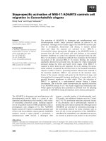
Figure 1.3. Scanning electron micrograph. (A) Schematic view of MDA-MB-231 cells
invading through type-I collagen fibres. Pericellular proteolysis mediated by
MT1-MMP allows tumour cells to remodel the matrix, supporting invasive
migration through the 3D fibrillar collagen network. (B) The higher
magnification image of the area in white, dotted box in (A). Cell is
pseudo-coloured in red and matrices in blue. The anterior part of the long
lamellipodial extension is covered by numerous small finger-like protrusions
that might correspond to sites of pericellular proteolysis and matrix
remodelling. Cells were plated for 6 hours on 3 mg/ml acid-extracted type-I
collagen before fixation for scanning electron microscopy. The figure is
adapted with permission from The Company of Biologists (Poincloux et al.
2009). 8
Figure 2.1. (A) Scratch wound assay: in a typical monolayer wound assay, a wound is created
in a confluent monolayer of cells at time = 0 (t0). Cell migration is then
evaluated at the end-point of the assay (t1), either by determining the speed
that the edges of the wound approach each other, or by counting the number of
cells observed in between the two lines that indicate the wound borders, as
determined at t0. The small arrow shows a cell in a mitotic state. (B) Ring
assay: illustration of the radial migration of cells from an initial cell cluster
(indicated by the circle) to the migration front (arrow), a distance which is the
maximum linear distance covered by the cells in the direction of the arrow.
Adapted with permission from Wiley Periodicals, Inc. (Decaestecker et al.
2007). 18
Figure 2.2. A µ-slide chemotaxis device. Principle (A), 3D view of the device (B). Here, two
reservoirs of media with different levels of chemoattractant (the higher
concentration is indicated by dots) are connected by a narrow observation area.
A concentration gradient is created along the observation area. Cell migration
in this area will be monitored through live microscopy. The figure is adopted
from
http://www. biophysics.com/ibidi/USLIDES/chemotaxisUSLIDE.html.19
Figure 2.3. Configuration and application of Dunn chamber. The Dunn chamber is shaped
with two concentric circular deep wells separated by a ring-shaped bridge that
is as shallow as 20 μm and is located below the top of the chamber (A). During
experiment, the inner well is filled with a control medium and the outer with a
medium plus a potential chemoattractant. The capillary effect will induce and
x
maintain a linear diffusion gradient along the radial direction of the
ring-shaped bridge. A square coverslip with cells adhered during culture is
inverted and put on top of the wells, with the periphery sealed with wax to
form a closed space. Microscopy imaging will record cell migration in the area
directly on top of the ring-shaped platform (B). Rose-plots in (C) and (D) are
examples of cell behaviour in random migration and directional chemotaxis
(with chemoattractant at the top of the plot) respectively. The length of the bar
at each direction in the rose-plots represents the number of cells migrating in
that particular direction. The arrow in (D) indicates the migration direction that
is significantly different from the others. The 3D view of a Dunn chamber
(E)
/>y/dunn/methods
20
. (A) – (D) are from (Eccles et al. 2005), adapted with
permission from Elsevier, Ltd
Figure 2.4. Schematic view of Boyden Chambers for chemokinesis, chemotaxis, haptotaxis
and invasion assays. Chemokinesis (A) measures the random migration
without gradient of chemoattractant between the lower and upper wells, where
there is also no difference in terms of ECM protein coating on the lower and
upper surface of the porous membrane; haptotaxis (B) is to evaluate directional
migration when only underside of the membrane is coated with ECM proteins,
which may function as a solid phase stimulant for migration; cell directional
migration under chemotaxis (C) is enabled with concentration gradient of
soluble factors between the two wells; a thick layer of ECM proteins or cell
monolayer on top of the membrane converted the assay into invasion or
transmigration assay (D). Adapted with permission from Elsevier, Ltd. (Eccles
et al. 2005). 22
Figure 2.5. 3D matrix invasion assays, with cells on top at the starting point (A); matrix
invasion combined with chemotaxis (B) and co-culture invasion assay, while
matrix layer pre-cultured with stromal host cells such as fibroblasts (C).
Adapted with permission from Elsevier, Ltd. (Eccles et al. 2005). 27
Figure 2.6. Schematic model of 3D gel invasion assays: top-down invasion (A): radial
invasion (B). Images from left to right suggest time-progress. Adapted with
permission from Wiley Periodicals, Inc. (Decaestecker et al. 2007). 28
Figure 2.7. Diagram of paired-nested collagen invasion, showing experiment configuration as
well as the collagen fibres alignment resulted from cell contraction. Adapted
with permission from Elsevier, Ltd. (Provenzano et al. 2008). 30
Figure 2.8. Schematic presentation of the similarities of a tumour (in vivo) and a multilayered
postconfluent cell culture and spheroids (in vitro) Adapted with permission
from Elsevier Ltd. (Padron et al. 2000). 36
Figure 2.9. Diagram showing the parameters for malignancy and drug effect evaluation.
Altered protein expression/ transcripts level, enzyme activity, cell viability,
morphology, proliferation, migration, and tissue morphogenesis as well as
angiogenesis are all parameters of interest for the evaluation of malignancy
manifestation. Effectiveness of drugs is evaluated not only by the above
parameters, but also through the time-dependent levels of drug uptake,
retention and metabolites. The selection of endpoint indices depends on
objectives of the study and specific targets of the drugs. 38
Figure 2.10. A drug-resistance variants of EMT-6 mammary tumour cells derived from mice
formed condensed spherical aggregates with a necrotic core and viable rims in
agarose gel, under the treatment of the same drug (eg., cyclophosphamide,
cis-diamminedichloroplatinum) that these cells are raised to resist, whereas
xi
parental EMT-6 cells formed loose cell aggregates of smaller sizes (Kobayashi
et al. 1993). Copyright (1993) National Academy of Sciences, U.S.A. 40
Figure 2.11. 3D endothelial network structures formed in flexible (A, C) and rigid (B, D)
collagen-I gels at day 7 of culture. Confocal images were recorded starting
from cells on the gel surface downwards at 5 µm intervals. In (A, B) the
images of the 3D networks were reconstructed using images taken at
depths>15 mm to avoid the cell monolayer. (C, D) were lateral views of the
3D networks. Scale bars, 100 µm. Adapted with permission from Mary Ann
Liebert, Inc.(Yamamura et al. 2007). 43
Figure 3.1. In the central well, the dotted region is the bead-gel, seeded with cells. On top of it,
the dome-shaped region is initially cell-free but susceptible to cell invasion. . 52
Figure 3.2. AR-G2 rheometer. Appearance (A) and plate-plate configuration & schematic
working mechanism (B). Courtesy of TA Instruments 2009. 54
Figure 3.3. Phase contrast image of cell distribution in a nested collagen gel after 12 days,
showing cells spreading out from the inner-gel (the dashed line enclosed area)
to the surrounding, originally cell-free outer gel (scale bar = 200 µm) both of
which were grafted in glass bottom well. Brighter areas are occupied by cells,
while the darker regions on the peripheral indicate the collagen gel region that
is cell-free. Each rectangle represents a field-of-view during confocal imaging.
The arrows point to the dominant directions of cell movement in the respective
rectangular field-of-view 57
Figure 3.4. Imaris
®
59
obtained cell tracks. Red: cells. Track colours indicate the time point: as
shown in the “Time” scale, dark purple labels the start of the entire period of
tracking, while bright yellow corresponds to the last time points.
Figure 3.5. A schematic plot of cell track. Euclidian Distance (ED) is the length of the vector
directly linking start and end of the cell paths, whereas Accumulative Distance
(AD) is calculated by adding up the length of cell movement vector at each
time step. 59
Figure 4.1. (A) Storage modulus G’; (B) loss modulus G”; (C) phase angle δ (tan δ = G”/G’),
obtained from the frequency sweep in rheometer measurement, with three
concentrations of collagen-I gel prepared in the three pH conditions as
indicated in the legend. It was measured at a constant strain of 0.1, and the
angular frequency ranged from 2π ×10
-3
65
to 100 rad/s. relaxation time prior to
measurement was selected to be 40 min.
Figure 4.2. Strain-stiffening effect, as indicated by storage modulus G’, under the rotational
measurement at an angular frequency of 1 rad/s. (A) When the gel
polymerized under neutral condition, strain-stiffening effect was more
pronounced comparing to that under a slightly acidic condition. (B)
Strain-stiffening effect was even more pronounced when the gel polymerized
at a slightly basic condition, since curve “pH9 - 2.5 mg/ml” is below curve
“pH7 - 4 mg/ml” when the strain is less than 10%, but it exceeded the latter
when the strain is above 10%. (C) For gels prepared at the same pH condition
but with different levels of collagen-I concentration, G’ curves show similar
shapes. 67
Figure 4.3. The strain levels when strain-stiffening effect starts. 68
Figure 4.4. SEM images of 2.5 mg/ml collagen polymerized at a range of pH conditions. (A)
pH6. There are some thicker and long fibres (yellow arrow head). (B) pH7.
Fibres are of intermediate thickness and formed a randomly organized network.
(C) pH9. Thin fibres formed a network of high mesh density. 72
xii
Figure 4.5. Histograms of fibre thickness distribution, corresponding to samples obtained at
collagen concentration = 2.5 mg/ml and pH= 6, 7, 9, respectively. 72
Figure 4.6. Effects of pH on average fibre diameters (bars indicate standard deviations),
which decreased as pH increased. Collagen concentration = 2.5 mg/ml. The
three conditions differ from each other significantly (*: p< 0.001). 73
Figure 4.7. SEM images showing collagen micro-structure. Magnification = 20k. Samples
were prepared with 3 levels of collagen concentration and 3 pH conditions
during polymerization, i.e., (A) pH7 - 1.5 mg/ml, (B) pH7 - 2.5 mg/ml, (C)
pH7 - 4 mg/ml, (D) pH6 - 2.5 mg/ml, (E) pH9 - 2.5 mg/ml. 74
Figure 4.8. (A) Histograms of the pore-size distribution of 1.5, 2.5 or 4.0 mg/ml collagen
matrix polymerized at pH7. (B) Enlarged section of histogram corresponding
to pore-size above 1 µm. Only the proportions of large pores varied with gel
concentration. Feret’s diameter of each pore was measured from segmented
SEM images, and is determined as the diameter of the smallest circle that can
contain each pore. 75
Figure 4.9. (A) Histograms of the pore-size distribution of 2.5 mg/ml collagen matrix
polymerized at pH6, 7 and 9, respectively. (B) Enlarged section of histogram
corresponding to pore-size above 1 µm. Only the proportions of large pores
varied with gel polymerization conditions. Feret’s diameter of each pore was
measured from segmented SEM images, and is determined as the diameter of
the smallest circle that can contain each pore. 76
Figure 5. 1. Statistics of cell speed in control conditions. Data from assays performed in
identical ECM conditions were clustered together for analysis. For example,
pH9-2.5 mg/ml is the collection of all control tests using gels of 2.5 mg/ml
collagen polymerized at pH9, including 1c, 6c, 11c and 16c in Table 5.1. (A)
The overall distribution of cell speed data. Relative frequency distribution
histograms show the proportion of cell tracks with a certain track-average
speed. The legend encodes polymerization pH and collagen concentration in
mg/ml, referring to cases with different collagen protein concentrations, or
with a constant 2.5 mg/ml collagen concentration but polymerized under
different pH conditions. Most curves differ from one condition to another,
except for the pair “pH7-1.5 mg/ml – pH6-2.5 mg/ml”. (B) Cell speed box plot.
The crosses inside the diamonds indicate the mean of each condition, while
stars and “×” signs mark the positions of 99 percentage and 1 percentage data,
respectively. The dots above the stars and below the “×” signs are the values of
the highest 1% and the lowest 1% data, respectively. The same applies to all
the following box plots for cell speed. The dotted curve suggests 99% of cell
speeds are negatively correlated to collagen gel stiffness. (C) Cell speed mean
and median plot. The error bars for the means indicate population standard
deviations. The trend-line suggests slight decrease of mean cell speed along
with elastic modulus. Hereinafter, “linear elastic modulus” refers to the G’
measured in the linear range. (D) The 99th percentile value of cell speed vs.
gel elastic modulus. The trend-line suggests the fastest migrating cells
decreased speed in stiffer gels. 84
Figure 5. 2. Statistics of cell directionality in control conditions. Data were collected with the
same methods as that in Figure 5. 1. (A) Distribution histograms of track
straightness index (as defined in Chapter 3.4.3) obtained from cases with
different collagen protein concentrations, or with a constant 2.5 mg/ml
collagen concentration but polymerized under different pH conditions. (B) Cell
track straightness index (0 – 1) box plot. The crosses indicate the means, while
stars and “×” signs mark the positions of 99 percentage and 1 percentage data,
respectively. The same applies to all the following box plots for cell track
xiii
straightness. The dotted curve suggests that 99 percentage of cell track
straightness data are negatively correlated to collagen gel stiffness. (C) The
mean and median of cell track straightness index. The trend-line suggests a
biphasic dependence of straightness on gel elastic modulus. (D) The 99th
percentile value of cell track straightness vs. gel elastic modulus. The
trend-line suggests that straightness decreased with gel elastic modulus,
particularly in stiffer gels. 87
Figure 5. 3. Cell morphology in pH9 - 2.5 mg/mL collagen gel, before (A) or after (B)
GM6001 treatment 91
Figure 5. 4. Paths of cell displacement (μm) over 8 hours in 3D collagen gel. X-Y
projections of cell tracks are in the top panel and X-Z projections are in the
bottom panel. The tracks of a population of cells (n>50) were adjusted to start
from the origin in the X-Y or X-Z coordinate system. Dotted lines: tracks of
cells in control conditions; Solid lines: cell tracks under drug treatment. The
arrow indicates the outer gel storage modulus decreased from 80 Pa for the
pH7 - 4.0 mg/ml gels to 8 Pa for the pH7 - 1.5 mg/ml gels.
92
Figure 5. 5. Cell speed: GM6001 treated cells compared with control. (A – E) shows the
histograms of relative frequency distributions. The height of each column
indicates the proportion of cell tracks of a certain track-average speed. The
legend encodes polymerization pH and collagen concentration in mg/ml. “cntr”
means control. (F) shows the box plots of cell track-speed, with (diamond) or
without (rectangle) 25 µM of GM6001 in the media. (G) plots the relative
change of mean and median speed of cells treated with GM6001 as compared
to the controls. 95
Figure 5. 6. (A) The box plots of cell track straightness, with or without 25 µM of GM6001 in
the media. (B) There is no clear trend showing dependence of the drug-induced
change of mean and median straightness on gel elastic modulus. 96
Figure 5. 7.Cell morphology in 2.5 mg/ml collagen gel polymerized at pH6 (A. control; B.
Y-27632-treated; C. Cytochalasin D -treated; D. nocodazole-treated). Scale
bars = 50 µm. 98
Figure 5. 8. Paths of cell displacement (μm) over 5 hours in 3D collagen gel. X-Y
projections of cell tracks are in the top panel and X-Z projections are in the
bottom panel. The tracks of a population of cells (n>50) were adjusted to start
from the origin in the X-Y or X-Z coordinate system. Dotted lines: tracks of
cells in control conditions; Solid lines: cell tracks under drug treatment. 99
Figure 5. 9. Cell speed, Y-27632 treated cells compared to the control. (A – E) show the
histograms of relative frequency distributions. The height of each column
indicates the proportion of cell tracks of a certain track-average speed. The
legend encodes polymerization pH and collagen concentration in mg/ml. “cntr”
means the control case. (F) shows the box plots of cell track-speed, with or
without 20 µM of Y-27632 in the media. (G) plots the relative change of mean
and median speed of cells treated with Y-27632, as compared to the controls. It
shows all conditions lead to over 30% reduction of cell speed, although softer
gels yielded even larger decrease. (H) Speed of the fastest cells, plotting 99
th
103
percentile cell speed vs. gel elastic modulus, showing the same trend with that
in chart (G). (I) compares the average cell speed with the strain value for
strain-stiffening to occur.
Figure 5. 10. (A) The box plots of cell track straightness, with or without 20 µM of Y-27632
in the media. (B) There are more significant drug-induced changes of mean
and median straightness in softer or stiffer gels. (C) The 99
th
104
percentile
straightness level vs. gel elastic modulus, showing the same trend with B.
xiv
Figure 5. 11. Examples of 3D cell tracks during 8 hours of migration in pH7-2.5 mg/ml
collagen gel, (A) before and (B) after CytoD treatment. Red: cells. Tracks are
colour-coded for time. 105
Figure 5. 12. Paths of cell displacement (μm) over 8 hours in 3D collagen gel. X-Y
projections of cell tracks are in the top panel and X-Z projections are in the
bottom panel. The tracks of a population of cells (n>50) were adjusted to start
from the origin in the X-Y or X-Z coordinate system. Dotted lines: tracks of
cells in control conditions; Solid lines: cell tracks under drug treatment. 106
Figure 5. 13. Cell speed, CytoD-treated cells compared to the control. (A – E) shows the
histograms of relative frequency distributions. The height of each column
indicates the proportion of cell tracks of a certain track-average speed. The
legend encodes polymerization pH and collagen concentration in mg/ml. “cntr”
means the control case. (F) shows the box plots of cell track-speed, with or
without 2 µM of CytoD in the media. (G) reveals that mean and median cell
speed decreased more significantly in softer gels. 110
Figure 5. 14. (A) The box plots of cell track straightness, with or without 2 µM of CytoD in
the media. (B) The relation between drug-induced change in straightness and
gel elastic modulus is complex. The decrease of straightness is significant in
stiffer gels. 111
Figure 5. 15. Paths of cell displacement (μm) over 6 hours in 3D collagen gel. X-Y
projections of cell tracks are in the top panel and X-Z projections are in the
bottom panel. The tracks of a population of cells (n>50) were adjusted to start
from the origin in the X-Y or X-Z coordinate system. Dotted lines: tracks of
cells in control conditions; Solid lines: cell tracks under drug treatment. 113
Figure 5. 16. Cell speed, nocodazole-treated cells compared to the control. (A – E) shows the
histograms of relative frequency distributions. The height of each column
indicates the proportion of cell tracks of a certain track-average speed. The
legend encodes polymerization pH and collagen concentration in mg/ml. “cntr”
means the control case. (F) shows the box plots of cell track-speed, with or
without nocodazole in the media. (G) reveals that there is little change in mean
and median cell speed after nocodazole treatment. 117
Figure 5. 17. (A)The box plots of cell track straightness, with or without 2 µM of nocodazole
in the media. (B) The trend line suggests nocodazole-induced changes in mean
straightness are more obvious in gels of lower stiffness. 118
Figure 6.1. Schematic relationship between cellular mechanisms and ECM physical properties.
124
Figure A.1. Cell speed calculated at each 10 min interval, over 8 hours of monitoring via
confocal fluorescence microscopy. The three lines represent three typical
tracks in one test. Cell speed did not show any trend of variation over time.
The three tracks maintained their relative positions across the entire period,
and their difference can be reflected by the average speed, as labelled on the
right of the chart. 138
Figure A.2. Averaged cell speed on different days into the assay. All data were collected from
one experiment using pH7-2.5 mg/ml collagen gel. Each sample contained
more than 450 data, and the variances were < 0.02 μm/hr, which are not
presented in the chart. 139
Figure B.1 Summary of K-S test of cell speed data. Column height = 1 means the data from
the paired drug treated– control conditions were from different distributions,
suggesting the drug effect is statistically significant. Column height = 0
xv
means the pair of data were not drawn from different distributions. All tests
were performed at a significance level = 0.05. 142
Figure B.2. Summary of K-S test of cell track straightness data. Column height = 1 means the
data from the paired drug treated–control conditions were from different
distributions, suggesting the drug effect is statistically significant. Column
height = 0 means the pair of data were not drawn from different distributions.
All tests were performed at a significance level = 0.05. 142
xvi
List of Symbols
AD
Accumulative Distance
Cell location at time
(
)
Mean squared cell displacement during period
Phase angle, = ”/’
The vector linking starting and end point of a cell track
The time interval
Euclidian Distance
Strain
The cumulative distribution function
′
Storage modulus
"
Loss modulus
The autocorrelation of velocity
Time point
The track straightness
The number of time points in a cell tracking duration
The persistence time of a cell track
The cell speed averaged by track
Time lag
Velocity
Data vector of a sample
Chapter 1 Introduction
1
Chapter 1: Introduction
Cancer is claiming millions of human lives on a yearly basis, and this figure continues to grow
worldwide. Research into cancer therapeutics has now become an urgent task. In this chapter,
we will first highlight the severity of cancer epidemics, the urgency of cancer research, and, in
particular, the significance of understanding cancer metastasis. We will then unveil why
anti-metastasis drug research requires quantitative in vitro three-dimensional (3D) cell
migration assays. Finally we will discuss the reasons why tissue mechanics is an important
variable in anti-migratory cancer drug assays, which leads to the motivation of my research. We
will conclude with a summary of the research objectives and scope of this thesis.
1.1 The significance of cancer metastasis research
Cancer is a set of diseases typically involving abnormal and unrestricted cell proliferation, and
the destruction of healthy tissue architecture, due to the loss of control in cell growth, apoptosis
and mobility. The major factors accounting for the onset of cancers are somatic mutations
and/or disordered tissue organization (Sonnenschein and Soto 2008). Cancer cells have two
forms of presence, i.e., in the circulatory systems (e.g., leukemia) or in solid malignant
tumours. Being a unique feature of malignant tumours, metastasis results in 90% of the deaths
of cancer patients with solid tumours (Sporn 1996), therefore, it is crucial to understand and to
curb cancer metastasis.
1.1.1. The urgency of cancer research
Each year there are 8 million cancer deaths globally, and 70% in developing countries [1]. In
Singapore, cancer is the No. 2 killer and takes away the life of every one in four Singaporeans
[2]. Between 2003 and 2007, the most common types of cancer affecting Singaporeans are
colon, lung and prostate cancer in men, and breast, lung and colo-rectum cancer in women [3].
To make the situation worse, cancer is becoming more and more prevalent. According to the
statistics from World Health Organization (WHO), global cancer deaths are projected to
Chapter 1 Introduction
2
increase by 45% from 7.9 million in 2007 to 11.5 million in 2030, along with the increase of
cancer cases from 11.3 million to 15.5 million in the same period.
One of the risk factors leading to the development of cancer is lifestyle. Around the world, the
size of affluent population is growing. As these people catch up with the west in terms of
lifestyle, it is not surprising to see a paralleled growth in the number of cancer cases. Another
factor is related to population aging. With an extended lifespan, people are subjected to more
carcinogenic exposures and thus, are prone to developing cancer [2].
If the trend continues as projected, combating cancer will continue to be a major challenge to
the healthcare practitioners world-wide. As such, advances in cancer research are urgently
needed so that patients can receive earlier diagnosis and better therapeutics.
1.1.2.
As most failures in cancer treatment are related to metastasized tumours, metastasis is drawing
more and more attention from the scientific community.
Cancer metastasis research
Metastasis is a term used to describe the dissemination of malignant cells from their original
habitat – primary tumour – into secondary sites of tumour growth. Taking the metastasis of
epithelial cancer cells for example, it involves a series of cancer cell activities: breaking down
of the basement membrane underlying original tumour, dissemination in interstitial tissue
compartment, intravasation into the circulatory system, followed by transport in blood or
lymphatic system until their arrest in a new organ, extravasation, reseeding and finally
developing into new secondary tumours (Chambers et al. 2002; Lee and Lim 2007) (Figure
1.1). Here, every step is critical to the success of cancer metastasis.
Unfortunately, cancer cell invasion and metastasis are still not well understood due to the
complexity of the process. Not only are the cells altered genetically and biochemically in
metastatic cancers, the physical interactions of cells with their surroundings and the enzymatic
activities in the extracellular matrices have also changed (Hanahan and Weinberg 2000).
Chapter 1 Introduction
3
Figure 1.1. A schematic diagram showing different stages in cancer metastasis, during which
malignant cells spread from a primary tumour, get transported and finally residing at a distant
site. Adapted with permission from Elsevier Ltd. (Lee and Lim 2007).
As assays are developed to specifically address each aspect of metastasis, such as angiogenesis,
tissue invasion, metastatic suppressor genes, etc, metastasis research has been accelerated
(Paweletz et al. 2001). Another factor that speeds up metastasis research lies in the
technological innovations. For example, there are several types of tests available to reveal the
effects of drugs on actin at different levels, such as examining the kinetics of actin
polymerization in tubes (by means of spectro-fluorimetry) and the dynamics of actin
cytoskeleton in the context of the whole cell (by means of fluorescence microscopy). There
are also cell level tests such as video-microscopy–enabled tracking of cell movement, as a
quantitative, whole-cell–based assay, which is useful to evaluate the overall effect of an
anti-metastatic drug on cellular level (Hayot et al. 2006).
Metastasis research is not only concerned with understanding the mechanisms that drive the
spread of cancer cells, but also with developing functionally directed treatments aiming at
Chapter 1 Introduction
4
repressing cancer dissemination and eventually winning the war against cancer. One of the
therapeutic targets is cancer cell movement through connective tissues, which plays an
important role in cancer progression, because it actively contributes to the first and last steps in
metastasis (Scanlon and Murthy 1991). However, thus far, there is no established, standardized
assays to evaluate anti-migratory drugs that have the potential to curb metastasis (Decaestecker
et al. 2007). This thesis attempts to contribute to this issue, by developing effective assays for
the study of cancer cell migration through in vitro connective tissue models.
1.2 Quantitative in vitro 3D cell migration assays
Here we will analyze the rationale of using quantitative in vitro 3D cell migration assays for
anti-migratory drug tests, by looking at the following: 1) the value of in vitro drug assays; 2)
advantages of 3D cell-based drug assay; 3) fundamentals of cell migration and 4) the
advantages of 3D cell invasive migration assays.
1.2.1. Values of in vitro drug assays
Among the various types of drug effect assays, animal models and in vitro assays are most
common.
Some rodents share 99% of genes with human (Gibbs et al. 2004; Lander et al. 2001; Waterston
et al. 2002), thus they are broadly adopted in medical research. However, animal models come
with a number of problems. For applications in drug effect assays, species-specific metabolism
pathways and signalling mechanisms bring challenges to the use of animal models to predict
the effects in human body.
When applied for testing anti-metastatic drugs, animal models have another limitation, since
the researchers cannot control either the progress that cells go through, or the tissue
environments that cells encounter.
In vitro cell-based assays are not restricted by these issues, thus they are not only a good
supplement to in vivo assays, but are also advantageous in some particular aspects.
Chapter 1 Introduction
5
1.2.2. Advantages of 3D cell-based drug assay
In laboratory settings, cells are commonly grown as monolayer cultures on flat 2D surfaces,
which inevitably have several drawbacks. Although convenient to set up, 2D cultures cannot
reproduce geometrically the environments that house cells in vivo, nor provide cells with cues
from all three dimensions that are found in real tissues. In vivo, most cells require such cues to
recapitulate the differentiated phenotypes and functions in the host organs. Besides, regarding
cellular adhesion and multi-cellular organizations are manifested differently in monolayer
culture as compared to that in vivo. As a result, many artefacts arise from 2D whole cell-based
drugs assays. Some more details will be further discussed in Chapter 2.
As an alternative, cell-based drug assays based on 3D cell culture systems have gained
increasing popularity in recent years, as they show great potential in recapitulating
physiological relevance of living tissues without cross-species issues involved in animal
models. In Chapter 2 we will recognize the merits of 3D assays through a comprehensive
review of various types of cell migration assays. To provide a relevant background, the
mechanisms of cancer cell invasion and metastasis will be explained in the following sessions
of this chapter.
1.2.3. Fundamentals of cell migration
Cell migration is an essential process during embryonic development, organogenesis, wound
healing, immune-cell trafficking and metastasis of cancers (Friedl and Brocker 2000). Cells
activate a variety of mechanisms for migration depending on the mode of cell migration as well
as the extracellular microenvironment they encounter. It is necessary to introduce the cellular
intrinsic factors and the external environments involved in cell migration, respectively.
Some studies categorize cell migration into mesenchymal mode and “amoeboid” mode (Friedl
and Brocker 2000; Sahai and Marshall 2003). As typical examples, fibroblast cell migration is
dominantly in the mesenchymal mode, while immune cells migrate in the amoeboid mode
(Friedl and Brocker 2000). Under mesenchymal mode, the typical cell migration speed is slow
Chapter 1 Introduction
6
(0.1~0.5 µm/min) and the migration process can be characterized by four steps: exploration of
the substrate by the leading edge protrusions, development of adhesions, advancement of the
cell body by cytoskeletal contraction, and release of adhesions to pull the rear forward (Friedl et
al. 1998). Various mechanisms are involved in the above processes. For step 1, the cell
membrane protrusions on the leading edge are formed from the polymerization of actin
filaments. In step 2, cells adhere to the substrate typically through adherent protein molecules
on the cell membrane, e.g., integrins. Next, cytoskeleton contraction is mediated by cellular
motor proteins that control the sliding of actin bundles over one another. Meanwhile, the
intermediate filaments and microtubules stabilize the overall structure of cell body, resembling
cables and beams in a mechanical structure.
In contrast, cells migrating in amoeboid mode adopt a constantly changing morphology and
faster movement, as they may achieve 25 µm/min (Friedl et al. 1998). Although amoeboid
mode migration depends less on cell-substrate adhesion, it requires substantial cytoskeleton
contractions (Sahai and Marshall 2003).
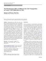
Figure 1.2. In connective tissues, collagen fibres are in irregular orientations and form
networks of different densities. (A) Loose connective tissue of mesentery, which is the
membrane-like lining of the abdominal cavity, showing collagen fibres (arrow heads), elastic
fibres (small, filled arrows) and fibroblasts nucleus (unfilled arrows)(stained for elastin, x40).
(B) Irregular, dense connective tissue of nipple skin (hematoxylin and eosin staining, x20).
Cell nucleus are the dark dots. Collagen fibres (light gray) are oriented irregularly but are
densely packed. Glycoproteins and ground substances are lost during dehydration, so they are
not visible in the stained samples. Source: Blue Histology on Web. School of Anatomy and
Human Biology, University of Western Australia.
URL:
As the scaffold of extracellular microenvironment, extracellular matrices (ECMs) are built
from multiple molecules into diversified forms of networks depending on the functions of
BA
Chapter 1 Introduction
7
tissues. As will be discussed next, ECMs largely determine the fate of metastatic cells. Because
metastatic cancer cells closely interact with ECM environments, such as basement membrane
and connective tissue, it is necessary to understand the components and micro-architecture of
these ECM environments. Basement membrane is a thin layer of high density matrix that the
epithelia / endothelia attach to. This layer maintains the proper orientation of epithelial /
endothelial cells and separates the epithelial / endothelial cells from the connective tissue. The
connective tissue is sparsely distributed with cells and mainly composed of four major types of
ECM proteins, i.e., glycoproteins, proteoglycans, collagen and elastin (Jones and De Clerck
1982). The proteoglycans are glycoproteins with higher proportions of sugar side chains.
Owning to the fact that sugar chains often bind a great amount of water molecules,
glycoproteins and proteoglycans mainly sustain the compression of the tissue. Besides
providing a structural support, glycoproteins have multiple functions such as binding to fibres,
cells, and ground substance of the tissue. Being the main protein component of connective
tissue, collagen is the also the most abundant protein, as it makes up around 30% of proteins in
the human body. Among a total of 19 subtypes, collagen type I (collagen-I) has the largest
quantity (90%), becoming the major protein of connective tissue. As collagen-I has a
triple-helices conformation and is further polymerized into fibrillar networks, it is responsible
for load-bearing functions of bone, ligament etc (Di Lullo et al. 2002). Collagen-I is also the
major protein component in breast stroma (Nelson and Bissell 2005). Elastin is a class of elastic
fibres, which include elastin, elaunin and oxytalan. They are in naturally relaxed states most of
the time, while bearing tensile load when stretched. The structural organization of elatic fibres
and the way that elastic fibres interweave with collagen allow them to buffer the expansion and
prevent tearing [4]. These fibrillar components are the major solid phase structures that form a
connective tissue (Figure 1.2), and largely determine the porous nature as well as mechanical
properties of the tissue.