Characterization of pin1 function in zebrafish development
Bạn đang xem bản rút gọn của tài liệu. Xem và tải ngay bản đầy đủ của tài liệu tại đây (20.54 MB, 166 trang )
CHARACTERIZATION OF ZPIN1 FUNCTION IN
ZEBRAFISH DEVELOPMENT
ZHAO LIQUN
NATIONAL UNIVERSITY OF SINGAPORE
2008
CHARACTERIZATION OF ZPIN1 FUNCTION IN
ZEBRAFISH DEVELOPMENT
ZHAO LIQUN
B.Sc., Shandong University, China;
M.Sc.,
Peking Union Medical College, China
A THESIS SUBMITTED
FOR THE DEGREE OF DOCTOR OF PHILOSOPHY
DEPARTMENT OF BIOLOGICAL SCIENCES
NATIONAL UNIVERSITY OF SINGAPORE
2008
i
Acknowledgements
I greatly appreciate the help and support from my supervisor, and labmates during
my graduate study in Department of Biological Sciences, National University of
Singapore. Without them, I would not be able to accomplish my research work.
First of all, I would like to express my sincere gratitude to my supervisor, Dr. Liou
Yih-Cherng. He is such a knowledgeable, and wise person who is always guiding me
and offering me wonderful advices whenever I have difficulties in my research. I am
grateful for his consistent encouragement and support which help me finish my
research project, and make my graduate career more enjoyable.
I would like to thank Dr. Vladimir Korzh and Dr. Steven Fong. With generous help
from Dr. Korzh, I was able to continue my research project in IMCB for later two
years of my graduate career. There, I received systematic training on manipulating
embryos, and taking excellent pictures from Dr. Steven. Furthermore, they introduced
me to the wonderful world of neurobiology. Without their tremendous help both on
techniques and on knowledge, I can not accomplish the latter part of this project.
I want to express my sincere gratitude to my QE committee members: Dr. Wang
Shu, Dr. Low Boon Chuan and Dr. Vladimir Korzh. They kindly give me helpful
suggestions which are beneficial for my research work for my whole Ph.D period.
I also want to thank Dr. Faroq, Dr. Liu Lihui, Dr. Zhu Shizen, Dr. Zhan Huiqing
and Dr. Cathleen Teh for their valuable suggestions and enthusiastic help.
I appreciate the help from my labmates: Dr. Wang Yu, Zhou Wei, Liu Jun, Xia Yun,
Yang Qiaoyun, Tan Weiwei, Ye Fan and Lai Cherng-Yu. In my entire graduate career,
they are very helpful in effective troubleshooting by discussing and solving problems.
Lastly, my innermost gratitude goes to my family. Whenever I feel worried or
depressed, it is their selfless love and encouragements that make me calm down and
ii
continue my research work. I especially need to thank my husband, Li Hao. He is my
spirit support through four years. Therefore, I would like to dedicate my thesis to my
parents, my brother, and my husband.
iii
Table of contents
Acknowledgements i
Table of contents iii
List of Tables ix
List of Figures x
List of Abbreviations xii
Chapter 1 Introduction 1
1.1 The function of Pin1 1
1.1.1 The relationship between Pin1 and protein phosphorylation 1
1.1.2 The characteristics of Pin1 4
1.1.3 Pin1 function in cell cycle and cancer 5
1.1.3.1 Pin1 function in M phase 5
1.1.3.2 Pin1 function in G1 and S phase 7
1.1.3.3 Pin1 function in oncogenesis 9
1.1.4 Pin1 function in apoptosis 10
1.1.5 Pin1 function in Alzheimer’s disease 11
1.1.6 Pin1 function in development 16
1.1.7 Pin1 function on protein stability 19
1.1.7.1 The ubiquitin-proteasome pathway 19
1.1.7.2 The role of Pin1 in protein stability 19
1.1.8 The summary of Pin1 function 23
1.2.1 Introduction of bHLH factors 25
1.2.2 Overview of NeuroD function 26
1.2.3 Phenotypes for NeuroD-null mice 27
1.2.4 Protein interactors of NeuroD 28
iv
1.2.5 Post-translational regulation of NeuroD 30
1.2.6 The upstream and downstream regulators of NeuroD 32
1.3 Advantages of zebrafish model 34
1.4 Introduction of zebrafish lateral line 35
1.4.1 Zebrafish lateral line developmental process 35
1.5 Objectives of this study 37
Chapter 2 Materials and Methods 39
2.1 Molecular technology 39
2.1.1 Polymerase chain reaction (PCR) 39
2.1.2 PCR product purification 39
2.1.3 DNA ligation and transformation 40
2.1.4 DNA sequencing 41
2.1.5 Rapid amplification of cDNA ends (RACE) 41
2.1.6 Southern Blotting 42
2.1.6.1 Probe synthesis 42
2.1.6.2 Isolation of genomic DNA 42
2.1.6.3 Digestion of genomic DNA 43
2.1.6.4 Neutralization, transfer and fixation 43
2.1.6.5 Prehybridization and hybridization 44
2.1.6.6 Washing and blocking 44
2.1.6.7 Antibody incubation and detection 44
2.2 In vitro studies using cell lines 45
2.2.1 Cell lines and cell culture 45
2.2.2 Transfection and cell lysates collection 45
2.2.3 SDS-PAGE and Western blotting 46
v
2.2.4 Site-directed mutagenesis 47
2.2.5 Co-immunoprecipitation 48
2.2.6 Expression and purification of recombinant GST-Pin1 49
2.2.7 GST pull-down assay 49
2.2.8 Stability and rescue assay 50
2.2.8.1 Protein concentration measurement 50
2.2.8.2 Stability assay 50
2.2.8.3 Rescue assay 51
2.2.9 Immunostaining 51
2.3 In vivo studies using zebrafish embryos 51
2.3.1 Maintenance and staging of zebrafish strains 51
2.3.2 In vitro transcription 52
2.3.3 Microinjection 52
2.3.4 In situ hybridization 53
2.3.4.1 Synthesis of labeled RNA probe 53
2.3.4.2 Embryos collection and fixation 54
2.3.4.3 Proteinase K treatment 55
2.3.4.4 Prehybridization 55
2.3.4.5 Hybridization 55
2.3.4.6 Post-hybridization washes 56
2.3.4.7 Antibody incubation 56
2.3.4.8 Color development 57
2.3.4.9 Mounting and photographing 57
2.3.5 Immunohistochemical staining on embryos 58
2.3.6 Acridine Orange staining 58
vi
2.3.7 Total RNA extraction from zebrafish embryos 59
2.3.8 Reverse-transcriptase PCR (RT-PCR) 59
Chapter 3 Results 61
3.1 Molecular analysis of zebrafish Pin1 61
3.1.1 Characterization of zebrafish Pin1 61
3.1.2 Sequence alignment of Pin1 in various species 63
3.1.3 Expression pattern of zPin1 in zebrafish embryos 65
3.2.2 The interaction of zPin1 with NeuroD via pSer/Thr-Pro motif 73
3.3 zPin1 morpholino knockdown phenotypes 78
3.3.1 The knockdown efficiency of both zPin1 morpholinos 78
3.3.2 Developmental delay caused by zPin1 loss-of-function 80
3.3.3 Global apoptosis caused by zPin1 lost-of-function 84
3.3.4 M-phase arrest caused by zPin1 loss-of-function 85
3.3.5 Neuronal phenotypes caused by zPin1 lost-of-function 87
3.3.5.1 The defects on mature neurons 87
3.3.5.2 The defects for neuroD expression 89
3.3.5.3 Neuromasts hair cells defects 90
3.3.5.4 Neuromasts mantle cells defects 96
3.3.6 The specificity of neuromasts defects 96
3.4 Regulation of NeuroD stability by zPin1 100
3.5 Rescue of NeuroD stability by Pin1 101
3.6 The effects of zPin1 on insulin gene expression 105
Chapter 4 Discussion and Conclusion 107
4.1 Molecular analysis of zebrafish Pin1 107
4.2 The effects of zPin1 morpholinos 109
vii
4.3 Developmental delay caused by zPin1 knockdown 110
4.4 The interaction between zPin1 and NeuroD 112
4.5 Protein stabilizing effect of NeuroD by zPin1 regulation 114
4.6 The specificity of neuromasts defects 117
4.7 Transcriptional activity of NeuroD mediated by zPin1 120
4.8 The expression of marker genes in zPin1 morphants 122
4.9 Pin1 as a novel regulator of bHLH family 125
4.10 Conclusion 126
References 128
Appendices 141
viii
Summary
The vertebrate pSer/pThr-Pro specific peptidyl-prolyl isomerase Pin1 has been
shown to play important roles in cell cycle regulation, apoptosis, oncogenesis, and
neuronal degeneration. However, its role in early neuronal development is not clear.
With the use of zebrafish embryos, we examined zPin1’s effect on development. We
showed that zebrafish Pin1 was expressed maternally and in a ubiquitous manner
early in development, but by 48 hpf it was restricted to the brain and neuromasts. Co-
immunoprecipitation assays (CoIP) in cell lines showed that zPin1 could interact with
neuroD and ath1 but not ngn1. This binding was reduced when Ser/Thr
phosphorylation sites were mutated. Antisense morpholino oligonucleotide (MO)
knockdown of zPin1 led to delay in development. Accounting for the delay, neuroD
expression was significantly diminished in the hindbrain of morphants by 48 hpf
equivalent. Morphants in the background of Tol2/GFP enhancer trap lines with
specific expressions in hair cells (ET4) and mantle cells (ET20) also displayed defects
in neuromasts formation. It has been shown that specification of hair cells in
neuromasts is neuroD dependent but ngn1 independent. Using siRNA Pin1
knockdown 293T cells and Pin1 knockout MEFs cells, we showed that neuroD
protein was degraded more rapidly in the absence of Pin1. NeuroD stability was
restored when Pin1 was over-expressed. This is the first study to demonstrate the
functional regulation of a bHLH factor by cis-trans isomerization in neuronal
specification. In view of the role of Pin1 in neurodegenative diseases, our results may
have important pharmaco-therapeutic applications.
ix
List of Tables
Table 3.1 zPin1 MO led to delay in development 82
Table 3.2 Loss of zPin1 affects lateral line hair cell development 95
x
List of Figures
Figure 1.1 Prolyl isomerization catalyzed by peptidyl-prolyl cis/trans isomerase
(PPIases) 3
Figure 1.2 Overall architecture of human Pin1 5
Figure 1.3 Pin1 catalyzed APP and tau processing in healthy and Alzheimer’s
disease neurons 15
Figure 1.4 Model of cell cycle defects for PGCs 18
Figure 1.5 A spectrum of target activities by Pin1 isomerase 24
Figure 1.6 General interactions of bHLH proteins 26
Figure 1.7 Schematic of NeuroD protein 30
Figure 1.8 Composition of zebrafish lateral line 36
Figure 3.1 Full- length cDNA sequence of zebrafish Pin1 62
Figure 3.2 Amino acid sequence alignment of Pin1 in different species 64
Figure 3.3 zPin1 spatial and temporal expression patterns 67
Figure 3.4 In situ hybridization analysis of zpin1 expression at different stages
68
Figure 3.5 zPin1 interacts with NeuroD and Ath1 72
Figure 3.6 Identification of zPin1 potential Ser/Thr-Pro binding motifs on NeuroD
76
Figure 3.7 The reduction of zPin1 expression in zPin1 morphant embryos 79
Figure 3.8 Developmental delay in zPin1 morphant embryos 82
Figure 3.9 Neurogenin1 staining of the wild type embryos and catch up zPin1
morphant embryos 83
Figure 3.10 Global apoptosis phenotype in zPin1 morphant embryos 84
Figure 3.11 Phospho-H3 staining of the wild type embryos and zPin1 morphant
embryos 86
Figure 3.12 The neuronal defects in zPin1 morphant embryos 88
xi
Figure 3.13 Expression of marker genes in zPin1 morphant embryos 94
Figure 3.14 The impaired formation of posterior lateral line neuromasts in zPin1
morphant embryos 95
Figure.3.15 Accessory mantle cells phenotypes in zPin1 morphant embryos 98
Figure 3.16 Posterior lateral line ganglion in mi20 embryos 99
Figure 3.17 The increased stability of NeuroD mediated by zPin1 103
Figure 3.18 The recovery of NeuroD half-life by re-introduction of Pin1 104
Figure 3.19 Pancreas insulin gene transcripts in zPin1 morphant embryos 106
Figure 4.1 Xenopus versus zebrafish NeuroD protein sequence alignment 114
Figure 4.2 Sequential expression of HLH genes during neurogenesis 124
xii
List of Abbreviations
AD Alzheimer’s disease
AEP auditory evoked potential
ALL anterior lateral line
AP alkaline phosphatase
APC anaphase-promoting complex
APS ammonium persulphate
BETA2 beta-cell E box trans-activator 2
βGK β-glucokinase
bHLH basic helix-loop-helix
CDKs cyclin-dependent protein kinases
CIAP alkaline phosphatase
Co-IP co-immunoprecipitation
Cyps cyclophilins
cytC cytochrome c
DAPI 4'-6-Diamidino-2-phenylindole
DBD DNA binding domain
dpf days post-fertilization
EGFR epidermal growth factor receptor
Emi1 early mitotic inhibitor-1
ERKs extracellular signal-regulated kinases
FKBPs FK506-binding proteins
GFAP glial fibrillary acidic protein
G6Pase glucose-6-phosphatase
GSK glycogen synthase kinase-3
xiii
HAP1 huntington-associated protein
HAT histone acetyltransfereases
Htt Huntington
IA-1 insulinoma-associated antigen-1
IAP inhibitor of Apoptosis Protein
Id inhibitor of differentiation
I
κ
B
α
NF-κB inhibito
IPTG isopropyl-beta-D-thiogalactopyranoside
IRF3 interferon-regulatory factor 3
HRP horse radish peroxidase
JNKs N-terminal protein kinases
MAPKs mitogen-activated protein kinases
MAPs microtubule-associated proteins
MBT midblastula transition
Mcl-1 cell leukemia sequence-1
MEFs mouse embryonic fibroblasts
MLK2 mixed –lineage kinase 2
MO morpholino oligonucleotides
NFTs intracellular neurofibrillary tangles
NIMA never in mitosis A
ORF open reading frame
PCR polymerase chain reaction
PGCs primordial germ cells
Pin1 protein interacting with NIMA
PLKs Polo-like kinase
xiv
PLL posterior lateral line
PML promyelocytic leukemia protein
RGC retina ganglion cells
RT-PCR reverse-transcriptase PCR
SHP small heterodimer partner
SUR1 sulfonylurea receptor 1
TopoIIα topoisomerase
UTR untranslated region
Introduction
1
Chapter 1 Introduction
1.1 The function of Pin1
1.1.1 The relationship between Pin1 and protein phosphorylation
Protein phosphorylation is one of the most essential signaling modes for post-
translational modification. The main effect of protein phosphorylation is to induce
changes in protein conformation, which then further affects protein-protein
interaction, subcellular localization and dephosphorylation (Blume-Jensen et al.,
2001; Pawson et al., 2005). Therefore, it is valuable for us to study molecules
related to conformation changes induced by protein phosphorylation.
Serine or threonine residues preceding proline (Ser/Thr-Pro) motifs are exclusive
phosphorylation sites for many key protein kinases that play essential roles in cell
proliferation and signal transduction. These kinases include cyclin-dependent protein
kinases (CDKs), mitogen-activated protein kinases (MAPKs), extracellular signal-
regulated kinases (ERKs), N-terminal protein kinases (JNKs), glycogen synthase
kinase-3 (GSK-3) and Polo-like kinase (PLKs). This group of phosphorylation
depends on the presence of Pro residues that immediately follow Serine or
Threonine residues; therefore, it is also called Pro-directed phosphorylation,
indicating an essential role of Proline residues (Blume-Jensen et al., 2001; Nigg,
2001; Lu, 2004; Pawson et al., 2005). Proline residue is unique among all amino
acids due to its five-membered ring in its backbone. Usually, the other amino acids
overwhelmingly adopt trans conformation (99%) because of the free energy
consideration (Pal et al., 1999). However, when it comes to Proline residue, both
trans (70-90%) and cis (10-30%) conformation exist due to its structural constraints
(Brandts et al., 1975; Grathwohl et al., 1976; Juy et al., 1983). The transition
between cis and trans conformation is called isomerization. This transition process
Introduction
2
is rather slow if it happens spontaneously. With help from one enzyme family called
cis/trans peptidyl-prolyl isomerase (PPIase), this process can be accelerated greatly
(Hunter, 1998; Fischer et al., 2003). Until now, three PPIase family members have
been identified. Among them, cyclophilins (Cyps) and the FK506-binding proteins
(FKBPs) catalyze conformation changes of Serine/Theronine-Proline (Ser/Thr-Pro)
motifs, independent of phosphorylation (Harding et al., 1989; Fischer et al., 2003).
Only the third family member, parvuline can specifically catalyze conformation
changes of a subset of phosphorylated proteins (Ranganathan et al., 1997; Yaffe et
al., 1997). Human Pin1 (protein interacting with NIMA) and Pin1-like parvulines
are considered novel members of parvuline and catalyze only phosphorylated
Ser/Thr-Pro motifs in proteins so as to influence various cellular aspects such as cell
growth, genotoxic stress, germ cell development and neuron differentiation.
Deregulation of Pin1 is involved in various pathological conditions: cancer,
Alzheimer’s disease and asthma (Lu, 2004; Etzkorn, 2006; Goutagny et al., 2006;
Balastik et al., 2007; Yeh et al., 2007). The model of Pin1-catalyzed isomerization is
demonstrated in Fig. 1.1.
Introduction
3
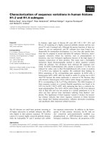
Figure 1.1 Prolyl isomerization catalyzed by peptidyl-prolyl cis/trans isomerases
(PPIases). Ser/Thr-Pro motif is a key regulatory phosphorylation motif in cells. Due to the
presence of Pro residue, there is a large energy barrier for motif to have spontaneous
isomerization. However, this isomerization can be greatly accelerated by PPIases. The
function of PPIases is to change conformation of Ser/Thr-Pro motif from cis to trans or trans
to cis. There are two conventional PPIases-cyclophilins and FKBPs which are
phosphorylation-independent enzymes. (a) They can only catalyze isomerization of Xxx-Pro
motifs, where Xxx indicates any amino acid except pSer or pThr. (b) Pin1 belongs to the third
family of PPIases. It is the only known phosphorylation-dependent enzyme to change the
conformation of pSer/Thr-Pro. Recent studies indicate that the cis and trans isomers of many
proteins have distinct functions, and their conversions by PPIases can function as a new
general class of protein regulatory mechanism. (This figure was adapted from Lu et al.,
2007).
Introduction
4
1.1.2 The characteristics of Pin1
Pin1 was first identified in 1996 in yeast using yeast two-hybrid system for its
ability to interact with the key mitotic kinase NIMA (Never In Mitosis A) and inhibits
its mitosis-promoting activity (Osmani et al., 1991; Pu et al., 1995; Lu et al., 1996).
Pin1 has been found to be evolutionarily conserved among eukaryotes (Zhou et al.,
1999; Huang et al., 2001; Metzner et al., 2001).
Pin1 is an 18 kDa protein with two domains: N-terminal WW domain (named after
two invariant Trp residues, amino acids 1-39) and C-terminal PPIase domain (amino
acids 45-163) (Lu et al., 1996). WW domain acts as a binding module to bind its
substrate via phosphoserine or phosphothreonine-proline motifs in its substrates. From
structural and functional stuides, four amino acids of the WW domain including Ser16,
Arg17, Tyr23 and Trp34 are responsible for its binding ability
(Yaffe et al., 1997;
Zhou et al., 2000; Wintjens et al., 2001). C-terminal PPIase domain is the catalytic
domain to isomerize conformational changes of specific pSer/Thr-Pro motifs (Lu et
al., 1996). In the PPIase domain, Lys63, Arg68 and Arg69 play an essential role for
its catalyzing ability (Ranganathan et al., 1997; Yaffe et al., 1997). A Pin1 structural
model is illustrated in Fig. 1.2.
Since the discovery of Pin1, its function in biological processes such as cell cycle
regulation, transcription, protein stability and pathological conditions including
oncogenesis, apoptosis, neuron degeneration as well as development have been
studied extensively and we will review these aspects in the following sections.
Introduction
5
Figure 1.2 Overall architecture of human Pin1. X-ray structure of human Pin1 complexed
with dipeptide Ala-Pro. Pin1 contains two domains: N-terminal WW domain and C-terminal
PPIase domain. The two domains are connected by a flexible linker. WW domain consists of
a tripel-stranded anti-parallel β sheets. PPIase domain has four β sheets as well as four α
helices. A sulfate ion is sequestered by a conserved basic cluster consisting of Arg68 and
Arg69 in close proximity to the β methyl group of the Ala residue in the bound Ala-Pro
dipeptide. (This figure was adapted from Lu et al., 2002).
1.1.3 Pin1 function in cell cycle and cancer
1.1.3.1 Pin1 function in M phase
NIMA kinase is the key mitotic kinase in Aspergillus nidulans. The early study in
Aspergillus nidulans and later study in eukaryotic organisms both indicate that NIMA
kinase is essential for cells entering mitosis and inactivation of NIMA is required for
exit from mitosis (Osmani et al., 1988; Osmani et al., 1991; Fry et al., 1995; Lu et al.,
1996). Based on these findings, Lu et al. hypothesized that Pin1 could play an
important role in cell cycle regulation especially in mitosis. Many studies have been
carried out to test this hypothesis.
Introduction
6
Lu et al. first gave evidence that depletion of Pin1 in budding yeast and tumor
HeLa cells caused mitotic arrest (Lu et al., 1996; Rippmann et al., 2000). Meanwhile,
overexpression of Pin1 in HeLa cells and Xenopus blocked cells in G2 by preventing
cells from entering mitosis (Crenshaw et al., 1998; Shen et al., 1998). These results
suggested a crucial role of Pin1 in mitotic regulation. More specifically, data indicated
that Pin1 functions as a negative regulator for mitosis entry and is also required for
exit from mitosis (Lu et al., 1996).
Thus far, many mitosis substrates of Pin1 have been identified. These substrates
include a large number of mitotic phosphoproteins such as Cdc25C, Plk1, Myt1,
Wee1 and Cdc27C. These substrates are also recognized by phosphospecific mitosis
marker MPM-2 antibody, suggesting that Pin1 must function essentially in mitosis
phase (Yaffe et al., 1997; Crenshaw et al., 1998; Shen et al., 1998).
Further studies have been carried on to study details about Cdc25C, Wee1,
Topoisomerase (TopoIIα) and early mitotic inhibitor-1 (Emi1). Cdc25C is an
phosphatase to activate an essential mitotic kinase Cdc2. Cdc25C itself needs to be
phosphorylated by mitosis-specific proteins for activation. Pin1 thus interacts with
phosphorylated Cdc25C to affect its activity to prevent premature entry into mitosis
(Crenshaw et al., 1998; Shen et al., 1998). Pin1 can also facilitate dephosphorylation
of Cdc25C by phosphatase PP2A (Zhou et al., 2000; Stukenberg et al., 2001). The
inhibitory kinase responsible for phosphorylating Cdc2 is Wee1. Wee1 has to be
downregulated after entering mitosis to release inhibitory effect on Cdc2. Pin1 helps
to inactivate Wee1 at M phase (Okamoto et al., 2007).
Emi1 is an early mitotic inhibitor-1 which prevents activation of cyclin A and B by
anaphase-promoting complex (APC) at G2 phase to guarantee the entry of S and M
phases at correct time. The binding of Pin1 with Emi1 stabilizes Emi1 and helps to
Introduction
7
inhibit cyclin A and B proteins. Therefore, cells can enter S and M phase smoothly
(Bernis et al., 2007).
Mitotic phosphorylated TopoIIα can also interact with Pin1. This binding localizes
Pin1 on chromatin and promotes chromatin condensation. Moreover, TopoIIα
phosphorylation and binding ability to DNA are both increased due to Pin1
association (Xu et al., 2007). Collectively, these findings indicated that Pin1 might act
as a switch from G2 to M phase to guarantee progression of cell cycle.
1.1.3.2 Pin1 function in G1 and S phase
It is revealed that Pin1
-/-
mouse embryonic fibroblasts (MEFs) lose their capability
to enter G1 and S phase from G0 phase of cell cycle (Fujimori et al., 1999).
Furthermore, Pin1 knockout mice display defects in primordial germ cells which
result from extended S phase (Atchison et al., 2003). These discoveries suggest that
Pin1 is involved in G0/G1-S transition process.
Accumulating evidence indicated that Pin1 functions essentially in G1/S transition.
The best known substrate in this process is cyclin D1 which is a key regulator of G1/S
progression (Ryo et al., 2001; Wulf et al., 2001; Liou et al., 2002). The first evidence
to confirm function of Pin1 in G1/S transition is phenotypes of Pin1 knockout mice.
Those mice display many severe phenotypes, including decreased body weight, retinal
degeneration, mammary gland retardation and testicular atrophy. Most of these
phenotypes are remarkably similar to those of cyclin D1-deficient mouse phenotypes.
Afterwards, it has been reported that Pin1 can increase cyclin D1 mRNA level and
protein expression through several pathways. Firstly, Pin1 can bind phosphorylated c-
Jun to enhance its transcriptional activity towards cyclin D1 promoter via AP-1
binding site on the promoter region. This upregulation of transcriptional activity is
further potentiated by cotransfection of Ras or JNK (Whitmarsh et al., 1996; Wulf et
Introduction
8
al., 2001). Secondly, Pin1 enhances cyclin D1 mRNA level through β-catenin. β-
catenin is an important oncogenic transcriptional activator that can be regulated by
tumor suppressor adenomatous polyposis coli protein (APC). APC binds β-catenin to
form a complex and keeps β-catenin in cytoplasm for ubiquitin-mediated degradation
(Polakis, 2000). Pin1 binds phosphorylated β-catenin to prevent its interaction with
APC and thus affects degradation of β-catenin. The consequence is that Pin1 can
accumulate more β-catenin in nucleus and increase its transcriptional activity towards
its most important downstream target: cyclin D1 (Ryo et al., 2001). Thirdly, Pin1 also
plays its role in regulating NF-κB (a heterodimeric complex of p50 and p65/RelA)
signaling pathway. It has been characterized that Pin1 specifically binds to p65
subunit of NF-κB. This association of Pin1 with p65 disrupts binding of NF-κB
inhibitor (I
κ
B
α
) with p65 to decrease
degradation of NF-κB (Ryo et al., 2003). As a
result, NF-κB becomes more stable and its transcriptional activity as well as the level
of downstream target: cyclin D1 is increased. Lastly, Pin1 can directly bind and
stabilize cyclin D1 (Liou et al., 2002).
Besides cyclin D1, Pin1 modulates another two essential proteins implicated in
G1/S phase and they are cyclin E as well as c-Myc. Pin1 can bind both cyclin E and c-
Myc and negatively regulate them by promoting their degradation through recruiting
E3 ubiquitin ligase FBXW7 (Yeh et al., 2004; van Drogen et al., 2006; Yeh et al.,
2006).
Pin1 is also involved in S phase centrosome duplication. Centrosome functions to
segregate duplicated chromosomes to ensure proper cell division and cytokinesis.
Duplication of centrosome begins at G1/S transition and is complete during S phase
before mitotic phase (Doxsey et al., 2005). It was revealed that Pin1 is localized to
centrosome and its expression level is correlated with centrosome amplification.
Introduction
9
Detailed investigation shows that loss of Pin1 delays centrosome amplification; while
overexpression promotes centrosome amplification (Suizu et al., 2006). These results
indicate that Pin1 plays its role in S phase by affecting centrosome duplication
without disturbing DNA synthesis.
1.1.3.3 Pin1 function in oncogenesis
Pin1 expression level is found to be increased in various kinds of cancers. As early
as in 2001, it was reported that Pin1 expression level was upregulated in human breast
cancer (Wulf et al., 2001). In prostate cancer, Pin1 expression level is an indicator of
clinical stage and has prognostic value (Ayala et al., 2003). Later, Bao et al. carried
out an extensive study in 2004 to investigate relationship between Pin1 expression
and occurrence of tumors. For this study, 38 types of cancers such as breast, prostate,
lung (60 types altogether) had phenomenon (around 10%) of Pin1 overexpression
compared with normal healthy tissues (Bao et al., 2004). The molecular mechanism
raveling Pin1’s implication in cancer will be described below.
From sequence analysis, it has been revealed that Pin1 has three E2F binding sites
and is subjected to E2F transcriptional regulation (Ryo et al., 2002). This finding is
valuable when we try to link Pin1 expression with cancer. Given the crucial role of
E2F/Rb deregulation in oncogenesis, it may be critical to upregulate Pin1 expression
level (Macleod, 1999). Pin1 overexpression can enhance transformed phenotypes
induced by oncogenic E2F upstream regulators Ras or Neu. Correspondingly,
depletion of Pin1 can suppress transformed phenotype (Ryo et al., 2002).
It was reviewed previously that Pin1 can regulate essential oncogene cyclin D1
from multiple oncogenic signaling pathways. Not only deregulation of cylcin D1, but
also centrosome defects and resulting chromosome instability by Pin1 overexpression
have been suggested to play an important role in oncogenesis (Suizu et al., 2006).