Expression dynamics of the hepatic mitochondrial proteome of the sod2+ mouse in response to troglitazone administration
Bạn đang xem bản rút gọn của tài liệu. Xem và tải ngay bản đầy đủ của tài liệu tại đây (10.32 MB, 226 trang )
EXPRESSION DYNAMICS OF THE HEPATIC
MITOCHONDRIAL PROTEOME OF THE SOD2
+/-
MOUSE
IN RESPONSE TO TROGLITAZONE ADMINISTRATION
LEE YIE HOU
HT051163E
A THESIS SUBMITTED FOR THE DEGREE OF DOCTOR OF
PHILOSOPHY
DEPARTMENT OF BIOCHEMISTRY,
YONG LOO LIN SCHOOL OF MEDICINE,
NATIONAL UNIVERSITY OF SINGAPORE
2009
i
ACKNOWLEDGEMENTS
For many, obtaining a postgraduate degree involves conducting experiments
decided by our supervisors and doing well in courses. If done right, this is enough to earn
one a Ph.D. Needless to say, it isn’t always the case. Scientific research is immersion into
the unknown, and when factors such as fund (in)sufficiency, ensuring publishing within
journals’ already limited room for articles and foreseeing unlimited possibilities of
problems, doing good research is no longer that easy. Confronting this vast number of
daunting tasks alone, while remaining productive in a multi-disciplinary project was not a
simple task. That realization was discouraging, but also liberating because of who my
academic advisor is.
I am indebted to my academic supervisor, Professor Maxey Chung Ching Ming.
Professor Chung was my mentor, teacher, role model and friend. I was always motivated
and inspired by his attitude, outlook and vision. He reached so many people as a result of
his unwavering belief in individuals and their strengths, as he did to my Ph.D and life.
Professor Chung has always been positive, and he gave me many opportunities,
supported and encouraged me in bad times. And that was how I was touched by his
sincerity and patience, creating a climate of friendliness and emotional support as I
muddled through my way to doing productive good science. On the research front, I was
granted ample freedom to steer my project, but of course with his constant guidance.
With much appreciation and respect, his guidance has changed my Ph.D course
ii
tremendously, and I would not be where I am today if not for Professor Chung’s patience
and mentorship that saw me through. Professor Chung has left a mark in my life.
My sincere gratitude towards Professor Urs Alex Boelsterli for hatching this
brilliant research proposal and imparting the many skills required in this field. Professor
Urs made me rediscover research – that science is more than just benchwork, requiring an
intimate interplay of soft skills that are essential in this field, as in any other.
Colleagues from Protein and Proteomics Centre, Professor Lin Qingsong, Dr Tan
Hwee Tong, Lim Teck Kwang, Cynthia Liang, Tan Gek San, Zubaida, and others whom I
fail to mention, thank you for your warmth, friendliness and generosity. Among them,
Professor Lin maintained my desire for questioning the unlimited boundaries of
knowledge, and facing them with strong analytical skills and sound, systematic thinking.
I would like to thank the National University of Singapore for the award of my research
scholarship and the various institutions for the grants they have provided, without which
this project could not have been completed.
Lastly, I especially want to thank my family and many close friends who had
stood by me and supported me all the while. I appreciate your every presence in my life.
The years spent doing my Ph.D has been fulfilling, challenging and at times daunting.
Were the support from my family, friends and colleagues placed elsewhere, I wonder if
the outcome will be entirely different.
iii
TABLE OF CONTENTS
SUMMARY viii
LIST OF TABLES x
LIST OF FIGURES xi
LIST OF ABBREVIATIONS xiv
INTRODUCTION 1
1.1. Idiosyncratic drug-induced liver injury 1
1.1.1. Susceptibility factors and mechanisms of idiosyncratic DILI 3
1.2. Troglitazone as a model drug for the study of idiosyncratic DILI 5
1.2.1. Mitochondrial dysfunction and threshold effect as a possible mechanism for
idiosyncratic DILI 11
1.2.2. Mitochondria and idiosyncratic troglitazone DILI 18
1.3. Heterozygous Sod2
+/-
mouse 22
1.3.1. The utility of HET mouse in toxicological studies 27
1.4. Proteomics 32
1.4.1. Toxicoproteomics using the HET mouse in the mechanistic study of
troglitazone toxicity 40
1.5. Objectives 44
2. METHODS AND MATERIALS 46
2.1. Chemicals 46
2.2. Nomenclature 46
2.3. Animals, drug treatment and experimental design 47
2.4. Assessment of liver injury 50
iv
2.5. Isolation of liver mitochondria 50
2.6. Determination of mitochondrial GSH 52
2.7. Determination of nitrite/nitrate levels 53
2.8. Detection of total mitochondrial protein carbonyls and 3-nitrotyrosine
adducts 53
2.9. Two-dimensional Difference Gel Electrophoresis 55
2.9.1. Labelling with cyanine dyes 55
2.9.2. Isoelectric focusing and two-dimensional gel electrophoresis 57
2.10. Image visualization and analysis 58
2.11. Protein identification by MALDI-TOF/TOF MS/MS 59
2.12. iTRAQ™ labelling 61
2.12.1. Two-dimensional Liquid Chromatography-MS/MS of iTRAQ™ samples 64
2.12.2. Mass Spectrometry for iTRAQ™ 65
2.13. Immunblotting 66
2.14. Aconitase-2 aggregation and degradation study 69
2.15. Immunohistochemistry 70
2.16. In silico analysis 71
2.16.1. Mass spectra analysis – ProteinPilot
™
71
2.16.2. Gene Ontology over-representation and pathway analysis 73
2.17. Statistical evaluation 74
3. RESULTS 77
3.1. High level of mitochondrial purity 77
3.1.1. Comparative proteomics of HET liver mitoproteome by 2D-DIGE 78
v
3.1.2. Quantitative proteomics HET liver mitoproteome by 4-plex iTRAQ™ 87
3.1.3. Combined proteomic analysis using 2D-DIGE and iTRAQ™ labelling 92
3.2. Prolonged troglitazone administration causes oxidative stress in
mitochondria and moderate liver injury in HET mice 95
3.3. HET Mitochondrial Proteome Dynamics induced by prolonged
troglitazone treatment 101
3.3.1. 2D-DIGE Analysis of Troglitazone-induced HET Mitoproteome 101
3.3.2. Analysis of different ACO2 fates under different oxidative stress
conditions 104
3.3.3. 8-plex iTRAQ™ Analysis of Troglitazone-induced HET Mitoproteome 108
3.3.3.1. ETC components show bimodal response to acute and chronic troglitazone
treatment 119
3.3.3.2. Modulation of PPAR-agonist targets 123
3.3.3.3. Parallel proteome shift suggests ROS-induced mitochondrial stress 126
3.3.4. Prolonged troglitazone treatment activates FOXO3a through oxidative
stress-mediated signals 129
3.3.5. Transcriptional regulation of SOD2 and the HET hepatic mitoproteome 133
4. DISCUSSION 136
4.1. Characterization of the HET liver mitoproteome 136
4.1.1. Introduction 136
4.1.2. Purity of mitochondria preparation 136
4.1.3. The HET liver mitoproteome 137
4.1.3.1. Redox proteins 140
vi
4.1.3.2. OXPHOS 141
4.1.3.3. Urea cycle 143
4.1.3.4. β-Oxidation 144
4.1.3.5. α-ketoglutarate dehydrogenase (KGDH) 144
4.1.4. Summary 146
4.2. Toxicoproteomics of Troglitazone-induced Mitoproteome Alterations 148
4.2.1. Introduction 148
4.2.2. Mitochondrial proteome expression dynamics induced by prolonged
troglitazone treatment 150
4.2.2.1. Functional clustering of mitochondrial proteome 153
4.2.2.2. Mitochondrial glutathione transport 154
4.2.2.3. PPAR-agonist mitochondrial targets 158
4.2.2.4. OXHPOS 161
4.2.2.5. Valine metabolism 162
4.2.2.6. Redox and Stress Response Proteins 163
4.2.3. Summary 164
4.3. Aconitase-2 as a Potential Biomarker to Early Prediction of Toxicity 167
4.4. Mechanistic toxicology of troglitazone-induced DILI 169
5. CONCLUSIONS 171
5.1. Implications of Studying the HET Hepatic Mitoproteome in Drug Safety
Evaluation 171
5.2. Summary 172
6. FUTURE WORK 175
vii
7. APPENDIX 179
7.1. MS/MS spectrum 179
7.2. Protein Tables 182
7.3. List of PPAR-responsive genes 194
7.4. iTRAQ™ supplementary data 197
7.5. List of publications 198
7.6. Posters and Presentations 198
8. BIBLIOGRAPHY 199
9. Supplemental Protein Table Found in Inserted CD
viii
SUMMARY
Idiosyncratic drug-induced liver injuries (DILI) are rare adverse events that inflict
susceptible patients exposed to certain normally-mild drugs. A major obstacle in
understanding idiosyncratic DILI etiology includes the lack of ideal animal models for its
reproduction in the laboratory. Recently, ROS has been implicated in idiosyncratic DILI
and the heterozygous superoxide dismutase 2 or Sod2
+/-
mouse (HET) is an ideal mutant
model for studying DILI arising from diminished mitochondrial antioxidant defence.
Using highly purified mitochondrial proteins from the HET
liver, we performed
comparative proteomics. The up-regulation of antioxidants such GPX1, GSTK1 and
MGST1 suggested the increased effort to restore redox equilibrium. Our proteomic
analysis indicated that HET
mice exhibit a mild mitochondrial oxidative stress which is
partly compensated by the antioxidant defense system linked to the tricarboxylic acid
(TCA) cycle, urea cycle, β-oxidation, and oxidative phosphorylation (OXPHOS). This
discreet and phenotypically silent mitochondrial proteome alteration represents a “1
st
hit”
which is compatible with studying pathological DILI conditions (“2
nd
hit”).
Applying integrative proteomics on the HET
hepatic mitochondria treated with
troglitazone, a withdrawn drug due to unacceptable hepatic liability, we generated a
comprehensive view of proteomic changes that correlated well with toxicological and
histological endpoints. 2D-DIGE and iTRAQ™ coupled to MALDI-TOF/TOF MS/MS
analysis revealed a two-stage mitochondrial response upon short-term and long-term
troglitazone administration, similar to the delayed hepatotoxicity observed in humans.
ix
The small number of proteins common to both time-points (3 out of 70 proteins) reflected
distinct changes that occurred at the molecular level. Early changes involved the
induction of a mitochondrial stress response such as seen by increased levels of heat
shock protein family members (mortalin, HSP7C), Lon protease, and catalase. In
contrast, after 4 weeks, a number of critical proteins including ATP synthase β-subunit,
aconitase-2 (ACO2), and mitochondrial dicarboxylate carrier (DIC) exhibited decreased
abundance. In addition, mitochondrial protein carbonyls and nitrotyrosine adducts were
significantly increased, suggesting uncompensated oxidative damage. Even in the
presence of increased SOD2 levels, the threshold for toxicity has been reached and liver
injury ensued. Building on clinical and biological evidence of mitochondrial ROS
perturbation on troglitazone DILI, we observed that impairment of mitochondrial
glutathione transport may play a role in precipitating the toxic effects of troglitazone
under compromised mitochondrial ROS defence. This further confirms the contribution
of glutathione and inheritable mitochondrial dysfunction in idiosyncratic DILI
susceptibility. ACO2 was decreased at both time points, making this protein a potential
sensitive and early biomarker for mitochondrial oxidative stress. ACO2 was decreased at
both time points, making this protein a potential sensitive and early biomarker for
mitochondrial oxidant stress. This integrative approach could signify a new paradigm in
advancing and predicting mechanistic toxicity of idiosyncratic DILI.
x
LIST OF TABLES
Table 1. Selected drugs causing idiosyncratic DILI experimentally incriminated with
mitochondrial dysfunction 15
Table 2. Summary of clinical evidence linking DILI with mitochondrial dysfunction 16
Table 3. Summary of functional characterization studies in Sod2
-/-
and Sod2
+/-
deficient
mice 31
Table 4. Advantages and disadvantages of major proteomic platforms. 38
Table 5. Experimental design of vehicle and drug-treatments of HET mice. 50
Table 6. Gel setup for 2D-DIGE experiments for HET hepatic mitochondrial proteome
characterisation. 56
Table 7. Gel setup for 2D-DIGE experiments for analysis of troglitazone-induced
changes in HET hepatic mitochondrial proteome. 56
Table 8. Hepatotoxicity score of HET mice treated with troglitazone for 2 or 4 weeks. . 97
Table 9. Biochemical and clinical chemistry properties of female HET mice 98
Table 10. Functional clustering of detected mitochondrial proteins. 128
Table 11. List of detected proteins used to calculate MCV 182
Table 12. List of PPAR-responsive genes/products using bioinformatics 194
xi
LIST OF FIGURES
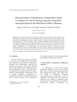
Figure 1. Drug toxicities leading to market withdrawals in the period 1976 to 2005. 3
Figure 2. Chemical structure of troglitazone 8
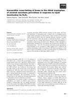
Figure 3. Crystal structure of PPARγ and RXR 8
Figure 4. Chart showing relationships between troglitazone exposure and risk of
troglitazone-induced liver injury. 20
Figure 5. Clinically silent mitochondrial abnormality and threshold effect. 21
Figure 6. Physiologically relevant ROS/RNS 23
Figure 7. Areas of research that utilizes the Sod2 mutant mouse. 26
Figure 8. Change in investment of successful new drug launch over time. 28
Figure 9. Increase in cost, time and drug amounts with drug development progession 28
Figure 10. A schematic diagram of the level of complexity from genome to the proteome.
33
Figure 11. Experimental setup of a typical 2D-DIGE experiment 39
Figure 12. Schematic of discontinuous Percoll gradient. 52
Figure 13. A flow-chart summary of the iTRAQ™ experiment design of 4-plex and 8-
plex systems. 63
Figure 14. Histogram of mean signal area (intensity) of reporter channels. 73
Figure 15. Assessment of genotype and mitochondria purification 78
Figure 16. Representative proteome map of mouse liver mitochondria. 81
Figure 17.Tandem mass spectrum of enoyl-CoA hydratase generated from MALDI-
TOF/TOF MS/MS. 82
Figure 18. Comparison of SOD1, SOD2, and GPX1 abundance levels by DIGE, silver-
staining of DIGE gel, and immunoblotting. 83
Figure 19. Immunoblotting of thioredoxin-2 and aconitase-2 85
Figure 20. 2D-DIGE observations of HET
hepatic mitochondrial proteome. 86
Figure 21. Global analysis HET mouse hepatic mitochondrial proteome by 4-plex
iTRAQ™. 89
Figure 22. Enrichment of function of proteins responsive to Sod2 haplodeficiency. 90
xii
Figure 23. Classification of HET hepatic mitochondrial proteins based on GO annotation
94
Figure 24. Liver histopathology in troglitazone-treated HET mice. 96
Figure 25. Prolonged troglitazone exposure leads to elevated
•
NO and mitochondrial
oxidative stress. 100
Figure 26. 2DE profile of HET mouse hepatic mitochondrial protein expression with
solutol or troglitazone treatment. 105
Figure 27. Validation using 2D immunoblotting. 106
Figure 28. Varying fates of ACO2 107
Figure 29. Sod2 haplodeficiency delays troglitazone hepatotoxicity as revealed by
quantitative proteomics. 110
Figure 30. Bias analysis of protein attributes. 111
Figure 31. Pie charts of GO slim analysis. 112
Figure 32. Non-intersecting GO terms of proteins in 2 and 4 weeks treatment 114
Figure 33. Schematic diagram of mitochondrial dysfunction after 4 –weeks of
troglitazone administration. 116
Figure 34. Histogram of “Toxic Pathways” affected by troglitazone treatment 117
Figure 35. Cluster analysis of detected of proteins show bimodal expression 118
Figure 36. Impact of troglitazone on HET electron transport chain complexes 121
Figure 37. Mass spectrometric quantification of mt-COX1 and NDUFS3. 122
Figure 38. Boxplots of PPAR-responsive proteins with differential expression upon
troglitazone administration 125
Figure 39. Prolonged troglitazone exposure causes ASK1-dependent JNK and p38
MAPK activation. 132
Figure 40. Transcriptional regulation over mitoproteome under elevated oxidative stress
and troglitazone administration 135
Figure 41. Workflow of toxicoproteomics 149
Figure 42. Optimising of collision energy for 8-plex iTRAQ™. 151
Figure 43. Proposed model of troglitazone-induced liver injury in the Sod2
+/-
mouse 166
Figure 44. Supplemental data of best scoring MS/MS of 3-hydroxyisobutyrate
dehydrogenase 179
xiii
Figure 45. Supplemental data of best scoring MS/MS of enoyl-CoA hydratase 180
Figure 46. Supplemental data of best scoring MS/MS of hydroxymethylglutaryl-CoA
synthase 181
Figure 47. Scatterplot of fold change ratios against peptides 197
xiv
LIST OF ABBREVIATIONS
∆
Ψ
m
Transmembrane potential
[Fe-S] Iron-sulfur
1D 1-dimensional
2DE 2-dimensional gel electrophoresis
2D-DIGE 2-dimensional difference gel electrophoresis
2D-LC 2-dimensional liquid chromatography
3-NT 3-nitrotyrosine
3-MGC 3-methylglutaconic aciduria
8-OHdG 8-hydroxydeoxyguanosine
8-oxodG 8-oxo-hydrodeoxyguanosine
ALT Alanine aminotransferase
AST Asparate aminotransferase
AUC Area under curve
CO
3
•-
Carbonate radical anion
ChIP Chromatin Immunoprecipitation
DAVID Database for Annotation, Visualization and Integrated Discovery
DILI Drug-induced liver injury
ELISA Enzyme-linked immunosorbent assay
EMSA Electrophoretic Mobility Shift Assay
ESI Electrospray ionization
ETC Electron transport chain
FDA U.S. Food and drug administration
FDR False discovery rate
GO Gene Ontology
GSH Glutathione
H
2
O
2
Hydrogen peroxide
HET Sod2
+/-
HPLC High performance liquid chromatography
IEF Isoelectric focusing
IPG Immobiline pH gradient
IPI International Protein Index
iTRAQ™ Isobaric tag for relative and absolute quantitation
KEGG Kyoto Encyclopaedia of Genes and Genomes
LC Liquid chromatography
LPS Liposaacharide
MALDI Matrix-assisted laser desorption/ionization
MnTBAP Manganese 5, 10, 15, 20-tetrakis (4-benzoic acid) porphyrin
mPT Mitochondrial permeability transition
MRM Multiple reaction monitoring
MS Mass spectrometry
MS/MS Tandem mass spectrometry
MudPIT Mulitdimensional Protein Identification Technology
mtDNA Mitochondrial DNA
nDNA Nuclear DNA
xv
•
NO Nitric oxide
NO
x
-
Nitrate/ nitrite
•
NO
2
Nitrogen dioxide
s/n Signal-to-noise ratio
SOD Superoxide dismutase
O
2
•-
Superoxide anion
OH
•
Hydroxyl radical
ONOO
-
Peroxynitrite
OXPHOS Oxidative phosphorylation
PPARγ Peroxisome proliferator-activated receptor gamma
PPRE PPAR response element
PTM Post-translational modification
ROS Reactive oxygen species
RNS Reactive nitrogen species
RXR Retinoid X receptor
SILAC Stable isotope labelling by amino acids in cell culture
TOF Time of flight
TCA Tricarboxylic acid
ULN Upper limit of normal
WT Wild-type
1
INTRODUCTION
1.1. Idiosyncratic drug-induced liver injury
Drug-induced liver injury (DILI) is a major cause for the withdrawal of drugs
from the market, regulatory actions and restriction of prescribing indications (US Food
and Drug Administration. Draft guidance for industry. Drug-Induced Liver Injury:
Premarketing Clinical Evaluation
accessed 13 March 2009). Figure 1 shows that from 1976 to 2005, hepatotoxicity formed
the single most common toxicity as to why drugs were removed from the market. As
such, there has been immense attention to address the challenges of detecting drugs early
that can potentially cause DILI and mitigate their adverse consequential effects.
Idiosyncratic DILI, by definition, is difficult to understand. It is unpredictable,
rare occurring at the frequency of about 1:10
4
or more, delayed onset, dose-independent
and may have an immune component (although the last two points are arguable). It is
highly likely that genetic risk factors are also involved. The term “idiosyncratic reaction”
can be defined as “toxic responses determined by individual susceptibility to (host)
factors that increase the penetrance and expressivity of the intrinsic toxicity of a drug or a
drug metabolite” (Boelsterli, 2003b). This would imply that these factors encompass the
penetrance (the proportion of individuals affected) and the expressivity (consistency or
severity of the DILI phenotype) of such a drug. A distinct feature of idiosyncratic DILI is
that these drugs do not cause liver injury in the vast majority of patients. It only manifests
the injuries in a very small fraction of patients featuring susceptibility factors coupled
2
with by drug exposure over time. It is likely that a combination of susceptibility factors
within an individual, rather than a single factor, that will trigger idiosyncratic DILI
(Ulrich, 2007). Clinically, idiosyncratic DILI can be manifested by parenchymal necrosis,
hepatocellular or cholestatic injury in the absence of necrosis, or a combination of both
(Kaplowitz, 2005). In certain cases, delayed hypersensitivity or inflammatory responses
may accompany the insult and drug rechallenge. Several clinical signatures can be
recognized from serum chemistries – (i) marked increases in serum aminotransferases
and bilirubin, and mild increases in alkaline phosphatases which resembles hepatitis, (ii)
prominent elevations in alkaline phosphatase levels, more than serum aminotransferases
which resembles cholestasis or (iii) a mix of hepatocellular and cholestatic features
(Navarro & Senior, 2006). Typically, increases in serum alanine aminotransferase (ALT)
levels and overt liver injury set in after a variable latency period (weeks to months and
even after more than 1 year of treatment). Once started, the progression of the liver
disease can often precipitate abruptly. When symptoms are present, drug-induced
hepatotoxicity can be diagnosed and drug treatment halted. In most instances, the patient
situation would improve. However liver injury can worsen in some cases even with
progressive falls in ALT levels, the latter usually taken as a sign of liver recovery
(Navarro & Senior, 2006). On rare occasions, the hepatic injury can result in acute liver
failure and death. The reasons for these typical hallmarks of idiosyncratic DILI have
remained poorly understood so far.
3
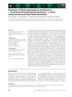
Figure 1. Drug toxicities leading to market withdrawals in the period 1976 to 2005.
Hepatotoxicity or DILI (21%) formed the majority cause for drug withdrawals.
Cardiotoxicity refers to heart-related toxicities other than torsades de pointes. Torsades is
a life-threatening arrhythmia and may present as sudden cardiac death in patients with
structurally normal hearts. Rhabdomyolysis is the breakdown of muscle fibres resulting
in the release of muscle fibre contents (myoglobin) into the bloodstream. ‘Other’ refers to
haemolytic anaemia (1), skin disease (1), immune toxicity (2), gastrointestinal toxicity
(1), respiratory toxicity (1), fatal (1), neurotoxicity (1), blood-related toxicity (1) and
birth defects (1). Percentage of total and number of cases shown in brackets. Figure taken
from Nature Reviews: Drug Discovery (2007), 6: 904-916.
1.1.1. Susceptibility factors and mechanisms of idiosyncratic DILI
Many attempts have been made to describe the mechanisms or hypotheses that
underlie idiosyncratic DILI. The occasional susceptibility of patients to adverse effects of
otherwise mild drugs means there is no intuitive consensus as to how idiosyncrasy
occurs.
Drug-allergic reactions have been suspected to play a role in various idiosyncratic
drug-induced hepatotoxicities (Uetrecht, 2007). Fever, rash, eosinophilia, auto-antibodies
accompanying hepatotoxicity and the rapid recurrence of liver injury upon drug re-
challenge (Gunawan & Kaplowitz, 2004) are features supporting the hypothesis of
4
immune-mediated idiosyncratic DILI. However, not all idiosyncratic DILI-causing drugs
have an allergic-mediated component (Kaplowitz, 2005). The frequent encounter of drugs
that elicit hypersensitivity and non-allergic reactions prompted Kaplowitz to classify
idiosyncratic reactions into allergic and non-allergic drug-induced reactions (Kaplowitz,
2005). Yet it is difficult to exclude allergic reactions based solely on the presentation of
clinical evidence noted above. Furthermore, the development of hapten, a reactive drug
metabolite that covalently binds to proteins, elicits an immune response one to five weeks
after drug exposure. In contrast, the clinical latency of idiosyncratic DILI usually occurs
several months to more than a year after the first drug exposure. In this regard,
Zimmerman classified this as metabolic idiosyncrasy (Zimmerman, 1976), although no
metabolic pathway or mechanism has yet been associated with the cause for idiosyncratic
DILI. Another hypothesis that has been put forward is the inflammagen hypothesis
(Uetrecht, 2008). This is based on a combination of drugs in doses normally tolerated and
inflammagens such as liposaacharide (LPS) that lead to acute hepatic injury in mice. In
contrast to an acute inflammatory phase, the onset of idiosyncratic drug reactions is
characteristically delayed and chronic. LPS itself is a confounding factor, thus making it
difficult to differentiate if the hepatic injuries were potentiated or caused by LPS, or the
drug was amplifying the liver toxic effects of LPS. Therefore it can be argued that
immune-mediated toxic response and inflammagens cannot satisfactorily explain the
uniqueness and pathogenesis of idiosyncratic DILI.
Genetic risk factors may increase the toxic potency of drugs by shifting the dose-
response curve (effectively LC
50
) to the left. However, presently, clinical evidence
5
supporting the presence of polymorphisms in causing idiosyncratic DILI has been
sporadic. Therefore, current hypotheses do not adequately describe possible mechanisms
that predispose susceptible patients to the adverse effects of drug and new paradigms are
urgently needed to explain the unpredictable nature of idiosyncratic DILI.
1.2. Troglitazone as a model drug for the study of idiosyncratic DILI
Troglitazone (Rezulin™, Pfzier; Figure 2), a first-generation thiazolidinedione
drug used in the treatment of type-2 diabetes mellitus was withdrawn from the market
due to an unacceptable risk of idiosyncratic hepatotoxicity (Graham et al., 2002). In early
drug safety assessments, even in long-term studies, troglitazone did not cause
hepatotoxicity in normal healthy rodents and monkeys, (Matsunuma et al., 1993,
Mayfield et al., 1993, Rothwell et al., 1997). Moreover, while subsequent post-market
withdrawal experiments showed that troglitazone caused mitochondrial injury in vitro at
high concentrations, troglitazone was allowed to progress to the clinical testing phase
stage.
A hallmark of troglitazone-induced hepatic injury is the seemingly random and
delayed onset of liver injury, which could abruptly progress to life-threatening and
irreversible liver failure ranging from one month to more than a year’s interval (Graham,
et al., 2002, Iwase et al., 1999). This idiosyncratic hepatotoxicity of troglitazone was not
repeated in 13 double-blind clinical trial studies with the other thiazolidinedione
members of safer profiles, namely rosglitazone and pioglitazone. In this clinical trial
study, 1.91% of 2510 patients (versus 0.6% in the placebo group), 0.26% of 1526
6
patients, and 0.17% of 3503 patients who received troglitazone, pioglitazone, and
rosiglitazone respectively had three times the upper limit of normal (ULN) of ALT
(Lebovitz et al., 2002). ALT is released from dead or injured hepatocytes and is generally
used as indicators to measure liver health
1
1
Healthy ALT range is placed at around 5 IU/L to 50 IU/L but this range changes slightly with the
ethnicity of the population and gender with males slightly higher. IU, international units
. However, it must be stated that ALT above
three times ULN alone does not always predict severe liver toxicity and therefore may
require use of additional clinical parameters (Kaplowitz, 2005). Out of these patients, two
individuals were hospitalised with drug-induced hepatitis while another two individuals
had jaundice although no cases of acute liver failure was reported (Graham et al., 2001,
Watkins & Whitcomb, 1998). Despite the mild elevations in ALT, such irregularity was
not necessarily indicative of subsequent cases of equal or worse severity and hence
troglitazone was brought into the market in 1997. Soon thereafter, several cases of acute
liver failure associated with troglitazone prescription was reported and by 2000,
troglitazone was removed from the market culminating in 94 reported cases of
troglitazone-associated liver failure (U.S. Department of Health and Human Services,
March 21, 2000 ). Ever since, numerous attempts to study the underlying mechanisms
troglitazone-induced liver toxicity have been made, but the in vitro results and ensuing
hypotheses provided little mechanistic relevance to address clinical troglitazone-induced
DILI (Chojkier, 2005, Smith, 2003). Studying the mechanisms behind the idiosyncratic
toxicity of troglitazone not only explain why only a subset of patients develop liver
injury, but also bring us closer to explain how a spectrum of drugs can induce
idiosyncratic DILI. With better understanding of the idiosyncractic DILI mechanisms,
7
potential idiosyncratic liabilities of drugs in preclinical development can be identified
early.
Being lipophilic, troglitazone readily enters the cell and nucleus and bind to
PPARγ with K
d
2
around 40 nM (Lehmann et al., 1995). When liganded, this causes a
conformational change of PPARγ and its heterodimer partner, retinoid X receptor (RXR)
and binds to specific PPAR-response element (PPRE) in or near the transcriptional start
site of target genes (Germain et al., 2002, Kliewer et al., 1992). The conformational
change of PPAR also causes the recruitment of co-activator and co-repressor proteins that
influences the set of transcribed genes (Heinaniemi & Carlberg, 2008). PPRE consists of
two hexameric half-sites of the consensus motif AGGTCA in a direct repeat interspaced
by a nucleotide. PPARγ binds to the first PPRE site while RXR binds the second,
resulting in the initiation of DNA transcription and expression of PPARγ-responsive
genes (Chandra et al., 2008). However, the PPRE sequences are not PPAR isoform-
specific (Lemay & Hwang, 2006). In a similar fashion, troglitazone binds to and activate
PPARγ to elicit its therapeutic effects in tissues. It is therefore interesting to understand
how a normally-mild and beneficial drug used for ameliorating diabetic symptoms can
cause severe hepatotoxicity in certain groups of patients.
2
K
d
is the concentration of a drug that results in binding to 50% of the receptors
8
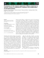
Figure 2. Chemical structure of troglitazone
Figure 3. Crystal structure of PPARγ and RXR
Crystal structure at 3.1 to 3.2Å resolution of PPARγ (red) and RXRα (blue) binding to
PPRE to initiate DNA transcription. The optimal PPRE consensus motif AGGTCA-A-
AGGTCAG.The spacer nucleotide which also forms the minor groove of PPRE
consensus sequence interacts with the DNA-binding domains of PPARγ and RXRα and
shields the highly polar side chains of the interacting residues (Asn 160 from PPARγ, and
Arg 209 and Gln 206 of RXRα) from an aqueous environment. Figure taken from Nature
(2008), 456: 350-356
11
1.2.1. Mitochondrial dysfunction and threshold effect as a possible mechanism
for idiosyncratic DILI
There is growing evidence showing the linkage of mutations in mitochondrial
proteins to reactive oxygen species / reactive nitrogen species (ROS /RNS) in the
pathogenesis of both rare and common human diseases (Droge, 2002). Mitochondrial
diseases due to mutations in nDNA and mtDNA encoding for mitochondrial proteins are
complex, and are confounded by a heterogeneous mix of clinical symptoms and
inheritance patterns (Wallace, 1999). Elevated levels in free radicals under non-regulated
conditions have been implicated with pathophysiological conditions that include
neurodegenerative diseases, aging, ischemia / reperfusion cycles, cancer and
mitochondrial diseases arising from mtDNA mutations and drug-induced toxicities
(Wallace, 1999).
mtDNA has high mutation rates, presumably due to their proximity to sites of
mitochondrial ROS production and the lack of protective histones (Ott et al., 2007). As
some mtDNA gets mutated, each cell could possess a mix of mtDNA variants, some
mutant and some WT, a condition known as heteroplasmy. During cell division of
heteroplasmic cells, cytokinesis will lead to the distribution of mutant mtDNA to other
daughter cells, and as a consequence may lead to an expansion of homoplasmic cells of
mutant mtDNA. Likewise, mtDNAs copy numbers can increase within cells during
clonally expansion, fission and fusion of mitochondria. A mutant mtDNA variant could
be tolerated at low copies, but once a dominance of detrimental mtDNA has been