Isolation and characterization of allergens from curvularia lunata 1
Bạn đang xem bản rút gọn của tài liệu. Xem và tải ngay bản đầy đủ của tài liệu tại đây (278.39 KB, 24 trang )
Chapter 1
19
CHAPTER 1:
BACKGROUND AND INTRODUCTION
Chapter 1
20
1.1 HYPERSENSITIVITY REACTIONS AND ALLERGY
1.1.1 Hypersensitivity Reactions
A normal immune system is beneficial to the human body in order to differentiate self
from non-self and to neutralize potentially pathogenic organisms or substances.
Hypersensitivity refers to undesirable (damaging, discomfort-producing and
sometimes fatal) reactions produced by the normal immune system. In other words,
hypersensitivity refers to a pre-sensitized state of an individual being abnormally
sensitive to the foreign substances causing inflammation and cellular damage.
Hypersensitivity reactions were classified into four types: Type I, II, III and IV, based
on the mechanisms involved (Gel and Coombs, 1975). Later Type V and VI reactions
were added (Rajan, 2003) to the above classification scheme. Details of various
hypersensitivity reactions are highlighted in Table 1.1.
Table 1.1: Various types of hypersensitivity reactions (modified from Gel and
Coombs, 1975).
Type
Mechanism Example/s
I
IgE-mediated immediate hypersensitivity
Systemic anaphylaxis, Asthma,
Eczema, Hay fever
II
Antibody-mediated cytotoxic
hypersensitivity
Haemolytic disease of newborn,
Goodpasture`s syndrome
III
Immune-complex mediated
hypersensitivity
Systemic lupus erythematosus,
arthritis, glomerulo nephritis
IV
Cell-mediated delayed hypersensitivity
Contact dermatitis, Tubercular
lesions
V
Stimulated antibody mediated
hypersensitivity Graves disease
VI
Antibody dependent cell mediated
cytotoxic hypersensitivity Parasitic helminthes infections
Chapter 1
21
1.1.2 Allergy
The term as well as the concept of “allergy” was first introduced by a Viennese
pediatrician, von Pirquet in 1906 (Bendiner, 1981). Allergy is used to refer to a Type I
hypersensitivity reaction. Out of the four major hypersensitivity reactions, allergy has
the most clearly defined and unambiguous immunological as well as pathological
correlation.
Allergy is characterized as a hyper response of IgE antibody to environmental
substances like pollen, dust mites, animal dander, fungal spores, insect venom and
food. Allergic conditions include allergic rhinitis, conjunctivitis, asthma, etc., causing
clinical symptoms like sneezing, coughing, wheezing and breathlessness with
reversible airway obstruction, urticaria and anaphylaxis (Stewart and Thompson,
1996). The initiation of the process is brought up by presentation of processed
environmental antigens to naïve Th precursor cells (ThP) by antigen presenting cells
(APCs) bringing selective proliferation of Th2-polarised memory cells (Th2M),
eventually causing production of antigen specific IgE by the B cells. Re-exposure to
this particular antigen elicits acute phase response brought by cross-linking of IgE
receptors (FcεRI) on mast cells or basophils causing them to degranulate. This results
in release of pro-inflammatory mediators like histamine, leukotrienes and
prostaglandins. These in turn cause symptoms of immediate allergic reactions as
mentioned above. The mast cells can also cause delayed type reactions, 4 -8 hours
after the immediate responses (Holt et al., Holgate, 1999). The mediators released by
mast cells induce release of cytokines and proteases causing tissue damage. The late
Chapter 1
22
phase or delayed type of reaction brings about nasal congestion in allergic rhinitis and
bronchial obstruction in asthma which may lead to airway hyperresponsiveness (AHR)
in future. The major cell types, molecules implicated in allergic reaction and the
overall mechanism underlying allergic reaction is described in Figure 1.1.
Allergy is often explained in terms of “atopy”. The term atopy refers to a hereditary
disorder marked by the tendency to develop immediate hypersensitivity reactions to
specific antigens. Hence it is also referred as “atopic allergy”. As atopy is a hereditary
disorder, the atopic individual shows a predisposition for a Th2-polarised response
which is further enhanced by factors like lack of pathogens in environment,
vaccination, industrialization, clean housing and bedding (Figure 1.2). The
development of atopy is a two-step process. As shown in Figure 1.3, phase 1 of atopic
asthma involves antigen specific immunological memory. This occurs normally in
childhood and results in Th0/Th2-polarized memory, increasing risk for respiratory
disease. The first phase is not sufficient for the disease presentation. The second phase
occurs only in the individuals with persistent inflammation (Holt et al., 1999).
1.2 ALLERGENS
1.2.1 Definition and various allergen types
The word “Allergen” is defined as an antigen that induces IgE antibody synthesis in
atopic patients in response to the allergen, leading to release of histamine and other
pharmacological mediators of immediate hypersensitivity from mast cells and
basophils (Kurup and Banerjee, 2000). Commonly, the allergens are classified into two
Chapter 1
23
Figure 1.1: Molecular and Cellular mechanism of allergy (Adapted from Holt et
al.,1999)
Chapter 1
24
Figure 1.2: Factors responsible for atopy (adapted from Umetsu et al., 2002)
Figure 1.3: Progression of allergic sensitization from early childhood to atopy in
adulthood (adapted from Holt et al., 1999)
Chapter 1
25
types: Indoor and outdoor allergens (Boulet et al, 1997; Kerkhoff et al., 2003). Plant
pollens and fungi are the two major groups of outdoor allergens (Burge, 2000;
Kerkhoff et al., 2003). Indoor allergens are from house dust mites, dander of pets (cats
and dogs), cockroaches and fungi (Burge, 2000; Kerkhoff et al., 2003). In
industrialized nations, atopic diseases affect up to 20% of the population (Kurup and
Banerjee, 2000).
Biochemically, allergens are proteins, carbohydrates or glycoproteins which stimulate
the immune system of the atopic individual and bind specifically to IgE produced in
response to stimulation. To date there are over 300 reported allergens which comprise
molecules of various physiological and biochemical functions (Scheiner, 1995).
Various allergens from pollens, house dust mites and cockroaches have been well
studied, but the same is not true for fungal allergens (Scheiner, 1995). Although fungal
allergens are important (as they are found both indoors and outdoors), very few fungal
species and fungal allergens have been studied in detail for possible allergenicity.
1.2.2 Recombinant allergens in allergy
Classically, allergologists used natural products such as total protein extracts for the
diagnosis and treatment of allergies. However, allergens prepared this way were highly
heterogeneous in the mixture due to the varying amounts of allergenic and non-
allergenic proteins. Moreover, natural extracts had various drawbacks such as chances
of contamination from other allergen sources being prone to proteolysis, degradation
and at times containing various lipopolysaccharides and endotoxins (Linhart and
Valenta, 2005). With the development of molecular biology and recombinant DNA
Chapter 1
26
technology, several recombinant allergens were cloned, expressed, purified and tested.
More than 300 allergen (nucleotide/protein) sequences are now available in Genbank
(www.ncbi.nlm.nih.gov) and various databases. These recombinant allergens would
soon be used in various diagnosis and treatments of allergy (Chapman et al., 2000).
These recombinant allergens may also be used to improve various forms of specific
immunotherapy – SIT (Norman, 1993).
1.3 FUNGAL ALLERGY AND FUNGAL ALLERGENS
1.3.1 Fungi as environmental allergens
Fungi are eukaryotic, achlorophyllus, chitinous cell walled, unicellular/multicellular
organisms which form a separate kingdom in classification (Whittaker, 1969). Fungi
form a large group of organisms found in every ecological niche (Hawksworth, 2001).
Around 1.5 million species of fungi are present worldwide (Alexopoulos et al., 1996).
Based on the spore type produced, the life cycle of a typical fungus is divided into
perfect (sexual) and imperfect (asexual) phases. In modern terms, these states are
referred as the teleomorph and anamorph, respectively and the fungus showing both
states, known as holomorph. Conidium is a term used for asexual spores produced by
anamorphs of filamentous fungi. Most fungi reproduce sexually by meiosis, producing
spores on specialized structures such as basidia or in a specialized structure called the
ascus. These types of fungi are referred as Fungi Perfecti. Fungi liberate spores and
respirable mycelial fragments in large numbers. Fungal species that produce airborne
Chapter 1
27
spores are found under the phyla Dikaryomycota, Zygomycota and Oomycota (Horner
et al., 1995). Details of the classification of the fungal species under these phyla are
shown in Table 1.2.
Fungi cause a number of infectious diseases. Many fungi produce toxins (Kendrick,
1985), some of which are potent carcinogens, e.g., Aflatoxins produced by Aspergillus
flavus. Fungal spores have been identified as one of the sources of indoor and outdoor
allergies (Platts-Mills et al., 1996). Given their smaller size (>10µm), fungal spores
can penetrate the lower respiratory tract causing allergies (Pepys, 1965; Dankaart et
al., 1991; Reponen et al., 2001). The immunological manifestations of fungal allergies
range from dermatitis, sinusitis and asthma, to bronchopulmonary mycoses,
pneumonitis and allergic alveolitis (Lehrer et al., 1983; Fink, 1998). The immune
responses in fungal allergies follow the same pattern as that of other inhalant allergens
such as pollens or house dust mites (Kauffman et al., 1995).
The most commonly found allergic fungi are Alternaria spp., Cladosporium spp.,
Epicoccum nigrum, Fusarium spp., Ganoderma spp., Penicillium spp., Aspergillus
spp., etc., (Beaumont et al., 1985; Solomon and Matthews, 1988). Many yeasts and
mushrooms capable of producing allergic reactions have also been reported (Horner et
al., 1995 and 1998). Generally, Aspergillus spp. and Penicillium spp. are considered as
indoor fungi and are less commonly seen outdoors (Beaumont et al., 1985; Licorish et
al., 1985). Outdoor fungal spore counts are seen to be correlated with clinical
symptoms (Malling, 1986). Most of the allergenic fungal genera belong to the class
Ascomycetes.
Chapter 1
28
Table 1.2: Taxonomic distribution of various airborne spores-producing fungal
genera (adapted from Horner et al., 1995)
Phylum Zygomycota
Class Zygomycetes
Order Mucorales………………………………Mucor, Rhizopus
Phylum Dikaryomycota
Subphylum Ascomycotina
Class Ascomycetes (including imperfect forms)
Order Dothidiales Alternaria, Cladosporium, Epicoccum
Order Eurotiales………………………….……Aspergillus, Penicillium
Order Pleosporales…………………………….Curvularia(Cochliobolus)
Order Helotiales……………………………….Botrytis
Order Hypocreales…………………………….Fusarium
Order Onyngeales…………………………… Trichophyton
Class Saccharomycetes……………………… …….Saccharomyces, Candida
Subphylum Basidiomycotina
Class Holobasidiomycetes
Order Agaricales………………………………Coprinus, Pleurotus, Psilocybe
Order Aphyllophorales……………………… Ganoderma, Merulius
Order Lycoperdales………………………… Calvatia, Geaster
Class Teliomycetes
Order Uredinales………………………….……Rusts
Order Ustilaginales…………………………….Smuts, red yeasts (Sporobolomyces)
Phylum Oomycota
Class Oomycetes
Order Peronosporales…………………… ……Phytophthora, Plasmopara (mildews)
Chapter 1
29
1.3.2 Recombinant fungal allergens
As explained earlier, recombinant allergens are thought to offer towards allergy
diagnosis as well as therapeutics. Although, the breakthrough in recombinant allergens
is promising, compared to other allergens (like dust mites, pollen and foods),
recombinant fungal allergens are less documented and are less studied. To date, there
are only around 90 recombinant fungal allergens submitted to the International Union
of Immunological Societies (IUIS): Allergen nomenclature sub-committee which
maintains the list of available recombinant allergens (Table 1.3). Taking into account
the importance of fungi as environmental allergens and the uniqueness of fungal
airspora, it is of great importance to identify and study these recombinant fungal
allergens in detail.
1.3.3 Global prevalence of fungal allergy
Fungal spores are present worldwide and many species can be observed at most times
of the year (Horner et al., 1995; Chou et al., 2003). Worldwide, more than 80 genera
of the major fungal groups have been associated with symptoms of respiratory tract
allergy (Horner et al., 1995). Fungal spores are usually present in outdoor air
throughout the year in high numbers and frequently exceed pollen concentrations by
100 to 1,000-fold (Lehrer et al., 1983). Globally, fungal allergy is prevalent at 20 to
30% among atopic individuals and up to 6% in the general population (Portnoy et al.,
1987). Epidemiological study on 16,204 civilians in the U.S.A. showed that 3.6% of
the population was sensitized to the fungus Alternaria alternata (Gergen et al., 1987).
Generally, the fungal allergic subjects are seen to have IgEs to various fungal species.
Chapter 1
30
Table 1.3: Fungal allergens as approved by the allergen nomenclature
committee (adapted from www.allergen.org/List.htm)
Fungal allergen name Biochemical type
Mol.wt.
(kDa) Accession
Alternaria alternata
Alt a 1 28 U82633
Alt a 3 heat shock prot. 70
U87807,
U87808
Alt a 4 prot. disulfideisomerase 57 X84217
Alt a 5 acid ribosomal prot. P2 11
X78222,
U87806
Alt a 6 enolase 45 U82437
Alt a 7 YCP4 protein 22 X78225
Alt a 8 mannitol dehydrogenase 29 AY191815
Alt a 10 aldehyde dehydrogenase 53
X78227,
P42041
Alt a 12 acid ribosomal prot. P1 11 X84216
Alt a 13 glutathione-S-transferase 26 AY514673
Cladosporium herbarum
Cla h 2 Ag54 23
Cla h 5 acid ribosomal prot. P2 11 X78223
Cla h 6 enolase 46 X78226
Cla h 7 YCP4 protein 22 X78224
Cla h 8 mannitol dehydrogenase AY191816
Cla h 9 vacuolar serine protease 55 AY787775
Cla h 10 aldehyde dehydrogenase 53 X78228
Cla h 12 acid ribosomal prot. P1 11 X85180
Aspergillus flavus
Asp fl 13 alkaline serine protease 34
Aspergillus fumigatus
Asp f 1 18
M83781,
S39330
Asp f 2 37 U56938
Asp f 3 peroxisomal protein 19 U20722
Asp f 4 30 AJ001732
Asp f 5 metalloprotease 40 Z30424
Asp f 6 Mn superoxide dismut. 26.5 U53561
Asp f 7 12 AJ223315
Asp f 8 ribosomal prot. P2 11 AJ224333
Asp f 9 34 AJ223327
Asp f 10 aspartic protease 34 X85092
Chapter 1
31
Asp f 11 peptidyl-prolyl isomeras 24
Asp f 12 heat shock prot. P90 90
Asp f 13 alkaline serine protease 34
Asp f 15 16 AJ002026
Asp f 16 43 g3643813
Asp f 17 AJ224865
Asp f 18 vacuolar serine protease 34
Asp f 22w enolase 46 AF284645
Asp f 23 L3 ribosomal protein 44 AF464911
Asp f 27 cyclophilin 18
Asp f 28 thioredoxin 12
Asp f 29 thioredoxin 12
Aspergillus niger
Asp n 14 beta-xylosidase 105 AF108944
Asp n 18 vacuolar serine protease 34
Asp n 25 3-phytase B 66-100 P34754
Aspergillus oryzae
Asp o 13 alkaline serine protease 34 X17561
Asp o 21 TAKA-amylase A 53
D00434,
M33218
Penicillium brevicompactum
Pen b 13 alkaline serine protease 33
Pen b 26 acidic ribosomal prot. P1 11 AY786077
Penicillium chrysogenum
(formerly P.notatum)
Pen ch 13 alkaline serine protease 34
Pen ch 18 vacuolar serine protease 32
Pen ch 20 N-acetyl glucosaminidas 68
Penicillium citrinum
Pen c 3 peroxisomal mem. prot. 18
Pen c 13 alkaline serine protease 33
Pen c 19 heat shock prot. P70 70 U64207
Pen c 22w enolase 46 AF254643
Pen c 24 elongation factor 1 beta AY363911
Penicillium oxalicum
Pen o 18 vacuolar serine protease 34
Fusarium culmorum
Fus c 1 ribosomal prot. P2 11 AY077706
Fus c 2 thioredoxin-like prot. 13 AY077707
Trichophyton rubrum
Tri r 2
Tri r 4 serine protease
Chapter 1
32
Trichophyton tonsurans
Tri t 1 30
Tri t 4 serine protease 83
Candida albicans
Cand a 1 40
Cand a 3 peroxisomal protein 29 AY136739
Candida boidinii
Cand b 2 20 J04984, J04985
Psilocybe cubensis
Psi c 1
Psi c 2 cyclophilin 16
Coprinus comatus
Cop c 1 leucine zipper protein 11 AJ132235
Cop c 2 thioredoxin AJ242791
Cop c 3 AJ242792
Cop c 5 AJ242793
Cop c 7 AJ242794
Rhodotorula mucilaginosa
Rho m 1 enolase 47
Rho m 2 vacuolar serine protease 31 AY547285
Malassezia furfur
Mala f 2 MF1, peroxisomal 21 AB011804
membrane protein
Mala f 3 MF2, peroxisomal 20 AB011805
membrane protein
Mala f 4 mitochondrial malate dehydrogenase 35 AF084828
Malassezia sympodialis
Mala s 1 X96486
Mala s 5 18 AJ011955
Mala s 6 17 AJ011956
Mala s 7 AJ011957
Mala s 8 19 AJ011958
Mala s 9 37 AJ011959
Mala s 10 heat shock prot. 70 86 AJ428052
Mala s 11 Mn superoxide dismut. 23 AJ548421
Mala s 12 glucose-methanol-choline oxidoreductase 67 AJ871960
Mala s 13 thioredoxin 12
Epicoccum purpurascens
(formerly E.nigrum)
Epi p 1 serine protease 30 P83340
Chapter 1
33
1.3.4 Prevalence of fungal spores in Singapore environment
In line with the global prevalence of fungi, an aerobiology survey conducted in
Singapore showed abundant presence of fungal spores (Tan et al., 1992). Fungal
spores were found to occur perennially in the Singapore air. Numerically, the fungal
spores dominated around 86-89% of the total airspora, exceeding fern and pollen spore
counts (Lim et al., 1998). Cladosporium (48%) was the most abundant spore type,
followed by Didymosphaeria (31%) and Pithomyces (12%), Curvularia (5%) and
Drechslera (2%) (Lim et al., 1998). The abundance of Cladosporium and Curvularia
was consistent with that of the surveys carried out in different parts of the world but
the presence of Didymosphaeria and Pithomyces was unique as it had not been
reported elsewhere (Lim et al., 1998). The abundance of Pithomyces was different
from the fungal profile reported in the neighboring country and some other parts of the
world where it constituted less than 1% (Lim et al., 1998). This suggests that the
fungal airflora in Singapore was distinct and different on some aspects.
A five year survey (June 1990-June95) was also conducted to study the indoor as well
as to follow the sporulation patterns of various fungal spores. It was observed that
spores of Didymosphaeria, Pithomyces and Curvularia were present in the
environment for more than 80% of the days sampled (Figure 1.4). This data suggests
that the climatic conditions of Singapore favor growth of these fungi almost all year
round. Distinct seasonal variations in the spore densities were observed despite the
absence of climatic seasons in Singapore (Lim et al., 1998). An average spore count of
1688 spores m
-3
day
-1
was found while the maximum spore load was found around
19,000 spores m
-3
day
-1
(Lim et al., 1998).
Chapter 1
34
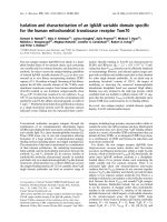
Figure 1.4: Five years` survey (June1990-June95) highlighting the occurrence of
various fungal spore types (%number of days of occurrence / total number of
surveyed days) in Singapore environment
0 20 40 60 80 100
Unknown fungi
Beltrania
Grallomyces
Pringsheimia
Alternaria
Smut fungus
Hiospira
Didymopleela
Pleospora
Torula
Tetraploa
Cladosporium
Drechslera
Curvularia
Didymosphaeria
Pithomyces
Fungal spore type
Occurence (% number of days)
Chapter 1
35
Lim et al. (1998) also observed two periods of high spore density each year, from
February to March and from October to November.
1.3.5 Studies on airborne fungal allergy in Singapore
Annually, an estimated 140,000 individuals suffer from asthma and more than 100
individuals die of this disease resulting in an estimated medical cost of US $33.93
million per annum in Singapore (Chew et al., 1999). As mentioned earlier, airspora
studies conducted in Singapore showed the presence of fungal spores, a number of
which are unique to this region. The high amount of fungal spore load in the
environment raises the question of possible sensitization in atopic patients to these
fungi. Hence a study (as part of an international effort to evaluate the effect of asthma
and allergy around the world) was carried out in school-going children in Singapore
(Goh et al., 1996). As part of the study, about 6000 school-going children (aged
between 6-7 years) and about 4000 children (aged between 12-15 years) were provided
with the International Study of Asthma and Allergies in Childhood (ISAAC)
questionnaire. The results showed that allergic disorders are common to Singaporean
children and the prevalence was comparable to some populations in west countries.
The overall cumulative prevalence of wheezing in children was found to be about 22%
in children of age group of 6-7 years, and 12% in those of age 12-15 years.
In the above study, demographics and socioeconomic factors influenced the prevalence
and severity of allergic disorders with a higher prevalence of rhinitis and wheezing in
male subjects with higher socio-economic status. It was also observed from the
questionnaire survey that there was under-recognition of childhood asthma in these
Chapter 1
36
school-going children, about 49% of 1856 children with asthma-like symptoms had
not been previously diagnosed with asthma (Chew et al., 1999).
In 2000, Chew et al. reported on the allergenicity of locally important fungal spore
types that were frequently found in Singapore’s environment as previously reported by
Lim et al., (1998). It was found that fungal spores induced allergic reactions in about
30% of the atopic Singaporean population on skin prick test (SPT) reactions.
Curvularia spp. which induced about 26-32% of the population showed the highest
response, followed by Drechslera-like spores accounting for around 30% response.
Overall, about 80% of the patients showed skin reactivity to at least one of the fungal
allergens tested (Table 1.4). The association of prevalent fungal spores with atopy
suggested that these fungi play a major role in allergic diseases in the tropics (Chew et
al., 2000). Curvularia was found to be the fungus of great importance locally, and a
better understanding of allergens from this genus would assist in better understanding
the allergic reactions and would also aid towards developing SIT against fungal
allergens.
1.4 Curvularia
Curvularia is a dematiaceous fungus. Most of the Curvularia species are facultative
pathogens, plants and cereals in tropical/subtropical areas and common in the soil.
They are commonly found as saprophytes on cereals (Domsch et al., 1980). The
conidia are multicellular, colored and develop in acropetal manner (youngest at the tip
Chapter 1
37
Table 1.4: Frequency of skin test sensitivity to fungi in Singaporean atopic
population. The positive counts were evaluated via Fisher`s exact test comparing
the patient groups with the healthy controls. * p<0.05, ** p<0.01, *** p<0.001
Healthy controls (n=102)
Atopic patients (n=289)
Allergen tested
Number (%) skin prick test positive
Aspergillus fumigatus 2 (2.0) 60 (20.8) * * *
Penicillium citrinum 7 (6.0) 52 (18.0) *
Healthy controls (n=76)
Atopic patients (n=231)
Allergen tested
Number (%) skin prick test positive
Dermatophagoides pteronyssinus 26 (34.2) 220 (95.2) * * *
Single species spore types
Cladosporium 7 (9.2) 38 (16.5)
Didymosphaeria 9 (11.8) 63 (27.3) * * *
Pithomyces 7 (9.2) 46 (19.9)
Tetraploa 5 (6.6) 37 (16.0)
Curvularia spp. Spores
Curvularia brachyspora 8 (10.5) 64 (27.7) * *
Curvularia fallax 6 (7.9) 54 (23.4) * *
Curvularia inequalis 0 (0.0) 60 (26.0) * * *
Curvularia lunata 5 (6.6) 60 (26.0) * * *
Curvularia pallescens 2 (2.6) 73 (31.6) * * *
Drechslera like spores
Bipolaris 5 (6.6) 71 (30.7) * * *
Corynespora 6 (7.9) 71 (30.7) * * *
Exserohilum 6 (7.9) 41 (17.1) *
Chapter 1
38
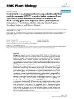
and oldest to the base of the conidiophore). Hyphae are brownish and mature conidia
have usually 3-4 segments (http://vtpb-
www.cvm.tamu.edu/vtpb/vet_micro/charts_fungi/). The colonies of Curvularia are
downy and brown to blackish brown. Figure 1.5 shows the culture as well as the
conidia of Curvularia lunata. According to the recent classification system,
Curvularia falls under phylum Ascomycota, class Euascomycetes, order Pleosporales
and family Pleosporaceae. Based on the information provided by the online guide on
various types of fungi, Mycology online (
there are around 35 species under the genus Curvularia. The conidia are adapted for
efficient aerial dispersal and the fungus is abundantly present in the environment
(Domsch et al., 1980). Though previously considered non-pathogenic, these fungi
have been increasingly reported for causing human disease (Yau et al., 1994).
Curvularia was first reported to be an important inhalant allergen in 1974 (Agarwal
and Shivpuri, 1974). The importance of Curvularia as an inhalant allergen was revised
and it was also thought to be a causative agent for respiratory mycoses (Trave et al.,
1991; Elliot and Taylor, 1997). Curvularia is now known as a causative agent for
various respiratory disorders such as asthma and rhinitis (Gupta et al., 1999). Globally,
there are few reports from America and Asian countries that show the prevalence of
Curvularia sensitization to be around 18-28%. Curvularia lunata was amongst the
most commonly isolated species in various Curvularia infections (Bartynski et al.,
1990). Clinical studies carried out with C. lunata extract in India showed positive skin
reactions (7-16%) in patients with sinusitis (Chakraborty et al., 2000). Curvularia
lunata has also been shown to be cross-reactive with other species within the
Chapter 1
39
Figure 1.5: a) Culture and b) Conidia of Curvularia lunata (Adapted from
)
a)
b)
Chapter 1
40
genus (Curvularia) as well as with other important allergenic fungal species such as
Aspergillus fumigatus, Alternaria alternata, Cladosporium herbarum, and with
various proteins ranging from 12-50 kDa (Gupta et al., 2000). Moreover, as discussed
earlier, various Curvularia species showed the highest response when tested over
atopic Singaporean population. Although C.lunata is an important fungus worldwide
as well as locally; its constitutive allergenic components are not been studied in detail.
Very few reports (as described in Chapter 2) have actually made an effort to
individually characterize them either by studying the native allergenic proteins or
studying them by generating recombinant proteins. In view of these, identification of
various allergenic components of Curvularia will contribute to a better understanding
of the repertoire of Curvularia allergens, and of the allergies caused by Curvularia.
This would then be useful in targeting various diagnostics as well as immunotherapy
tools towards Curvularia allergies.
Chapter 1
41
1.5 Objectives of the present study
The purpose of this study is to identify and characterize the allergens from Curvularia
lunata and to characterize these allergens and cross-compare it with other putative as
well as known allergen homologs. The obtained data will help in understanding the
allergens and allergenicity of Curvularia lunata as well as fungi in general and will
assist in development of improved fungal allergy diagnosis and will provide with a
background to develop fungal allergen specific immunotherapy.
Based on the background knowledge as mentioned earlier, this project aims to:
1) To generate Expressed Sequence Tag database of Curvularia lunata to find
putatively allergenic proteins.
2) To identify allergenic components of Curvularia lunata using 1D western
blots.
3) To identify the expressed allergen transcripts in proteome using two
dimensional sodium dodecyl poly acrylamide gel electrophoresis (2D SDS
PAGE).
4) To clone and express the obtained putatively allergenic proteins from
Curvularia lunata.
5) To clone and express the corresponding homologs of Curvularia lunata
allergenic proteins from Aspergillus fumigatus, Penicillium citrinum,
Alternaria alternata, Saccharomyces cerevisiae, Homo sapiens and Mus
musculus.
6) To characterize various allergen groups for:
Chapter 1
42
a) In-vitro IgE binding reactivity via immuno-dotblot patients` sera from
various populations.
b) IgE antibody cross-reactivity.
c) In-vivo IgE binding via skin-prick reactivity.
d) Specific allergen levels in the environment (outdoors in air as well as
indoors in the house dust).
e) Structural comparison of the allergens and locating possible residues
which are critical for allergenicity.