Studies on the hyaluronidase enzyme purified from the venom of chinese red scorpion buthus martensi karsch
Bạn đang xem bản rút gọn của tài liệu. Xem và tải ngay bản đầy đủ của tài liệu tại đây (6.59 MB, 202 trang )
STUDIES ON THE HYALURONIDASE ENZYME PURIFIED
FROM THE VENOM OF CHINESE RED SCORPION
BUTHUS
MARTENSI KARSCH
FENG LUO
NATIONAL UNIVERSITY OF SINGAPORE
2010
STUDIES ON THE HYALURONIDASE ENZYME PURIFIED
FROM THE VENOM OF CHINESE RED SCORPION
BUTHUS
MARTENSI KARSCH
A thesis submitted by
FENG LUO
(B.Med., M.Med.)
for the degree of
DOCTOR OF PHILOSOPHY
in the
NATIONAL UNIVERSITY OF SINGAPORE
Department of Anatomy
Yong Loo Lin School of Medicine
National University of Singapore
2010
Acknowledgements
I would like to take this opportunity to express my sincere appreciation to my supervisor
Prof. P. Gopalakrishnakone, Department of Anatomy, National University of
Singapore. During my study in Anatomy, I owed much to his great patience, academic
guidance and endlessly encouragement. It is my luck to study under his supervision.
I’d also like to thank Prof. Bay Boon Huat, the head of Department of Anatomy,
National University of Singapore, for his management to make the whole department as a
big family and hence I could enjoy the stay in the department.
I will not forget the great support from Dr. Gao Rong. I have learned much from him,
from the understanding of the science to the techniques of the experiments. I felt so
happy to meet such a big brother in the lab.
I would also thank Dr. M.M.Thwin, the senior member of Venom and Toxin Research
Programme, who helped me in writing the manuscript, patiently listened to my queries
and unselfishly shared his experience. He was always available when I needed the help.
I highly appreciate the kindly help of Mr. Meng Jun, Dr. R. Saminathan, Ms. Hema
D/O Jethanand for my research activities and thank as well to Dr. P. Saravanan, Dr. A.
Pachiappan, Dr. Perumal Samy, and all other Venom and Toxin Research Programme
members, for maintaining a favorable working environment.
I
I would like to thank Ms Yong Eng Siang, Ms Ng Geok Lan, for their efficient
organization to keep the lab clean and safe, Ms Violet Teo, Ms Carolyne Ang and Ms
Diljit Kour d/o Bachan Singh, for their secretarial assistance. I also wish to thank all the
Department staffs and students; I will never forget the life in the Department.
I would also like to show the gratitude to my family. My parents’ support and tolerance is
always the drive for me to step forward.
Last but not least, I would acknowledge the National University of Singapore, for
generously offering me a scholarship to complete this research work.
II
TABLE OF CONTENTS
Acknowledgements I
Table of Contents III
Summary VIII
Publications X
Abbreviations XII
CHAPTER 1: INTRODUCTION
1.1 Venomous animals and their venoms
1.2 Scorpion biology
1.3 Scorpion venom
1.3.1 Sodium channel toxins
1.3.2 Potassium channel toxins
1.3.3 Calcium channel toxins
1.3.4 Chloride channel toxins
1.3.5 Peptides not targeting on ion channels
1.4 Low molecular weight toxins from the venom of BmK scorpion
1.5 High molecular weight proteins from animal venoms
1.6 Hyaluronidase and its substrate hyaluronan (formerly hyaluronic acid)
1.7 Venom hyaluronidases
1.8 Structures of hyaluronidases
1.9 The biological and medical applications of hyaluronidases
1.10 Aims of the present study
1
1
5
7
8
9
9
10
11
13
18
20
23
29
31
III
CHAPTER 2: MATERIALS AND METHODS
2.1 The venom
2.2. Gel filtration of BmK crude venom and molecular weight distribution of fractions
2.3 The screening of the biological activities of BmK crude venom and its gel filtration
33
33
fractions
2.4 The purification of BmHYA1
34
38
2.4.1 Gel filtration
2.4.2 Anion exchange chromatography
2.4.3 Cation exchange chromatography
2.4.4 Reversed-phase high-performance liquid chromatography
2.5 Characterization of BmHYA1
2.5.1 SDS-PAGE
2.5.2 Mass spectrometry
2.5.3 N-terminal sequencing
2.5.4 Optimal pH and temperature
2.5.5 Thermostability
2.5.6 K
m
and V
max
determination
2.5.7 Deglycosylation of BmHYA1
2.5.8 Effect of inhibitors on hyaluronidase activity
2.5.9 Thin-layer chromatography for determination of the final degradation
product
2.6. BmHYA1 cloning and expression
43
43
44
41
42
40
41
40
40
39
39
38
39
38
38
IV
2.6.1 Total RNA extraction 44
2.6.2 First strand cDNA synthesis from Total RNA 47
2.6.3 3’ rapid amplification of cDNA ends 48
2.6.3.1 Design of degenerate GSP 48
2.6.3.2 Amplification of 3’ end cDNA of BmHYA1 with PCR 49
2.6.3.3 Agarose gel electrophoresis 51
2.6.3.4 Isolation of DNA from agarose gel 52
2.6.4 Enzymatic manipulation of DNA 53
2.6.4.1 DNA ligation 53
2.6.4.2 DNA digestion 53
2.6.4.3 Heat shock transformation and white/blue screening 54
2.6.4.4 Isolation of plasmids from the bacteria 55
2.6.4.5 Verification of the insert fragment 56
2.6.4.6 DNA sequencing 57
2.6.5 Protein sequence analyzing 57
2.6.6 Expression of BmHYA1 59
2.7. Biological activity test 60
2.7.1 Cell culture 60
2.7.2 Immunohistochemical staining for hyaluronan 61
2.7.3 Western blot analysis for investigating the effect of the enzyme on the
expression of cancer-related biological molecule 61
2.8 Statistical analysis 62
V
CHAPTER 3: RESULTS AND OBSERVATIONS
3.1 The crude venom 62
3.2 Preliminary separation of BmK crude venom and biological activity screening 62
3.2.1 Preliminary separation of BmK crude venom 62
3.2.2
L-amino acid oxidase activity 72
3.2.3 Fibrinogenolytic activity 72
3.2.4 Hemolytic activity 72
3.2.5 Antibacterial activity 78
3.2.6 Amidolytic activity 78
3.2.7 Phospholipase A
2
activity 78
3.2.8 Hyaluronidase activity 78
3.3. Purification of BmK venom hyaluronidase BmHYA1 84
3.3.1 Gel filtration chromatography 84
3.3.2 Anion exchange chromatography 84
3.3.3 Cation exchange chromatography 84
3.3.4 Reversed-phase high-performance liquid chromatography 84
3.4. Homogeneity and molecular weight of BmHYA1 85
3.5 N-terminal sequence of BmHYA1 92
3.6 Optimal pH profile 92
3.7 Optimal temperature profile 92
3.8 Thermostability 96
3.9 K
m
and V
max
determination 96
3.10 Inhibition assays 96
63
63
63
73
73
73
79
79
79
79
85
85
85
85
85
86
93
93
93
97
97
97
VI
3.11 Deglycosylation assays 96
3.12 End products of hydrolysis of hyaluronan by BmHYA1 101
3.13 The molecular biological study 101
3.13.1 RNA isolation and integrity test 101
3.13.2 RT-PCR and 3’ rapid amplification of cDNA ends 103
3.13.3 TA cloning of the 1.3 kb fragment 103
3.13.4 3’ end cDNA nucleotide and full length protein sequences of BmHYA1 105
3.13.5 Expression of BmHYA1 in E.coli system 114
3.13.5.1 Cloning of BmHYA1 cDNA in pET41a(+) vector 114
3.13.5.2 Expression of recombinant BmHYA1 115
3.14 The biological activity investigation of BmHAY1 116
3.14.1 Direct effect of BmHYA1 on cultured cancer cells 116
3.14.2 BmHYA1 and the expression of CD44 isoforms 119
CHAPTER 4: DISCUSSIONS
4.1 The protein content of the animal crude scorpion venom 122
4.2 The biological activities of BmK crude venom 122
4.3 The purification and characterization of BmHYA1 131
4.4 The N-terminal amino acids sequence of BmHYA1 135
4.5 The cloning and expression of BmHYA1 137
4.6 Biological activities of BmHYA1 145
4.7 Future directions 147
97
102
102
102
104
104
106
115
115
116
117
117
120
123
123
132
136
138
146
148
REFERENCES 151
VII
Summary
The present work includes 1) screening of the biological activities in scorpion Buthus
martensi Karsch (BmK) crude venom; 2) the purification and characterization of the
hyaluronidase enzyme (BmHYA1) from the venom of BmK; 3) the cDNA cloning and
expression of BmHYA1 and 4) the preliminary pharmacological study of BmHYA1.
Scorpion venom is a rich source for short neurotoxic peptides but this study indicates
it also contains various high molecular weight (M.W.) proteins. A number of enzymatic
activities have been detected in the present work including
L-amino acid oxidase (LAAO),
serine protease, and hyaluronidase. It is also possible to contain Phospholipase A
2
(PLA
2)
and
metalloproteinase. This work should be the pioneer in comprehensive investigation of
the enzymatic proteins in scorpion BmK venom.
The hyaluoridase from the crude venom of BmK, later named as BmHAY1, was
studied in detail. The enzyme was purified from the crude venom by a successive
chromatography of gel filtration, ion-exchange and reversed-phase high-performance
liquid chromatography (RP-HPLC). The homogeneity was manifested by dodecyl
sulfate-polyacrylamide gel electrophoresis (SDS-PAGE), matrix assisted laser desorption
ionisation time-of-flight mass spectrometry (MALDI-TOF MS) and Edman degradation.
MALDI-TOF result also showed its molecular weight of 48,696 Da. Its N-terminal amino
acids were determined by Edman degradation and showed homologies to other venom
hyaluronidases to some degree. BmHYA1 has an optimal temperature of 50
o
C and
VIII
optimal pH of 4.5. Its K
m
and V
max
are determined to be 95.3 µg/mL and 3.9 µg/min,
respectively. Additionally, the enzyme can hydrolyze the substrate hyaluronan into
tetrasacchrides.
Rapid amplification of cDNA ends- polymerase chain reaction (RACE PCR)
technique was used to clone the 3’ end BmHYA1 cDNA sequence. The 5’ degenerate
primer was designed based on known N-terminal sequence hence the whole mature
BmHYA1 sequence was deduced. This is also the first hyaluronidase full protein
sequence from the scorpion species. The alignment shows it has some homologies (up to
34%) to other Glycol-Hydro-56 family members. The phylogenetic analysis indicates
early divergence
and independent evolution of BmHYA1 from other hyaluronidase
family members. The recombinant BmHYA1 was expressed in E.coli but did not show
the activity.
The treatment with BmHYA1 to MDA-MB-231 breast cancer cells gave rise to the
removal of hyaluronan from the cell surfaces. The further study about its effect on CD44
molecules showed that the environmental hyaluronidase (BmHYA1) can modulate the
expression of CD44 variant 6.
IX
Publications
Peer Reviewed Papers:
1. Feng, L., Gao, R., Gopalakrishnakone, P., (2008) Isolation and characterization of a
hyaluronidase from the venom of Chinese red scorpion Buthus martensi. Comp Biochem
Physiol C Toxicol Pharmacol. 148:250-7.
2. Feng, L., Gao, R., Meng, J., Gopalakrishnakone, P., Cloning and molecular
characterization of BmHYA1, a novel hyaluronidase from the venom of Chinese red
scorpion Buthus martensi Karsch. Toxicon (In press) doi: 10.1016/j.toxicon.2010.04.009
3. Saminathan, R., Pachiappan, A., Feng, L., Rowan, E.G., Gopalakrishnakone, P., (2009)
Transcriptome profiling of neuronal model cell PC12 from rat pheochromocytoma
Cellular and Molecular Neurobiology 29:533-48.
Conference Abstracts:
1. Feng, L., Gao, R., Gopalakrishnakone, P., Characterization and biological activity
study of a novel hyaluronidase from the venom of Asian scorpion Buthus martensi
Karsch. 8th IST Asia-Pacific Congress on Animal, Plant and Microbial Toxins, Vietnam,
2008
2. Gao, R., Feng, L., Gopalakrishnakone, P., A novel serine protease isolated from the
venom of Asian scorpion Buthus martensi Karsch. 8th IST Asia-Pacific Congress on
Animal, Plant and Microbial Toxins, Vietnam, 2008
X
3. Feng L., Gao R., Gopalakrishnakone P. Cloning and molecular characterization of a
novel serine protease, BMK-CBP, from the venom of Chinese red scorpion Buthus
martensi Karsch. International Anatomical Sciences and Cell Biology Conference
(IASCBC), Singapore, 2010
XI
Abbreviations
AP adapter primer
AUAP abridged universal amplification primer
BmK Buthus martensi Karsch
BCV BmK crude venom
BVHYA bee venom hyaluronidase
BPH-20 bovine PH-20
cDNA complementary DNA
Da dalton
DAB 3,3'-diaminobenzidine
DMEM Dulbecco's Modified Eagle's Medium
DNA deoxy ribonucleic acid
dNTP deoxy nucleotide triphosphate
ECM extracellular matrix
EDTA ethylenediamine tetraacetic acid
FBS fetal bovine serum
GSP gene specific primer
GST tag glutathione S-transferase tag
HPLC high performance liquid chromatography
Human HYAL-1~4 human hyaluronidase-1~4
IPTG isopropyl β-
D-1-thiogalactopyranoside
kDa kilodalton
XII
LAAO
L-Amino Acid Oxidase
LB lysogeny broth
MALDI-TOF matrix assisted laser desorption ionisation time-of-flight
MS mass spectrometry
mRNA messenger ribonucleic acid
M.W. molecular weight
OD optical density
PAGE polyacrylamide gel electrophoresis
PBS phosphate buffered saline
PLA
2
phospholipase A
2
RACR PCR rapid amplification of cDNA ends- polymerase chain
reaction
RBC red blood cell
RNA ribonucleic acid
Rpm revolutions per minute
RT-PCR reverse transcription polymerase chain reaction
PR-HPLC reversed-phase high-performance liquid chromatography
S-2238 Bz-Ile-Glu-Gly-Arg-pNa
SDS-PAGE dodecyl sulfate-polyacrylamide gel electrophoresis
SOC super optimal broth
ss cDNA single-stranded cDNA
TFA trifluoroacetic acid
TIM triose phosphate isomerase
XIII
TLC thin layer chromatography
UAP universal amplification primer
VGSCs voltage-gated sodium channels
XIV
CHAPTER I
INTRODUCTION
Chapter 1: Introduction
1.1 Venomous animals and their venoms
Venomous animals produce lethal secretion known as venom from specialized
venom glands. In animal kingdom, there are many creatures, e.g., snakes, scorpions,
spiders and bees, etc., which may be different in morphology, habit, species and size,
but they all share one remarkable specialty of producing the venoms.
Venomous animals employ venoms for defensive or offensive purpose. The
venom can paralyze or even kill the victim in a very short time. The components in
animal venoms accounting for these biological effects are mainly proteins or peptides.
According to their molecular sizes, the venom proteins can be roughly classified into
two groups: high M.W. enzymes, which are involved in various biochemical
processes, and low M.W. peptides, which act mainly on numerous ion
channels/receptors.
These properties make animal venoms a rich source for biomedical scientists in
search of novel molecules to study biological phenomena or treat human disorders.
1.2 Scorpion biology
The scorpion is one of the most important sources of venom which has been
widely studied. They are one of the oldest creatures which have been in existence on
earth for millions of years since the middle Silurian (about 425~450 million years ago)
period. Scorpions are widely distributed, with over 1,500 species reported so far
1
Chapter 1: Introduction
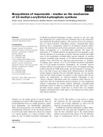
(Polis, 1990), and can be found in all the continents except Antarctica (Fig. 1.1).
Scorpions belong to arthropods and have a large family. Under the class Arachnida,
there are nine families: Bothriuridae, Buthidae, Chactidae, Chaerilidae,
Diplocentridae, Ischnuridae, Iuridae, Scorpionidae and Vaejovidae (Sissom, 1990).
The family Buthidae, containing 48 genera and more than 500 species, is supposed to
be the largest and most widespread species among these families (Sissom, 1990).
Buthidae is also considered as the medically important scorpion family (Fet and Lowe,
2000; Simard and Watt, 1990). Scorpion Buthus martensi Karsch (BmK), the Chinese
red scorpion (also called East Asian scorpion, note: scorpion Mesobuthus tamales is
usually called Indian red scorpion), which belongs to Buthidae family, is the most
commonly found scorpion in mainland China.
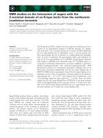
The BmK Scorpion is yellowish to brown in color, and with the length (including
the tail) of up to 6 cm, it is generally small in size as compared to other scorpion
species (Fig. 1.2). It is not aggressive, and its venom toxicity is considered to be
moderate and non-lethal to human (Goudet et al., 2002). The venom is produced and
secreted from the venom gland which is located in the telson (the last segment of the
metasoma. Fig. 1.2). In telson, there is a pair of venom glands each on either side of
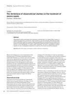
the middle septum (Fig. 1.3).
2
Chapter 1: Introduction
Fig. 1.1. Geographic distribution of scorpions whose venoms have been mostly
studied. Aah, Androctonus australis hector; Amm, Androctonus mauretanicus
mauretanicus; Be, Buthus epeus; Bom, Buthus occitanus mardochei; Bot, Buthus
occitanus tunetanus; Ce, Centruroides sculpturatus; Clt, Centruroides limpidus; Css,
Centruroides suffuses suffuses; Cn, Centruroides noxius; Lqq, Leirus quinquestriatus
quinquestriatus; Lqh, Leiurus quinquestriatus hebraeus; BmK, Buthus martensi
Karsch; Bt, Buthus tamulus; Ts, Tityus serrulatus. (After Loret and Hammock, 2001)
3
Chapter 1: Introduction
Telson
Fig. 1.2. Buthus martensi Karsch (Chinese red scorpion). Wild-specimen from
Xuzhou, Jiangsu Province, PR China. Inset: Segments of metasoma and telson
(venom gland inside).
4
Chapter 1: Introduction
The medical significance of BmK scorpion itself has been recorded for more than
a thousand years. In China, during the Song Dynasty (A.D. 960-1279), the medical
use of the BmK scorpion body was recorded in the official pharmaceutical book Kai
Bao Ben Cao (Kai Bao, referring to a time period from 968-975; Ben Cao means
“herbs”). In another pharmacopoeia, Ben Cao Gang Mu (Compendium of Materia
Medica, A.D.1578), which is probably the most famous Chinese pharmacopoeia book,
the medicinal use of scorpions was described in detail as anti-epilepsy, analgesic,
anticoagulant and anti-rheumatism agents.
1.3 Scorpion venom
Scorpion venom is produced and secreted by the venom glands. When needed,
the scorpion erects the tail and stings the victim with its telson to inject the venom
into the victim’s body. The venom can also be milked by electrical stimulation.
Generally, 1 gram of dry crude venom could be collected from 3000 scorpions
(information from the venom supplier). The first drop of the venom (called pre-venom)
is transparent and clear, but becomes milk-white and mucous later on. The
components of the pre-venom are different from the mature venom (Inceoglu et al.,
2003).
5
Chapter 1: Introduction
Fig. 1.3. Representative diagram of scorpion venom glands. ① cuticle ② skeletal
muscle ③ capsule ④ venom gland ⑤ lumen ⑥ scretory cells. (After Snodgrass,
1952)
6
Chapter 1: Introduction
The venom of scorpion is a very complex mixture, which is composed of mucus,
salts, various small neurotransmitters (e.g., serotonin, histamine, acetylcholine and
norepinephrine), low M.W. peptides (mainly 3~8 kDa neurotoxins) and high M.W.
enzymes (Polis, 1990; Martin-Eauclaire and Couraud, 1995; Nirthanan et al., 2002).
The proteins in the venoms attract the most attention due to their significant
medical/scientific applications.
Small peptides in scorpion venoms are mainly neurotoxins which may modulate
various ion channels on excitable cells. Possessing a broad spectrum of specificity for
ion channels, they are also valuable tools as molecular probes for the basic
neuroscience research. Scorpion neurotoxins can be classified according to the
different ion channels they target, though there are several other species of peptides
that do not target ion channels.
1.3.1 Sodium channel toxins
The sodium channel toxin was the first neurotoxin purified from the scorpion
venom (Rochat et al., 1967). They are usually long chain peptides (60~70 amino acids)
with four disulfide bonds (Possani et al., 1999). On the basis of different targeted sites
on sodium channel, they are further divided into α- and β-toxins (Couraud et al.,
1982).
Voltage-gated sodium channels (VGSCs) consist of an α subunit and two β
subunits (β1 and β2). Scorpion α-toxins interact with the domain in α subunit
(neurotoxin receptor site 3 of VGSCs), while β-toxins bind to the domain located in
7
Chapter 1: Introduction
β1 subunit (neurotoxin receptor site 4 of VGSCs) (Jover et al., 1988; Martin-Eauclaire
et al., 1995). Hence, there is no competitive relationship between these two groups.
The α-toxins can prolong the action potential, and therefore inhibit or slow down the
inactive process of sodium channel. The β-toxins may increase sodium current by
shifting the threshold of activation to hyperpolarized potentials (Couraud et al., 1982;
Marcotte et al., 1997). The sodium channel scorpion toxins that specifically act on
mammals and insects are classified into vertebrate sodium channel toxins and insect
sodium channel toxins, respectively. The latter can be further divided into two groups:
the excitatory and the depressant insect toxins. (Goudet et al., 2002; Martin-Eauclaire
et al.,1995).
1.3.2 Potassium channel toxins
Potassium channels have a large family. Voltage-gated potassium channel
blockers may inhibit cellular proliferation and suppress cellular activation, through
the modulation of calcium influx (Wulff et al., 2009). Scorpion toxins can specifically
target at three potassium channel types: the delayed rectifier potassium channels, the
transient (or A-type) potassium channels and the calcium-dependent potassium
channels (Hille, 1991). Based on structural homology, they fall into 17 sub-families,
termed α-KTx
1-17
(Tytgat et al., 1999; Zhang et al., 2004a; Wang et al., 2005). Most
scorpion potassium channel blockers are relatively short peptides (30-40 amino acids)
with three or four disulfide bridges (Garcia et al., 2001). On the other hand, some long
chain potassium toxins have been purified or cloned from scorpion venoms and may
8