Modelling and simulation of ion implantation induced damage
Bạn đang xem bản rút gọn của tài liệu. Xem và tải ngay bản đầy đủ của tài liệu tại đây (4.14 MB, 191 trang )
MODELING AND SIMULATION OF
ION IMPLANTATION INDUCED DAMAGE
MOK KAI RINE, CAROLINE
NATIONAL UNIVERSITY OF SINGAPORE
2006
MODELING AND SIMULATION OF
ION IMPLANTATION INDUCED DAMAGE
MOK KAI RINE, CAROLINE
(B. Eng (Hons.), NUS)
A THESIS SUBMITTED
FOR THE DEGREE OF DOCTOR OF PHILOSOPHY
DEPARTMENT OF CHEMICAL AND BIOMOLECULAR
ENGINEERING
NATIONAL UNIVERSITY OF SINGAPORE
2006
Acknowledgements
In the course of this work, I am truly thankful and grateful to the many won-
derful people I have had the honor of meeting, interacting with and learning from.
First, I wish to thank my supervisor, Associate Professor M. P. Srinivasan for
his support, patience and kind guidance in my work at the National University of
Singapore (NUS). Without him and the support from the Department of Chemical
& Biomolecular Engineering, this work would not have been possible.
Secondly, I would like to thank those from the industry at Chartered Semi-
conductor Manufacturing (CSM). I am grateful to Dr Lap Chan for encouraging
me to enter the intriguing world of microelectronics, to Dr Francis Benistant for
introducing me to TCAD and for showing me the practical purpose of my work,
to Dr Benjamin Colombeau for being helpful, supp ortive and for our many inter-
esting discussions. Their infectious enthusiasm has provided the motivation that
sustained me throughout this process.
In addition, this work would have been impossible without the guidance of Pro-
fessor Martin Jaraiz and the support of the Department of Electronics, University
of Valladolid (UVa). I am grateful to all for their hospitality and especially to
members of the DADOS team for help with the software. I am grateful to Dr
P. Castrillo, Dr J. E. Rubio, Dr R. Pinacho and Dr I. Martin-Bragado. I have
benefited a lot from their deep knowledge in the field of modeling and simulation.
Furthermore, my scholarship from the Agency of Science, Technology and Re-
search (A*STAR, Singapore) and supp ort from Dr Jin Hongmei from the Ins titute
of High Performance Computing (iHPC, A*STAR) are also gratefully acknowl-
edged.
Validation of models and simulation results would have been impossible without
i
ii
experimental data. I thank the many experimentalists, whose experimental data I
have used to validate my work, especially to J. J. Hamilton, for exchange of ideas
and for providing his experimental SIMS data.
I am grateful to all fellow students at NUS, UVa and CSM for all the fun
and joy, especially to Chan H. Y., Serene and Yeong S. H., Allen, fellow chemical
engineering students working with CSM. I thank them for teaching me, working
with me, whining with me and keeping me sane.
Lastly, I would like to express my love and gratitude to my parents for support-
ing me in whatever I do.
Table of Contents
Page
Acknowledgements . . . . . . . . . . . . . . . . . . . . . . . . . . . i
Table of Contents . . . . . . . . . . . . . . . . . . . . . . . . . . . . iii
Summary . . . . . . . . . . . . . . . . . . . . . . . . . . . . . . . . . . vii
List of Tables . . . . . . . . . . . . . . . . . . . . . . . . . . . . . . ix
List of Figures . . . . . . . . . . . . . . . . . . . . . . . . . . . . . . x
List of Symbols . . . . . . . . . . . . . . . . . . . . . . . . . . . . . . xvii
1 Introduction . . . . . . . . . . . . . . . . . . . . . . . . . . . . . 1
1.1 Motivation . . . . . . . . . . . . . . . . . . . . . . . . . . . 2
1.1.1 Challenges of Ultra-Shallow Junction Forma-
tion . . . . . . . . . . . . . . . . . . . . . . . . . . . 2
1.1.2 Modeling and Simulation of Ion Implantation
Induced Damage . . . . . . . . . . . . . . . . . . . . 6
1.2 Objectives . . . . . . . . . . . . . . . . . . . . . . . . . . . . 8
1.3 Overview . . . . . . . . . . . . . . . . . . . . . . . . . . . . . 10
2 Background Literature . . . . . . . . . . . . . . . . . . . . . . 12
2.1 Defects in silicon . . . . . . . . . . . . . . . . . . . . . . . 12
2.1.1 Point Defects . . . . . . . . . . . . . . . . . . . . . 13
2.1.2 Amorphous Pockets . . . . . . . . . . . . . . . . . . 13
2.1.3 Extended Defects . . . . . . . . . . . . . . . . . . . 18
2.2 Experimental observations of ion-implantation induced
damage accumulation . . . . . . . . . . . . . . . . . . . . . 22
iii
iv
2.3 Simulation techniques . . . . . . . . . . . . . . . . . . . . 28
2.3.1 Binary Collision Approximation . . . . . . . . . . 28
2.3.2 Molecular Dynamics . . . . . . . . . . . . . . . . . 32
2.3.3 Continuum Approach . . . . . . . . . . . . . . . . . 33
2.3.4 Kinetic Monte Carlo . . . . . . . . . . . . . . . . . 34
2.4 Existing models of damage accumulation . . . . . . . . . 38
2.4.1 Homogeneous Amorphization Mechanism . . . . . 38
2.4.2 Heterogeneous Amorphization Mechanism . . . . 43
2.4.3 Dynamic Annealing . . . . . . . . . . . . . . . . . . 44
2.5 Conclusion . . . . . . . . . . . . . . . . . . . . . . . . . . . 47
3 Model Description . . . . . . . . . . . . . . . . . . . . . . . . . . 48
3.1 Simulation Technique . . . . . . . . . . . . . . . . . . . . . 48
3.2 Amorphous pockets . . . . . . . . . . . . . . . . . . . . . . 52
3.2.1 Structure . . . . . . . . . . . . . . . . . . . . . . . . 52
3.2.2 Rat e of Amorphous Pocket Recrystallization . 53
3.2.3 Amorphous Pockets and Clusters . . . . . . . . . 54
3.3 Amorphization . . . . . . . . . . . . . . . . . . . . . . . . . 57
3.4 Recrystallization . . . . . . . . . . . . . . . . . . . . . . . 61
3.4.1 Damage Smoothing . . . . . . . . . . . . . . . . . . 65
4 Effect of Defect Spatial Correlation . . . . . . . . . . . . . 67
4.1 Introduction . . . . . . . . . . . . . . . . . . . . . . . . . . 68
4.2 Model . . . . . . . . . . . . . . . . . . . . . . . . . . . . . . 68
4.3 Results . . . . . . . . . . . . . . . . . . . . . . . . . . . . . . 69
4.4 Conclusion . . . . . . . . . . . . . . . . . . . . . . . . . . . 76
5 Model Validation . . . . . . . . . . . . . . . . . . . . . . . . . . 77
5.1 Amorphous-Crystalline Transition Temperatures . . . 78
5.2 Noble Gas Effect . . . . . . . . . . . . . . . . . . . . . . . 81
v
5.3 Amorphous Layer Thickness . . . . . . . . . . . . . . . . . 83
5.3.1 Ge Preamorphization Implants . . . . . . . . . . . 84
5.4 Dose Effect . . . . . . . . . . . . . . . . . . . . . . . . . . . 90
5.5 Temperature dependence . . . . . . . . . . . . . . . . . . . 94
5.6 Conclusion . . . . . . . . . . . . . . . . . . . . . . . . . . . 99
6 Bimodal Distribution of Damage Morphology . . . . . . . . . 100
6.1 Introduction . . . . . . . . . . . . . . . . . . . . . . . . . . 101
6.2 Model . . . . . . . . . . . . . . . . . . . . . . . . . . . . . . 101
6.3 Results . . . . . . . . . . . . . . . . . . . . . . . . . . . . . . 103
6.4 Conclusions . . . . . . . . . . . . . . . . . . . . . . . . . . . 107
7 Extended Defects Simulations . . . . . . . . . . . . . . . . . . 108
7.1 Introduction . . . . . . . . . . . . . . . . . . . . . . . . . . 109
7.2 Model . . . . . . . . . . . . . . . . . . . . . . . . . . . . . . 111
7.3 Results . . . . . . . . . . . . . . . . . . . . . . . . . . . . . . 117
7.3.1 {311} defects . . . . . . . . . . . . . . . . . . . . . . 117
7.3.2 Non-amorphizing Si implantation . . . . . . . . . . 121
7.3.3 Amorphizing Si implantation . . . . . . . . . . . . 122
7.3.4 Damage Evolution of a Buried Amorphous Layer 126
7.4 Conclusion . . . . . . . . . . . . . . . . . . . . . . . . . . . 130
8 Influence of SOI Structure on Defects . . . . . . . . . . . . 131
8.1 Introduction . . . . . . . . . . . . . . . . . . . . . . . . . . 131
8.2 Results . . . . . . . . . . . . . . . . . . . . . . . . . . . . . . 135
8.2.1 Damage evolution . . . . . . . . . . . . . . . . . . . 135
8.2.2 Dopant Concentration Profile . . . . . . . . . . 136
8.2.3 Sheet Resistance . . . . . . . . . . . . . . . . . . . 139
8.3 Discussion . . . . . . . . . . . . . . . . . . . . . . . . . . . . 142
8.4 Conclusion . . . . . . . . . . . . . . . . . . . . . . . . . . . 145
vi
9 Conclusion . . . . . . . . . . . . . . . . . . . . . . . . . . . . . . 146
9.1 Summary of work . . . . . . . . . . . . . . . . . . . . . . . 146
9.2 Recommendations for future work . . . . . . . . . . . . 149
9.2.1 Effect of dopant atoms on damage . . . . . . . . 149
9.2.2 Effect of stress on damage . . . . . . . . . . . . . 150
9.2.3 Effect of SOI on damage accumulation . . . . . 151
Bibliography . . . . . . . . . . . . . . . . . . . . . . . . . . . . . . . 152
Appendix . . . . . . . . . . . . . . . . . . . . . . . . . . . . . . . . . . 164
Summary
Ion implantation is a well-established processing technique in integrated circuit
fabrication. However, this process induces extensive damage to the silicon crystal
structure. Understanding of ion implantation induced damage is crucial as it af-
fects device performance. In addition, modeling and simulation of ion implantation
induced damage is complicated due to the many interdependent parameters and
defect configurations. Defect production mechanisms, damage kinetics during ion
implantation, damage evolution, amorphization and recrystallization must be accu-
rately simulated in silicon and emerging new substrates such as silicon-on-insulator
(SOI).
In order to model damage accumulation taking into account dynamic annealing,
the most viable option is to use the binary collision approximation (BCA)/kinetic
Monte Carlo (kMC) coupled simulation technique. The software that is used in
this work incorporates an implant function that uses MARLOWE to generate the
coordinates of the interstitials (I) and the vacancies (V) for each cascade. The
coordinates of this damage are then fed into DADOS, a kMC simulator, which
simulates defect reactions.
Central to this model are defect structures known as the amorphous pockets
(AP). Instead of undergoing immediate recrystallization, I’s and V’s are assumed
to form a distinct, disordered region (AP), preventing their diffusion when they
are within a capture distance (second neighbor distance) of each other. Although
the AP recrystallization rate is only size dependent, it is essential to preserve the
I, V spatial correlation in the collision cascades to form the initial APs with a size
distribution dependent on ion mass.
The parameters used in this model have been obtained from experimental
vii
viii
amorphous-crystalline transition temperatures for a range of implanted ions (C
to Xe) to reproduce the experimentally observed dose rate effects. The thicknesses
of the amorphous layers have also been well-simulated in a range of amorphizing
conditions. In terms of the dose effect, the proportion of APs and amorphous re-
gions as a function of dose, and the two-layered damage distribution along the path
of a high-energy ion are consistent with experimental observations. Furthermore,
this model is able to show that dynamic annealing is more effective at removing
damage than p ost-cryogenic implantation annealing at the same temperature.
In addition, it was shown that different implant conditions can lead to different
damage morphology. Since APs and clusters have different thermal stability, with
clusters being more stable and hence more difficult to anneal, the same amount
of damage with different morphology consequently leads to different annealing be-
havior.
An important aspect of damage evolution during post-implantation thermal
annealing involves the transformation of extended defects from {311} defects to
dislocation loops. Based on a size-dependent energy barrier, the transformation
model has been successfully tested against experimental data.
Finally, it has been shown that the damage models developed in this work can
be successfully used in technologically relevant processes involved in the formation
of ultra-shallow junctions. Dopant concentration and activation calculated in terms
of sheet resistance have been simulated in both bulk silicon and SOI. It was demon-
strated that the buried oxide interface has an impact on both defect evolution and
dopant diffusion and activation.
The good agreement between simulation results and various experimental data
shows that the simulations are predictive and can provide valuable insights for
process optimization.
List of Tables
3.1 Parameter values for interstitials and vacancies diffusion. . . . . . . 51
3.2 Parameter values of amorphous pocket defect recrystallization. . . . 53
3.3 Experimental activation energies for 80keV implantation of silicon
with various ions. From Ref. [Goldberg et al., 1995]. . . . . . . . . 55
3.4 Parameter values for the recrystallization of an amorphous-crystalline
plane. . . . . . . . . . . . . . . . . . . . . . . . . . . . . . . . . . . 61
4.1 Details on implantation parameters used in the simulations to attain
the same level of damage induced by C (10 keV) and Xe (80 keV).
Dose rate and temperature were kept constant at 5×10
12
cm
−2
s
−1
and 100 K respectively. . . . . . . . . . . . . . . . . . . . . . . . . . 71
7.1 Parameter values for the transition of {311} defects to dislocation
loops. . . . . . . . . . . . . . . . . . . . . . . . . . . . . . . . . . . 113
ix
List of Figures
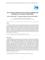
1.1 Cross-section of an NMOSFET doping regions. . . . . . . . . . . . . 3
1.2 The implantation-diffusion interaction matrix. . . . . . . . . . . . . 5
2.1 Some configurations for (a) vacancy (b) interstitial (I) (c) dopant-
interstitial pair (AI) when one of the dark spheres is a dopant atom
(A). From Ref. [Fahey et al., 1989]. . . . . . . . . . . . . . . . . . . 14
2.2 Molecular dynamics simulation of 5 keV Si ion cascades in crystalline
Si target. (a) 0.1 ps (b) 2.5 ps (c) 9 ps. The surface of the incoming
ion is the (100) surface plane of the silicon lattice. Perpendicular line
at the top surface of the box indicates the incoming direction of the
ion. The computational cell is 13.5 nm on each edge, and contains
1.6 × 10
5
Si atoms. Only atoms with potential energy larger than
0.2 eV are plotted. From Ref. [Law et al., 2000]. . . . . . . . . . . . 15
2.3 Molecular dynamics simulations show ing atoms with potential en-
ergy higher than 0.2 eV for (a) 3 keV B and (b) 2 keV As implant
in Si at 300 K after 10 ps. The damage energy is the same for both
ions (1.3 keV). From Ref. [Caturla et al., 1996]. . . . . . . . . . . . 16
2.4 Image of amorphous zones resulting from an impact of a single
200 keV Xe ion. (Total micrograph width = 30 nm) From Ref.
[Donnelly et al., 2003]. . . . . . . . . . . . . . . . . . . . . . . . . . 17
2.5 Cross-section high-resolution electron micrograph showing {311} de-
fects. From Ref. [Eaglesham et al., 1994]. . . . . . . . . . . . . . . . 19
2.6 Transmission electron microscopy micrographs showing faulted and
perfect dislocation loops. From Ref. [Cristiano et al., 2000]. . . . . 20
2.7 Channeling Rutherford backscattering spectra of the same silicon
implant at different substrate temperatures. Ref. [Schultz et al., 1991]. 23
2.8 Experimental crystalline-amorphous transition temperatures for (100)
Si implanted with 80 keV ions to a fluence of 1×10
15
ions/cm
2
for Si,
Ar, Ge, Kr and Xe ions. Data points for carbon are for irradiations
to 2 × 10
15
ions/cm
2
. From Ref. [Goldberg et al., 1995]. . . . . . . . 26
2.9 Schematic drawing of two-body scattering theory in center-of-mass
(CM) coordinates. . . . . . . . . . . . . . . . . . . . . . . . . . . . . 30
2.10 Schematic showing the energy barrier in a process that leads to a
(a) higher-energy state and (b) lower-energy state. . . . . . . . . . . 35
x
xi
2.11 Schematic showing the kMC algorithm that selects random events
according to their corresponding rates. . . . . . . . . . . . . . . . . 36
2.12 Critical point defect density determined from the position of the
amorphous-crystalline interface and simulated BCA point defect pro-
files. . . . . . . . . . . . . . . . . . . . . . . . . . . . . . . . . . . . 39
2.13 Critical point defect density (CPDD) extracted from the measured
depths of amorphous layers and BCA simulations. Figure from Ref.
[Hobler and Otto, 2003]. . . . . . . . . . . . . . . . . . . . . . . . . 40
2.14 Dose dependence of damage produced by 100 keV Si
+
ions in Si (100)
single crystal at room temperature. Damage curves corresponding to
both single alignment (SA) and double alignment (DA) Rutherford
Backscattering Spectra (RB S) measurements, as well as the different
damage components determined from the annealing results. From
Ref. [Holland et al., 1989]. . . . . . . . . . . . . . . . . . . . . . . . 41
2.15 <100> aligned spectra from Si(100) samples implanted at various
doses with 1.25 MeV self-ions. From Ref. [Holland and White, 1991]. 42
2.16 Scheme of damage topology. Each gray circle represents an IV pair
and the dashed lines their interaction radius. Isolated IV pairs (A)
annihilate first as they do not have any IV neighbor. The amorphous
pockets start recombining by the outer IV pairs (B and C) as the
inner ones (F or H) have more IV neighbors. When an IV pair
in a planar structure recombines, the whole layer regrows as the
surrounding defects (D or E) have less IV neighbors than the other
defects in the layer (G). From Ref. [Pelaz et al., 2003]. . . . . . . . 45
3.1 Schematic s howing the division of three-dimensional space in DA-
DOS into small BitBoxes. . . . . . . . . . . . . . . . . . . . . . . . 50
3.2 Amorphous pockets are irregularly-shaped agglomerates of intersti-
tials and vacancies, formed when the point defects are within an
interaction radius of each other. Dashed line represents the interac-
tion radius. . . . . . . . . . . . . . . . . . . . . . . . . . . . . . . . 52
3.3 Activation energy of recrystallization as a function of size of the
amorphous pockets. . . . . . . . . . . . . . . . . . . . . . . . . . . . 55
3.4 Amorphous pockets formed in simulation. (a) In terms of I (yellow)
and V(green) (b) In terms of the number of IV pairs. . . . . . . . . 56
3.5 Schematic showing the consistent treatment of APs and clusters.
Once an AP is completely recrystallized, remaining I or V behave
as pure clusters (point defect capture/emission). Pure cluster can
transform back into AP by capturing defect of the opposite type
within its capture radius. . . . . . . . . . . . . . . . . . . . . . . . . 56
xii
3.6 Simulated damage (total I+V) profiles for 200 keV B implant at
100 K and a dose rate of 1.25 × 10
12
cm
−2
s
−1
. (a) Up to the critical
dose of 1 ×10
15
cm
−2
. (b) Up to a dose of 5 ×10
14
cm
−2
. Dimension
of BitBox: 0.977 nm × 0.938 nm × 0.938 nm. . . . . . . . . . . . . . 59
3.7 Simulated damage (total I+V) profiles for 200 keV B implant at
100 K and a dose rate of 1.25 × 10
12
cm
−2
s
−1
. (a) Up to the critical
dose of 1 ×10
15
cm
−2
. (b) Up to a dose of 5 ×10
14
cm
−2
. Dimension
of BitBox: 0.977 nm × 1.875 nm × 1.875 nm. . . . . . . . . . . . . . 60
3.8 Source and gate time evolution of a transistor during a 600
◦
C an-
neal, following a typical Ge pre-amorphization implant at 20 keV,
1×10
15
cm
−2
(RT and a dose rate of 1×10
13
cm
−2
s
−1
). Snapshots
show annealing at (a) t=0 s, (b) t=20 s, (c) t=40 s, (d) t=60 s, (e)
t=80 s and (f) t=100 s. White represents amorphous damage, yellow
represents amorphous pockets and red represents {311} defects. . . 63
3.9 Source and gate time evolution of a transistor during a 600
◦
C anneal,
following a non-amorphizing Ge implant at 20 keV, 5×10
13
cm
−2
(RT
and a dose rate of 1×10
13
cm
−2
s
−1
). Snapshots show annealing at (a)
t=0 s, (b) t=20 s, (c) t=40 s, (d) t=60 s, (e) t=80 s and (f) t=100 s.
Yellow represents amorphous pockets. . . . . . . . . . . . . . . . . . 64
3.10 Simulated damage (total I+V) profiles for 200 keV B implant at
100 K and a dose rate of 1.25×10
12
cm
−2
s
−1
without damage smooth-
ing, up to the critical dose of 1 × 10
15
cm
−2
. Dimension of BitBox:
0.977 nm × 0.938 nm × 0.938 nm. . . . . . . . . . . . . . . . . . . . 66
4.1 AP histograms obtained from C (lines with symbols) and Xe (dot-
ted lines) implants using BCA at diff erent damage concentrations.
Implantations were simulated with conditions specified in Table 4.1. 70
4.2 Normalized AP distribution obtained from Xe implants using BCA
at different damage concentrations. Implantations were simulated
with conditions specified in Table 4.1. . . . . . . . . . . . . . . . . . 72
4.3 2D color maps showing the AP size distribution at a constant dam-
age of 1 × 10
20
cm
−3
generated by BCA implants of (a) C and (b)
Xe. . . . . . . . . . . . . . . . . . . . . . . . . . . . . . . . . . . . . 74
4.4 AP histograms at a constant damage concentration of 1×10
20
cm
−3
,
of BCA H, C and Xe implants and by loading damage profiles of C
and Xe. . . . . . . . . . . . . . . . . . . . . . . . . . . . . . . . . . 75
xiii
5.1 Simulation (lines) compared to experimental data (symb ols) from
Ref. [Goldberg et al., 1995] for amorphous-crystalline transition tem-
peratures as a function of dose rate, for (100) silicon implanted with
80 keV ions to a dose of 1 × 10
15
cm
−2
for Si, Ar, Ge, Kr and Xe,
and 2 × 10
15
cm
−2
for C. . . . . . . . . . . . . . . . . . . . . . . . . 79
5.2 Activation energy of recrystallization as a function of ion mass from
experimental data [Goldberg et al., 1995]. (Lines drawn to guide the
eyes.) . . . . . . . . . . . . . . . . . . . . . . . . . . . . . . . . . . . 82
5.3 Sequence of simulations showing the development of the amorphous
layer in silicon with increasing dose of 300 keV Si (a) 1 ×10
15
cm
−2
,
(b) 2 ×10
15
cm
−2
, (c) 5 ×10
15
cm
−2
, implanted at 300 K with a dose
rate of 1.5 ×10
12
cm
−2
s
−1
. White represents amorphous regions and
yellow represents amorphous pockets. . . . . . . . . . . . . . . . . . 86
5.4 Simulated amorphous layer thickness as a function of dose rate for
80 keV, 1 × 10
15
cm
−2
Si implant at room temperature. . . . . . . . 87
5.5 Amorphous layer thickness as a function of implant energy for Ge
PAI at a dose of 1 ×10
15
cm
−2
. Simulations were done at room tem-
perature and at a dose rate of 1 ×10
13
cm
−2
s
−1
. Filled symbols rep-
resent experimental data [Pawlak et al., 2002, Cristiano et al., 2004,
Lindsay et al., 2004, Hamilton et al., 2005], open symbols are from
simulations. Experimental characterization methods are indicated
when specified. . . . . . . . . . . . . . . . . . . . . . . . . . . . . . 88
5.6 Amorphous layer thickness as a function of dose for 150 keV Ge
PAI. Simulations were done at room temperature and at a dose rate
of 1 × 10
13
cm
−2
s
−1
. Filled symbols represent experimental data
measured by TEM [Colombeau et al., 2001], open symbols are from
simulations. . . . . . . . . . . . . . . . . . . . . . . . . . . . . . . . 89
5.7 Dose dependence of damage produced by 100 keV Si ions at room
temperature and a dose rate of 5 ×10
12
cm
2
. Symbols show different
damage components determined from the experimental annealing
results, dotted lines are results obtained from simulations. Experi-
mental data from Ref. [Holland et al., 1989]. See also Fig. 2.14. . . 91
5.8 Simulated damage profile resulting from 1 MeV Si implantation at
room temp erature at various doses. . . . . . . . . . . . . . . . . . . 93
5.9 Simulated normalized damage as a function of temperature for 80 keV
C implantations. Post-cryogenic implantation annealing represents
10 minutes annealing after cryogenic temperature implantation. . . . 95
xiv
5.10 2D histograms of APs composition at (a) cryogenic (-150
◦
C) C im-
plantation to a dose of 2×10
14
cm
−2
, (b) post-cryogenic implantation
annealing at 20
◦
C, (c) 20
◦
C C implantation to a dose of 1×10
15
cm
−2
(dynamic annealing). . . . . . . . . . . . . . . . . . . . . . . . . . . 98
6.1 2D histogram of AP composition of an amorphizing 80 keV Carbon
implant at (a) 10% and (b) 80% of the total dose of 2 × 10
15
cm
−2
.
Simulations were done at 20
◦
C and at a dose rate of 5 ×10
12
cm
−2
s
−1
.102
6.2 2D histograms of AP composition showing damage composition at
a constant damage level of 1 × 10
21
cm
−3
, resulting from 80 keV Si
implant at a dose rate of 5 ×10
12
cm
−2
s
−1
. (a) Implant temperature
of -100
◦
C and dose of 1×10
13
cm
−2
(b) Implant at room temperature
and a dose of 1 × 10
14
cm
−2
(c) Implant temperature of 200
◦
C and
dose of 1 × 10
15
cm
−2
. . . . . . . . . . . . . . . . . . . . . . . . . . . 105
6.3 Annealing behavior of damage induced by room temperature im-
plant and by 200
◦
C implant at 800
◦
C. . . . . . . . . . . . . . . . . 106
7.1 Formation energies of the different extrinsic defects as a function of
their size from Ref. [Calvo et al., 2004]. FDL: Faulted dislocation
loops. PDL: Perfect dislocation loops. . . . . . . . . . . . . . . . . . 112
7.2 Energy barrier for the transformation from {311} defects to disloca-
tion loops as a function of the size (number of interstitials, y) of the
{311} defects. . . . . . . . . . . . . . . . . . . . . . . . . . . . . . . 114
7.3 The resulting transition rate, R(y), at 800
◦
C. . . . . . . . . . . . . 114
7.4 Examples of size distribution, g(y) of two different defect popula-
tions, with mean size of 300 and mean size of 2500. . . . . . . . . . 115
7.5 Transformation rate as a function of the size of the {311} defects,
R(y)×g(y) at 800
◦
C. . . . . . . . . . . . . . . . . . . . . . . . . . . 115
7.6 Number of interstitials in {311} defects as a function of time at var-
ious temp eratures after 40 keV, 5 ×10
13
cm
−2
Si . Symbols represent
experimental data and lines represe nt simulations. Experimental
data is obtained from [Stolk et al., 1997]. . . . . . . . . . . . . . . . 118
7.7 Interstitial supersaturation as a function of annealing time at various
tempeartures after 40 keV, 2 × 10
13
cm
−2
Si implantation. Symbols
represent experimental data and lines represent simulations. Exper-
imental data from [Cowern et al., 1999c]. . . . . . . . . . . . . . . . 119
xv
7.8 Cross-sectional images showing the damage morphology after 40 keV,
6 × 10
13
cm
−2
Si implantation, followed by annealing for 30 min at
740
◦
C. (a)Experimental image from Ref. [Colombeau et al., 2003].
(b) Simulation from DADOS showing only {311} defects and no
dislocation loops. Scale for both (a) and (b): 200 nm × 200 nm. . . 120
7.9 Time evolution of the amount of interstitials in extended defects
for the 800
◦
C anneal that follows a non-amorphizing 100 keV, 2 ×
10
14
cm
−2
Si implant. Symbols represent experimental data from
Ref. [Li and Jones, 1998]. Simulation results are plotted as lines
and have been divided by 3. . . . . . . . . . . . . . . . . . . . . . . 123
7.10 Plan-view of {311} defects and dislocation loops corresponding to
the anneal of Fig. 7.9 after 10 minutes. (a) TEM image from Ref.
[Li et al., 1998]. (b) Simulated plan-view. Scale for both (a) and
(b): 100 nm x 100 nm. . . . . . . . . . . . . . . . . . . . . . . . . . 124
7.11 Time evolution of the amount of interstitials in extended defects
for the 750
◦
C anneal that follows a Si 20 keV, 1 × 10
15
cm
−2
amor-
phizing implant. Symbols represent experimental data from Ref.
[Robertson et al., 2000]. Simulation results are plotted as lines and
have been divided by 2. . . . . . . . . . . . . . . . . . . . . . . . . . 125
7.12 Comparison between experimental x-TEM [Pan and Tu, 1997] and
simulation for as-implanted samples. Letters s and a/c indicate the
implanted surface and the location of the as-implanted amorphous-
crystalline interface. The same scale is used for both images, whereby
the simulation width is 50 nm. In the simulation, white represents
amorphous damage and yellow represents amorphous pockets. . . . 128
7.13 Comparison between experimental x-TEM [Pan and Tu, 1997] and
simulation after 1 s, 800
◦
C. In the simulation, yellow represents
amorphous pockets and red represents {311} defects. . . . . . . . . 128
7.14 Comparison between experimental x-TEM [Pan and Tu, 1997] and
simulation after 60 s, 800
◦
C. . . . . . . . . . . . . . . . . . . . . . . 129
7.15 Comparison between experimental x-TEM [Pan and Tu, 1997] and
simulation after 120 s, 900
◦
C. A dislo cation loop is shown in the
simulation. . . . . . . . . . . . . . . . . . . . . . . . . . . . . . . . . 129
8.1 Simulated damage (interstitial) concentration profile of as-implanted
8 and 20 keV Ge, followed by 500 eV B. The saturated (≈ 1 ×
10
22
cm
−3
) level corresponds to amorphized material. . . . . . . . . 136
xvi
8.2 Simulated plan-view of defects in bulk Si corresponding to 60 s an-
neal at (a) 700
◦
C (b) 800
◦
C (c) 900
◦
C of damage induced by 20 keV
Ge preamorphization implant at a dose of 1×10
15
cm
−2
, followed by
500 eV B at a dose of 2 × 10
15
cm
−2
. Scale: 80 nm x 80 nm. Yellow
represents APs, red represents {311} defects and green represents
dislocation loops. . . . . . . . . . . . . . . . . . . . . . . . . . . . . 137
8.3 Simulated plan-view of defects in SOI corresponding to 60 s anneal
at (a) 700
◦
C (b) 800
◦
C (c) 900
◦
C of damage induced by 20 keV Ge
preamorphization implant at a dose of 1 × 10
15
cm
−2
, followed by
500 eV B at a dose of 2 × 10
15
cm
−2
. Scale: 80 nm x 80 nm. Yellow
represents APs, red represents {311} defects and green represents
dislocation loops. . . . . . . . . . . . . . . . . . . . . . . . . . . . . 138
8.4 B concentration profile for 20 keV Ge PAI samples after 60 s, 850
◦
C
anneal in (a) Bulk Si (b) SOI. Experimental SIMS obtained from
Ref. [Hamilton et al., 2005] . . . . . . . . . . . . . . . . . . . . . . 140
8.5 Sheet resistance as a function of annealing temperature, after 60 s
isochronal anneal. (a) 8 keV Ge PAI (b) 20 keV Ge PAI. Experi-
mental points from Ref. [Hamilton et al., 2005] . . . . . . . . . . . 141
8.6 Two-dimensional view of a simulated source-drain extension formed
in bulk Si and SOI (55 nm) with 20 keV 1 × 10
15
cm
−2
Ge PAI, fol-
lowed by 500 eV 2 × 10
15
cm
−2
B. (a) 750
◦
C anneal for 5 min (b)
spike anneal up to 1050
◦
C. (White represents B.) . . . . . . . . . . 144
List of symbols
α prefactor parameter of the amorphous pockets recystallization rate
β exponent parameter of the amorphous po ckets recrystallization rate
φ screening function
Φ angle of recoil in binary collision approximation
γ parameter of energy barrier for {311} defect to dislocation loop
λ second neighb or distance
µ hole mobility
ν
recrys
recrystallization velocity
Θ angle of scatter in binary collision approximation
ω
o
proportionality constant
a screening length
a
o
Bohr radius
a
u
universal screening length
C
B
carrier concentration
D
m
point defect diffusivity
D
mo
prefactor parameter of point defect diffusivity
E0 parameter of energy barrier for {311} defect to dislocation loop
transition
E1 parameter of energy barrier for {311} defect to dislocation loop
transition
E energy barrier for {311} defect to dislocation loop transition
E
act
activation energy
E
c
initial kinetic energy of ion in center-of-mass coordinates
E
ion
energy of ion
E
m
parameter of activation energy for point defect diffusivity
E
o
initial kinetic energy of ion
E
recrys
activation energy of recrystallization
xvii
xviii
g size distribution of defect population
k Boltzmann’s constant
k
e
constant in the electron energy loss calculation
K parameter in the Oen-Robinson formula
m number of vacancies in an amorphous pocket
M
1
, M
2
ion mass
n number of interstitials in an amorphous pocket
N total number of possible events in the kinetic Monte Carlo
algorithm
p impact parameter in binary collision approximation
P probability of event in the kinetic Monte Carlo algorithm
q electronic charge
R0 prefactor parameter in the transition rate of {311} defect to
dislocation loop
r interatomic separation
r
i
rate of individual events in the kinetic Monte Carlo algorithm
r
min
minimum interatomic separation
R transition rate of {311} defect to dislocation loop
R
N
cumulative rate in the kinetic Monte Carlo algorithm
R
s
sheet resistance
s effective size of amorphous pockets
S
E
electron energy loss
T temperature (Kelvin)
T
atom
energy transferred in a collision from an incident projectile
to the target
T
c
amorphous-crystalline transition temperature
V potential energy
V
0
initial velocity of projectile ion
V
C
system velocity in the center-of-mass coordinates
V
0,recrys
prefactor parameter of the recrystallization velocity
xix
x depth of implant
x
j
junction depth
y number of interstitials in {311} defects
y
o
crossover size between the formation energies of {311} defects
and dislocation loops
Z
1
, Z
2
atomic number
Chapter 1
Introduction
The phenomenal growth of the semiconductor industry is driven by technological
advances that fabricate ever smaller and faster transistors. These smaller devices
deliver better performance at lower cost per function. In the competitive world
of consumer electronics, the most advanced technologies are essential for both the
central processing unit (CPU) and memory requirements. This is only possible
with the drastic integration of complementary metal-oxide semiconductor (CMOS)
transistors. Today, 65 nm technology devices are already in production, while the
45 nm technology devices would soon be in 2007. These devices are being de-
veloped and manufactured not only on 300 mm bulk silicon wafers, but also on
silicon-on-insulator (SOI) wafers. The typical gate length for the 65 nm technology
is below 50 nm and for the 45 nm technology, it is below 35 nm. According to the
2005 edition of the International Technology Roadmap for Semiconductors (ITRS),
physical gate length of devices will scale further down to less than 10 nm in the
year 2016 [Semiconductor Industry Association, 2005]. The main issue faced when
fabricating such advanced technology devices is the cost of development. Due to
the escalating cost of fabrication, it becomes increasingly important for foundries
to use less silicon to optimize and yield their processes. The key to overcome this
is to rely on predictive process simulators. This is why there is great interest in
atomistic pro ces s simulation, which can incorporate the physics of the implanta-
tion and diffusion steps to accurately predict junction profiles and thus transistor
performance.
1
CHAPTER 1. INTRODUCTION 2
1.1 Motivation
1.1.1 Challenges of Ultra-Shallow Junction Formation
The typical front-end processing for metal-oxide semiconductor field-effect tran-
sistors (MOSFETs) includes etching, oxidation, ion implantation, diffusion and thin
film deposition. Among these processes, ion implantation and annealing are espe-
cially important, since the formation of ultra-shallow junctions is one of the keys
to achieving sub-50 nm MOSFETs [Koyanagi, 2000].
Ion implantation is a well-established processing technique in integrated cir-
cuits (IC) fabrication for the controlled doping of silicon. It is a process in which
energetic, charged atoms (or molecules) are directly introduced into a substrate.
A typical CMOS process employs dozens of ion implantation steps to form, for ex-
ample, retrograde wells, source/drain junctions and extensions, and halo implants.
Retrograde well is an approach to well formation whereby the highest concentration
of dopant in the well is located at a certain distance from the surface. Halo implants
are necessary to increase the abruptness of the source-drain extension junction dop-
ing profile, decrease the junction depth and increase the punch-through voltage.
In halo implants, dopant of the same type as the major well dopant is implanted
beneath the source-drain extension junction. Figure 1.1 shows the doped regions
of an NMOSFET.
However, implanting high-energy ions into a silicon substrate induces extensive
damage to the crystal structure through nuclear collisions between the incoming
ions or recoils (Si atoms that have been displaced by another high-energy projectile
ion) and the lattice atoms. The as-implanted damage exhibits several different
damage configurations ranging from isolated point defects, point defect clusters,
amorphous pockets surrounded by crystalline silicon to a continuous amorphous
layer. A subsequent high temperature annealing step is required to remove the
damage to maintain good electrical properties. In addition, since most as-implanted
CHAPTER 1. INTRODUCTION 3
Figure 1.1: Cross-section of an NMOSFET doping regions.
dopant atoms do not occupy substitutional sites, this annealing step also serves to
bring about electrical activation.
Understanding of ion implantation induced damage is crucial as it affects fi-
nal junction properties, such as dopant profile and dopant activation. Defects
induced by ion implantation are known to interact with dopant atoms, contribut-
ing to dopant clustering and transient enhanced diffusion (TED) of the dopant
[Stolk et al., 1997]. For the case of boron, damage induced by ion implantation
is known to retard boron activation to concentrations well below the equilibrium
solid solubility [Solmi et al., 1991].
TED is a phenomenon observed during the post-implantation annealing step.
As dopant diffusion is defect-mediated, the implanted dopants experience enhanced
diffusion in the presence of defects ge nerated by the ion implantation step. Since
ion implantation generates a net excess of interstitials, TED significantly affects
dopants whose diffusion mechanisms are predominantly interstitial-mediated. Boron
is a classical example of interstitial-mediated TED in silicon.
CHAPTER 1. INTRODUCTION 4
Mechanisms of boron TED are such that a silicon interstitial “kicks out” the
substitutional boron atom to an interstitial site where it can diffuse easily. Alter-
natively, silicon interstitials and boron atoms form highly mobile pairs. In both
cases, silicon interstitials are required for the diffusion of boron [Stolk et al., 1997,
Jain et al., 2002]. The silicon interstitials for dopant TED may come directly from
the ion implantation process or during the formation and dissolution of extended
defects, like the {311}s defects [Eaglesham et al., 1994] and the dislocation loops
[Bonafos et al., 1997]. Detailed explanations of these defects will be provided in
the following chapter. The impact of ion implantation induced damage becomes
more crucial with shrinking devices, as TED dominates dopant diffusion and limits
the reduction in junction depths.
The key to the formation of ultra shallow junctions is an optimum trade-off
between minimizing dopant diffusion while sufficiently activating the implanted
dopant and removing the damage. The complex interaction between dopants and
defects is schematically shown in Fig. 1.2 [Jones and Rozgonyi, 1993]. Activation
is related to the fraction of dopant atoms that are on substitutional lattice sites
acting as donors or acceptors. Thus, maximum electrical activation could be at-
tained up to the dopant solubility limit. In order to achieve dopant activation and
remove damage, high annealing temperatures are required, which in turn leads to
significant diffusion.
One main method of forming highly-active, ultra-shallow junctions is to im-
plant dopants into preamorphized silicon, followed by a low temperature solid phase
epitaxial regrowth (SPER) process [Jin et al., 2002, Lindsay et al., 2004]. Germa-
nium is often employed in preamorphization implants. At sufficiently high dose, the
implantation-induced damage can result in crystalline to amorphous phase transi-
tion. T he amorphous silicon reduces dopant channeling, resulting in abrupt, shal-
low profiles. Subsequent SPER at low temperature recrystallizes the amorphous
layer, allows only slight dopant diffusion and incorporates dopant atoms in the