Lipid alterations in excitotoxic brain injury
Bạn đang xem bản rút gọn của tài liệu. Xem và tải ngay bản đầy đủ của tài liệu tại đây (3.81 MB, 196 trang )
LIPID ALTERATIONS IN EXCITOTOXIC BRAIN INJURY
HE XIN
(MSc)
Supervisor: Associate Professor Ong Wei Yi
A THESIS SUBMITTED FOR THE DEGREE OF
DOCTOR OF PHILOSOPHY
DEPARTMENT OF ANATOMY
YONG LOO LIN SCHOOL OF MEDICINE
NATIONAL UNIVERSITY OF SINGAPORE
2006
ACKNOWLEDGMENTS
I wish to express my deepest appreciation and heartfelt thanks to my
supervisor, Associate Professor Ong Wei Yi, Department of Anatomy, National
University of Singapore, for suggesting this study topic, and for his constant and
patient guidance and encouragement throughout the course of the study. He has
not only introduced me to an entirely new basic research field but also has been
a role model for hardwork and commitment to research. His deep and sustained
interest, immense patience and stimulating discussions have been most
invaluable in the accomplishment of this thesis.
I am very grateful to Professor Ling Eng Ang, Head, Department of
Anatomy, National University of Singapore, for his constant encouragement,
kindness and unfailing support to execute this research. I am greatly indebted to
Assistant Professor Andrew M. Jenner, Department of Biochemistry, National
University of Singapore, for strong guidance in cholesterol and oxysterol analysis,
and all-round expertise and opinions helped me through many problems. My
deep indebtedness goes to Assistant Professor Markus R. Wenk, Department
of Biochemistry, National University of Singapore, for his invaluable suggestions
and friendly help during this study. I thank Ranbaxy Malaysia Sdn Bhd for
generous supply of lovastatin, and Professor David W. Russell, Department of
Molecular Genetics, University of Texas Southwestern Medical Center, USA, for
I
generous gift of cholesterol 24-hydroxylase antibody and helpful comments on
the manuscript.
I must also acknowledge my gratitude to Mrs Ng Geok Lan and Mrs
Yong Eng Siang for their excellent technical assistance; Miss Chan Yee Gek
and Mdm Wu Ya Jun for Electron Microscopy work; Mr Yick Tuck Yong for his
constant assistance in computer work; Mr Lim Beng Hock for looking after the
experimental animals; Mdm Ang Lye Gek Carolyne and Mdm Teo Li Ching
Violet for their secretarial assistance.
I sincerely thank my co-worker Miss Guan Xue Li, Department of
Biochemistry, National University of Singapore, for her invaluable help in
sphingolipid analysis. I would like to thank all other staff members and my fellow
honous and postgraduate students at Department of Anatomy who help me in
one-way or another.
A major credit also goes to my parents, my brother and my husband, Mr.
Li Quan Sheng, for their full and endless support for my study.
Last , but not least, my many thanks are due to the National University of
Singapore for supporting me with a Research Scholarship to bring this study to
reality.
II
This thesis is dedicated to
my beloved family
III
TABLE OF CONTENTS
ACKNOWLEDGEMENTS …………………………………… …… …………… I
TABLE OF CONTENTS………………………………….……………… ………… IV
PUBLICATIONS…………………………………………… …………….…………. XI
ABBREVIATIONS……………………………………………….…… … ……… XIII
SUMMARY…………………………………………………….……… ……….… XVII
CHAPTER I INTRODUCTION …………………………………………… … ……. 1
1. General introduction … …….……… … …… 2
2. Cell lipids.…………………… ………… 3
2. 1. Phospholipids.…….………… … …….…… 3
2. 1. 1. Structure and functions 3
2. 1 .2. Phospholipids in the brain 4
2. 1. 3 Phospholipids in neurological disorders 7
2. 2. Cholesterol…… ……………….…… …………… …… 9
2. 2. 1. Distribution and functions……………………….…………… 9
2. 2. 2. Cholesterol in the brain 12
2. 2. 2. 1. Cholesterol synthesis and elimination in the brain 12
2. 2. 2. 2. Cholesterol binding/transport proteins in the brain 15
2. 2. 2. 3. Apolipoprotein D 17
2. 2 .3. Cholesterol in neurological disorders 20
2. 3. Ceramide 22
IV
2. 3. 1. Structure and functions 22
2. 3. 2. Ceramide generation and metabolism 24
2. 3. 3. Ceramide in the brain 27
2. 3. 4. Ceramide in neurological disorders 29
3. Kainate-induced excitotoxic neuronal injury ……………………… 31
4. Aims of the present study ………………… ……………… …… ……… 34
4. 1. Dysregulation of cholesterol metabolism after kainate injury 35
4. 2. Dysregulation of ceramide metabolism after kainate injury 35
4. 3. Effect of apolipoprotein D on the neuronal injury after kainate injury 36
CHAPTER II EXPRIMENTAL STUDIES …………….…………………………… 38
I. Lovastatin modulates increased cholesterol and oxysterol levels and has
a neuroprotective effect on rat hippocampal neurons after kainate injury 39
1. Introduction ………… ……………………………………………… ………… 40
2. Materials and methods …………………… …………… ………………… 41
2. 1. Animals and intracerebroventricular kainate injection 41
2. 2. Western blots …… ……………………… …….… 42
2. 3. Immunohistochemical analyses … 43
2. 3. 1. Immunoperoxidase labeling 43
2. 3. 2. Quantitation of labeled cells 44
2. 3. 3. Electron microscopy 45
2. 3. 4. Double immunofluorescence labeling 45
2. 4. Hippocampal slice cultures …… …… 46
V
2. 5. Gas chromatographic/mass spectrometric (GC/MS) analysis 47
2. 5. 1. Kainate and lovastatin treatment 47
2. 5. 2. Lipid extraction 49
2. 5. 3. Lipid hydrolysis 49
2. 5. 4. Cholesterol and oxysterol extraction 49
2. 5. 5. GC/MS measurement 50
2. 5. 6. Cholesterol analysis 51
2. 5. 7. Oxysterol analysis 51
2. 6. In vivo effect of lovastatin on neuronal survival after kainate injury 52
2. 7. In vitro effect of lovastatin on neuronal survival after kainate injury 53
2. 8. In vitro effect of oxysterols on neuronal survival 54
2. 9. Statistical analysis 54
3. Results 55
3. 1. Western blot analysis 55
3. 2. Immunohistochemical analyses of cholesterol 24-hydroxylase after
kainate lesions 55
3. 2. 1. Light microscopy 55
3. 2. 2. Electron microscopy 57
3. 2. 3. Double immunofluorescence labeling 58
3. 3. GC/MS analysis of cholesterol and oxysterols in the kainate-injected rat
hippocampus 58
3. 4. Effect of lovastatin on cholesterol and oxysterol concentrations after
kainate injury 59
VI
3. 4. 1. In vivo analyses 59
3. 4. 2. In vitro analyses 60
3. 5. Effect of lovastatin on neuronal survival after kainate injury 60
3. 5. 1. In vivo analyses 60
3. 5. 2. In vitro analyses 61
3. 6. Effect of 24-hydroxycholesterol on neuronal injury 61
4. Discussion 62
II. Expression, activity, and role of serine palmitoyltransferase in the rat
hippocampus after kainate injury 68
1. Introduction 69
2. Materials and methods 70
2. 1. Animals and intracerebroventricular kainate injection 71
2. 2. SPT expression by Western blot analyses 71
2. 3. SPT activity assay 71
2. 4. SPT immunohistochemistry 72
2. 4. 1. Immunoperoxidase labeling 72
2. 4. 2. Double immunofluorescence labeling 73
2. 4. 3. Electron microscopy 74
2. 5. Hippocampal slice cultures 74
2. 6. Electrospray ionization mass spectrometry (ESI-MS) 74
2. 7. Quantitation of cellular injury by microtubule associated protein (MAP2)
immunolabeling 75
VII
2. 8. Quantitation of cellular injury by lactate dehydrogenase (LDH) assay 76
3. Results 77
3. 1. SPT expression by Western blot analyses 77
3. 2. SPT activity assay 77
3. 3. SPT immunohistochemistry 77
3. 3. 1. Immunoperoxidase labeling 77
3. 3. 2. Double immunofluorescence labeling 79
3. 3. 3. Electron microscopy 79
3. 4. Role of SPT in kainate injury 79
3. 4. 1. Effect on ceramide and sphingomyelin concentrations 79
3. 4. 2. Effect on MAP2 immunolabeling 80
3. 4. 3. Effect on LDH release 80
4. Discussion 80
III. Effect of apolipoprotein D on neuronal survival, cholesterol and Lipid
oxidation product formation after kainate-induced neuronal injury 85
1. Introduction 86
2. Materials and methods 87
2. 1. Hippocampal slice cultures 87
2. 2. Quantitation of cellular injury by MAP2 immunolabeling 88
2. 3. Quantitation of cellular injury by LDH assay 88
2. 4. GC/MS analysis 88
2. 4. 1. Chemicals 88
VIII
2. 4. 2. Lipid extraction 89
2. 4. 3. Lipid hydrolysis 89
2. 4. 4. Mixed anion exchange solid phase extraction 89
2. 4. 5. Derivatization 90
2. 4. 6. GC/MS analysis of cholesterol and oxysterols 91
2. 4. 7. GC/MS analysis of F
2
-isoprostanes 91
2. 5. Statistical analysis 92
3. Results 92
3. 1. Effect of apoD on kainate-induced injury 92
3. 2. Effect of apoD on F
2
-isoprostanes, cholesterol, and oxysterol levels in
cultured hippocampal slices 93
3. 3. Effect of apoD on F
2
-isoprostanes, cholesterol and oxysterol levels in
cultured fibroblasts after hydrogen peroxide treatment 93
4. Discussion 94
CHAPTER III CONCLUSION 98
CHAPTER IV REFERENCES 104
CHAPTER V TABLE, TABLE CAPTION, FIGURES AND FIGURE
LEGENDS 134
IX
PUBLICATIONS
Various portions of the present study have been published or submitted
for publication.
International Refereed Journals
1. He X, Jenner AM, Ong WY, Farooqui AA, Patel SC (2006) Lovastatin
Modulates Increased Cholesterol and Oxysterol Levels and Has a
Neuroprotective Effect on Rat Hippocampal Neurons After Kainate Injury. J
Neuropathol Exp Neurol 5:652-663
2. He X, Guan XL, Ong WY, Farooqui AA, Wenk MR (2007) Increased
expression of serine palmitoyltransferase, and role of ceramide biosynthetic
activity in neuronal degeneration after kainate treatment. J Neuro Res 85:423-
432
3. Guan XL, He X, Ong WY, Yeo WK, Shui G, Wenk MR (2006) Non-targeted
profiling of lipids during kainate-induced neuronal injury. FASEB J 20:1152-1161
(GXL and HX contributed equally)
X
ABBREVIATIONS
ABC avidin-biotin complex
Acetyl- CoA acetyl-coenzyme A
ACAT acyl-coenzyme A: cholesterol acyltransferase
AD Alzheimer’s disease
ALS amyotrophic lateral sclerosis
AP alkaline phosphatase
ApoA-I apolipoprotein A
ApoA-IV apolipoprotein A-IV
ApoD apolipoprotein D
ApoE apolipoprotein E
ApoER2 apolipoprotein E receptor 2
ApoJ apolipoprotein J
Aβ amyloid β-peptide
ATP adenosine triphosphate
BACE1 β-site APP cleaving enzyme 1
BBB blood brain barrier
B-BFAP brain fatty acid binding protein
BHT butylated hydroxytoluene
BLG beta-lactoglobulin
BSTFA N,O-bis(trimethylsilyl)trifluoroacetamide
CA cornu amonis
CA1 hippocampal cornu amonis area 1
XI
CA3 hippocampal cornu amonis area 3
Cer ceramide
Chol cholesterol
CNS central nervous system
DAB 3, 3’-diaminobenzidine tetrahydrochloride
DIPEA N,N-diisopropylethylamine
DNA deoxyribonucleic acid
EM electron microscope
ESI/MS electrospray ionization mass spectrometry
ER endoplasmic reticulum
FA Fatty acid
FAN factor associated with NSmase activation
GC/MS Gas chromatography/mass spectrometry
GFAP glial fibrillary acidic protein
GluRs glutamate receptors
H
2
O
2
hydrogen peroxide
HCl hydrochloric acid
HD Huntington’s disease
HDL high density lipoprotein
H-FABP heart fatty acid binding protei
HMG-CoA 3-hydroxy-3-methylglutaryl coenzyme A
HNE hydroxynonenal
HRP horseradish peroxidase
XII
IHC immunohistochemistry
JNK jun kinase
KA kainic acid
kDa kilodalton
LCAT lecithin cholesterol acyltransferase
LDH lactate dehydrogenase
LDL low-density lipoprotein
LDL-R low-density lipoprotein receptor
LPA lysophospholipases
LRP low density lipoprotein receptor-related protein
MAX Mixed Anion Exchange
MW molecular weight
NGF nerve growth factor
NPC Niemann–Pick disease type C
PAGE polyacrylamide gels
PBS phosphate buffered saline
PBS-Tx phosphate buffered saline containing 0.1% Triton X
PC phosphatidylcholine
PD Parkinson’s disease
PE phosphatidylethanolamine
PFBBr pentafluorobenzylbromide
PG glycerol glycerophospholipid
PI phosphoinositide
XIII
PKC protein kinase C
PLA phospholipase
PP phospholipid
PS phosphatidylserine
PVDF polyvinylidene difluoride
RER rough endoplasmic reticulum
RNA ribonucleic acid
SCP-2 sterol carrier protein-2
SCP-X sterol carrier protein-X
SDS sodium dodecyl sulfate
SER smooth endoplasmic reticulum
SK sphingosine kinase
Sph sphingosine
SMase sphingomyelinase
SPT serine palmitoyltransferase
S1P sphingosine 1-phosphate
TBS Tris buffered saline
TCR T cell receptor
TEM transmission electron microscope
TEMED N,N,N,N -tetramethyl-ethylenediamine
TMCS trimethylchlorosilane
VLDL very low-density lipoprotein
VLDL-R very low-density lipoprotein receptor
XIV
SUMMARY
Lipids are necessary components of all cell membranes and are important
both as structural elements and as modulators of cell fluidity. Several lipid
molecular species are present in cells, including various types of phospholipids,
cholesterols and sphingolipids. Lipids are especially important in the central
nervous system (CNS). Homeostasis of membrane lipids in neurons and myelin
is essential to prevent the loss of synaptic plasticity, cell death and
neurodegeneration. Because membrane lipids are so important as structural
components in the CNS, changes in brain lipid levels due to their increased or
decreased synthesis or metabolism may result in homeostatic dysregulation and
ultimately neurodegeneration. This is important because neurodegeneration is a
characteristic component of all dementias. Inhibition of dysregulated lipid
metabolism may confer neuroprotection. This study used a kainate-induced
neurodegenerative model and suggests that dysregulation of two important
membrane lipids, cholesterol and ceramide, may lead to or accelerate
neurodegeneration and therefore may be important in the pathogenesis of
neurodegenerative diseases. The results also indicate the neuroprotective effect
of a lipid binding protein, apolipoprotein D (apoD).
The first part of the present study was carried out to elucidate alterations
in metabolism of cholesterol, a key lipid component of the cell membrane, after
neuronal injury induced by the excitotoxin, kainate. Increased immunolabeling of
the oxysterol biosynthetic enzyme, cholesterol 24-hydroxylase, was observed in
XV
the rat hippocampus after kainate lesions. This was accompanied by increased
levels of cholesterol, 24-hydroxycholesterol (product of cholesterol 24-
hydroxylase enzymatic activity) and 7-ketocholesterol in homogenates of the
degenerating hippocampus, as detected by gas chromatography / mass
spectrometry (GC/MS). Hippocampi from rats or organotypic slices that had been
treated with kainate plus lovastatin showed significantly lower levels of
cholesterol, 24-hydroxycholesterol, and 7-ketocholesterol, compared to those
treated with kainate only. Lovastatin also modulated hippocampal neuronal loss
after kainate treatment, in vivo and in vitro. The level of 24-hydroxycholesterol
detected in vivo after kainate treatment ( > 50 µM) was found to be neurotoxic in
hippocampal slice cultures. The above results suggest that increased brain
cholesterol biosynthesis and oxysterol formation play a role in propagation of
neuronal death after kainate injury and brain permeable statins such as lovastatin
could have a neuroprotective effect by limiting the levels of oxysterols in brain
areas undergoing neurodegeneration.
The second part of the study focused on changes in metabolism of
ceramide, another major lipid component of the cell membrane, after kainate-
induced neuronal injury. Ceramide is involved in many cellular events including
apoptosis, growth arrest, differentiation, senescence, mediating an immune
response, oxidative stress responses, and nitric oxide signaling. An increase in
ceramide species has recently been demonstrated by lipidomic analysis of the rat
hippocampus after kainate-induced excitotoxic injury. In addition, increased
expression of serine palmitoyltransferase (SPT), the first enzyme in the ceramide
XVI
biosynthetic pathway was observed in reactive astrocytes of the hippocampus
after kainate injections. The increase in enzyme expression was paralleled by
increased SPT enzyme activity in the hippocampus at two week post-kainate
injection. In vitro studies showed that treatment of hippocampal slice cultures
with SPT inhibitor ISP-1 (myriocin) or L-cycloserine modulated increases in 16:0,
18:0 and 20:0 ceramide species and partially reduced kainate-induced cell death.
The above findings indicate a role of SPT in ceramide increase after kainate
injury. They also suggest that increased SPT activity and biosynthetic ceramide
might contribute to neuronal injury after kainate excitotoxicity.
The third part of this study was carried out to examine potential effects of
a lipid binding protein, apoD on neuronal survival after kainate injury. ApoD
belongs to the lipocalin superfamily of transporter proteins that carry various
small hydrophobic ligands, such as arachidonic acid and cholesterol. A marked
increase of apoD has been shown in the rat hippocampus after neuronal injury
induced by kainate. Addition of purified human apoD to kainate treated
hippocampal slice cultures resulted in reduction in neuronal death, and
modulation of increased arachidonic oxidation product (F
2
-isoprostane),
cholesterol, and cholesterol oxidation product (24-hydroxycholesterol and 7-
ketocholesterol) levels in the kainate treated slices. The results showed that the
neuroprotective effect of apoD may be due to its ability to bind arachidonic acid,
thus resulting in reduction of lipid peroxidation products, and its ability to prevent
the formation of neurotoxic cholesterol oxidation products by regulating the
cholesterol metabolism. Fibroblasts from apoD knockout mice showed increased
XVII
F
2-
isoprostane and 7-ketocholesterol levels after hydrogen peroxide induced
oxidative stress, suggesting that this lipocalin may be an important antioxidant
protein in the brain.
Taken together, the above findings indicate that deleterious changes in
lipid homeostasis and signaling may be a key factor in the onset and progression
of pathologies of the brain. They also provide clues to the development of
pharmaceutical strategies to treat neurodegenerative disorders by regulating the
lipid metabolism, in which cholesterol and ceramide metabolic enzyme, and
apoD may play important roles.
XVIII
CHAPTER I
INTRODUCTION
1
1. General introduction
Lipids are important for the brain, as it contains the second highest
concentration of lipids exceeded only by adipose tissue (Adibhatla et al. 2006).
Besides this quantitative importance, lipids in the brain show bewildering diversity
(Wenk 2005). A large number of proteins are associated with synaptic
membranes. In addition, a number of key enzymes involved in the metabolism of
lipids have been discovered and characterized in nerve terminals (Cremona and
De Camilli 2001).
The majority of cellular lipids are organized in membranes (van Meer
2001). This is a fluid patchwork of lipid and protein molecules in constant motion.
Carbohydrates attached to the proteins and phospholipids form glycoproteins
and glycolipids (Alberts et al. 1994). The most abundant membrane lipids are the
phospholipids (PPs). In addition, sphingolipids form a static, solid membrane,
which is fluidized by cholesterol (reviewed in Fahy et al. 2005).
Functional responses of ion channels, synaptic function and cellular
signaling cascades may be affected by the lipid composition of the cell
membrane. It has been suggested that neuronal cell function can be modified to
meet physiologic demand through appropriate alterations in the type, nature and
organization of lipids in specific cell membrane compartments (reviewed in Gross
et al. 2005).
Deleterious changes in lipid homeostasis are viewed as important factors
in the pathogenesis of many neurological disorders such as Alzheimer’s disease
(AD) (Cutler et al. 2004b), Parkinson’s disease (PD) (Sharon et al. 2003),
2
Niemann–Pick disease type C disease (NPC) (Sturley et al. 2004), and cerebral
ischemic injury (Farooqui et al. 2004; Rao et al. 2000; Nakane et al 2000).
2. Cell lipids
2. 1. Phospholipids
2. 1. 1. Structure and functions
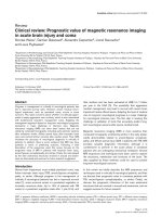
Phospholipids are composed of a glycerol (3 carbon chain) backbone with
fatty acids esterified at the sn-1 and sn-2 positions (Fig. 1). The fatty acids can
vary in length from 14 to 22 carbons and can have from 0 to 6 double bonds
(Schiller et al. 2004). The sn-3 position has a phosphate group attached to a
polar head group (Fig. 1). The amphiphilic nature of phospholipids, owing to the
polar head group and non-polar fatty acid tails, causes them to come together as
a bilayer (reviewed in Peterson and Cummings 2006). There are several groups
of phospholipids based on the polar head group: choline glycerophospholipids
(PC), ethanolamine glycerophospholipids (PE), inositol glycerophospholipids (PI),
glycerol glycerophospholipids (PG) or serine glycerophospholipids (PS) (Paltauf
1994; Farooqui et al. 2000a). The cell membrane has an asymmetrical
distribution of phospholipids. The outer leaflet is mainly composed of PC while
PE and PS are the primary phospholipids found in the inner cytosolic membrane
(Bevers et al.1998). Besides the above glycerophospholipids, membranes also
contain plasmalogens (PlsC and PlsE), which are glycerophospholipids of neural
membranes containing vinyl ether bonds (Farooqui and Horrocks 2001).
3
Figure 1. Basic structure of phospholipids. Consists of a glycerol backbone with fatty acids (R1
and R2) linked at the sn-1 and sn-2 positions. Various polar head groups (X) link to the
phosphate group at the sn-3 position (Peterson and Cummings 2006).
Phospholipid bilayer membranes are highly structured, dynamic and
penetrated to varying degree by receptors, enzymes, and ion channels. The
latter protrude differentially through the membrane or localize predominantly on
the intracellular or extracellular membrane surface. The changes in phospholipid
metabolism can regulate activities of membrane-bound enzymes, receptors, and
ion channels (Farooqui and Horrocks 1985). Different pools of phospholipid
molecular species may have different metabolic and physical properties
depending upon their localization in different types of cell membranes (Farooqui
et al. 2004).
2. 1. 2. Phospholipids in the brain
Brain tissue contains relatively high amounts of phospholipids. Together
with cholesterol and glycolipids, they represent 50–60% of the total membrane
mass of neural membranes (Farooqui et al. 2000b). Human brain neural
membranes contain a variety of phospholipids including PC, PE, PlsE, PS, PI,
and sphingomyelin (Horrocks et al. 1981). PC, PlsE, and PE are major
4
phospholipid components of neural membranes in all regions. This is followed by
sphingomyelin, which is most enriched in white matter (Söderberg et al. 1990).
Among the membranes of the brain, myelin contains the highest content of
phospholipids. The phospholipid composition of myelin is similar to that of white
matter and very different from that of grey matter (Farooqui et al. 2004).
Neural membrane phospholipids are predominantly synthesized in the
endoplasmic reticulum (ER). Significant synthesis of PC and PI also occurs in
Golgi membranes (Farooqui et al. 2000a). Following synthesis, phospholipids are
transported to membranes by phospholipid transfer-exchange proteins (Voelker
et al. 2003). Neural membrane phospholipids are degraded by receptor-mediated
hydrolytic process involving phospholipases (PLA), lysophospholipases (LPA),
and lipases (Farooqui 2000b).
The polyunsaturated fatty acids at the sn-2 position of phospholipids are
susceptible to free radical attack at the α-methylene carbon. The lipid
hydroperoxides thus formed are not completely stable in vivo and, in the
presence of iron, can further decompose to radicals that can propagate the chain
reactions started by an initial free radical attack. Lipid hydroperoxides also
generate aldehydes that can in turn cross-link enzymes and proteins making
them inactive (Farooqui et al. 2000a, Halliwell 1994).
The damage to neural membranes induced by lipid peroxidation can
result in the following effects: (a) changes in physicochemical properties of neural
membranes (microviscosity) resulting in alterations in the orientation of optimal
domains for the interaction of functional membrane proteins such as receptors,
5
enzymes, and ion-channels; (b) changes in the number of receptors and their
affinity for neurotransmitters and drugs; and (c) inhibition of ion pumps resulting
in changes in ion homeostasis (Farooqui et al. 2000a).
The presence of peroxidized phospholipids in neural membranes may also
produce a membrane-packing defect, making the sn-2 ester bond more
accessible to the action of PLA
2
. The hydrolysis of peroxidized phospholipids
results in removal of peroxidized fatty acyl chains, which are reduced and re-
esterified. Thus, the action of PLA
2
repairs and restores the physiological
physicochemical state of neural membranes (Farooqui et al. 2000a). Healthy
neural cells contain tight packing of phospholipids in the outer leaflet. The
disruption of phospholipid asymmetry leads to looser phospholipid packing in the
outer leaflet, thus allowing Ca
2+
entry. The alteration in Ca
2+
homeostasis and its
short duration may lead to neuronal degeneration by the activation of PLA
2
(Farooqui et al. 2000b).
The activation of PLA
2
releases arachidonic acid from neural membrane
phospholipids and sets in motion an uncontrolled ‘‘arachidonic acid cascade’’.
That includes the synthesis and accumulation of prostaglandins, leukotrienes,
thromboxanes, and 4-hydroxy-2-nonenal (4-HNE), a peroxidized product of
arachidonic acid. High concentration of arachidonic acid has a profound adverse
effect on the ATP producing capacity of the brain mitochondria. It uncouples
oxidative phosphorylation and induces efflux of Ca
2+
and K
+
from mitochondria
(Katsuki and Okuda 1995). 4-HNE impairs the activities of key metabolic
enzymes, including Na
+
, K
+
-ATPase, glucose-6-phosphate dehydrogenase, and
6