Molecular mechanisms underlying the regulation of the positioning and formation of the cleavage furrow in cytokinesis in mammalian cells
Bạn đang xem bản rút gọn của tài liệu. Xem và tải ngay bản đầy đủ của tài liệu tại đây (2.71 MB, 140 trang )
MOLECULAR MECHANISMS UNDERLYING THE REGULATION OF THE
POSITIONING AND FORMATION OF THE CLEAVAGE FURROW IN
CYTOKINESIS IN MAMMALIAN CELLS
XIAODONG ZHU
(M.Sc., University of Science and Technology of China)
A THESIS SUBMITTED
FOR THE DEGREE OF DOCTOR OF PHILOSOPHY
TEMASEK LIFE SCIENCES LABORATORY AND
DEPARTMENT OF BIOLOGICAL SCIENCES
NATIONAL UNIVERSITY OF SINGAPORE
2009
i
THIS THESIS IS DEDICATED TO MY FAMILY
ii
ACKNOWLEDGEMENTS
I would like to express my deepest gratitude to my highly respected supervisor Dr.
Maki Murata-Hori. It is my great honor to work with Dr. Maki as her first Ph.D
student. She offered me the rigorous scientific training from the fundamental
techniques to the creation of the great ideas. Her encouragement and guidance
helped me move through all the up and downs of my research. Without her, I would
have been lost and this work would not be possible.
I wish to express my sincerely gratitude to my co-supervisor Associate Professor Dr.
Sohail Ahmed. His deep knowledge of the small GTPases helped my work
immensely. He always gave me encouragement and confidence to pursue my PhD
research in the past 5 years.
I would sincerely like to thank my committee members, Professor Mohan K.
Balasubramanian and Dr. Snezhana Oliferenko for their constructive criticism,
warm encouragement and valuable suggestions.
I would like to thank Ms. Er Poh Nee for technical assistance and help with confocal
microscopy. I would like to extend my thanks to the confocal facility, sequence
facility and all my friends in the TLL for their support.
I also would like to thank all the past and current members of mammalian cell
biology group at the TLL, specially Dr. Svetlana Mukhina, Ms. Shethva Sankaran,
Ms. Charlene Foong, Dr. Ramirez Hernandez Tzutzuy, Mr. Vinayaka Srinivas, Ms.
Shyan Huey Low, Ms. Shazmina Rafee, and Mr. Sriramkumar Sundaramoorthy.
iii
They provide a stimulating environment for me to carry out my research.
Particularly, I would like to thank Ms. Charlene Foong and Dr. Hiroshi Hosoya and
his colleagues (Hiroshima University, Japan) for collaborating with me.
I am especially indebted to my family, my parents, sisters, brother-in-laws, and my
nephew, nieces for their endless love and support. This thesis is dedicated to them.
iv
TABLE OF CONTENTS:
1. Introduction…………………………………………………………… 1
1.1. The cell cycle and cell division…………………………………… … 1
1.2. Cytokinesis………………………………………………………….… 4
1.2.1. Cleavage plane determination ………………………………… 4
1.2.1.1. Roles of microtubules in positioning of the cleavage
furrow……………………………………………………… 5
1.2.1.2. Molecular mechanisms for the determination of the
position of the cleavage furrow …………………… 8
1.2.2. Cleavage furrow formation….………………… 16
1.2.3 Cleavage furrow ingression…………………………………… 19
1.2.4 Abscission………………………………………………….…… 20
2 Materials and Methods…………………………………………………… 23
2.1 Cell Biology……………………………………………………………23
2.1.1 Cell line…………………………………………………… 23
2.1.2 Reagents…………………………………………………… 23
2.1.2.1 Solution……………………………………………………23
2.1.2.2 Drugs………………………………………………………24
2.1.2.3 Antibodies…………………………………………………26
2.1.2.4 F-actin and DNA markers…………………………………27
2.1.3 Cell culture condition……………………………… ………… 28
2.1.4 Transfection………………………………………… …… 29
2.1.5 Microinjection………………………………………… … 29
2.2 Molecular biology………………………………………………………29
2.2.1 E.coli strain……………………………………………… …… 29
2.2.2 Transformation of E. coli……………………………………… 30
2.2.3 Growth and maintenance of E.coli………………… 30
2.2.4 Plasmid construction…………………………… …… 30
2.3 Microscopic imaging………………………………………… … 31
2.3.1 Sample preparation for live-cell imaging………… 31
2.3.2 Sample preparation for immunofluorescence staining……… 31
2.3.3 Image acquisition ………………………….…………… … 32
2.3.4 Fluorescence recovery after photobleaching (FRAP)……… 32
2.4 Image analyses……………………………………………………….… 32
2.4.1 Quantification of fluorescence intensity……………… 32
2.4.2 Kymographic analysis………………………………… 33
2.4.3 Determination of the turnover rate……………………………… 33
3 Molecular mechanism for the regulation of cleavage furrow
positioning………………………………………………………………… 34
3.1 Introduction…………………………………………………………… 34
3.2 Results……………………… ………………………………………….38
3.2.1 Effects of inhibition of aurora B kinase activity on
cytokinesis……………………………………………………… 38
3.2.2 Effects of inhibition of aurora B kinase activity on cytokinetic
regulators………………………………………………….…… 40
3.2.3 Effects of inhibition of aurora B kinase on the microtubule
dynamics………………………………………………………… 42
v
3.2.4 Effects of inhibition of aurora B kinase on localization of the spindle
midzone components………………………………………….…46
3.3 Discussion………………………………………………………… 48
4 Molecular mechanism of the regulation of cleavage furrow
formation…………………………………………………………………….52
4.1 Introduction…………………………………………………………….52
4.2 Results………………………………………………………………….59
4.2.1 Effects of modulation of Cdc42 activity on
cytokinesis……………………………………………………….59
4.2.2 Effects of modulation of Cdc42 activity on RhoA
localization………………………………………………………61
4.2.3 Effects of modulation of Cdc42 activity on the dynamics and
organization of actin cytoskeleton………………………………63
4.2.3.1 Effects of inhibition of Cdc42 activity on actin dynamics and
organization…………… 63
4.2.3.2 Effects of overstimulation of Cdc42 activity on actin
dynamics and organization………………… 67
4.2.4 Involvement of N-WASP in de novo actin assembly at the
equator……………………………………………… 70
4.2.5 Localization of Cdc42 during cytokinesis…………………… 73
4.3 Discussion……………………………………………………… 75
5 Discussion……………………………………………………………… 84
6 References………………………………………………………………… 87
vi
SUMMARY
Cytokinesis is the final step of cell division crucial for cell growth and development.
A clear understanding of the spatial and temporal regulatory mechanisms of
cytokinesis is important not only for basic knowledge of the cellular function but
also for developing effective countermeasures against various diseases such as
cancer and birth defects.
Animal cell cytokinesis consists of four steps: cleavage plane determination,
cleavage furrow formation, ingression and abscission. The mitotic spindle is
responsible for the determination of the position of the cleavage plane. After the
division plane is determined, cytokinetic machinery such as actin and myosin II are
assembled and form the actomyosin contractile ring. Constriction of the contractile
ring drives furrow ingression. Abscission occurs after a furrow has fully ingressed.
Numerous studies have focused on the functions of the proteins that localized at the
cleavage furrow in order to elucidate the molecular mechanisms that regulate
cytokinesis. However, there is an increasing body of research suggesting that
cytokinesis in animal cells likely involves entire cortex in addition to equatorial
cortex. Thus, it is important to identify the functions of each protein involved in
cytokinesis at a high spatial and temporal resolution.
In this thesis, I have studied the molecular mechanisms that regulate the
determination of the position of the cleavage furrow and the cleavage furrow
formation in cytokinesis in mammalian cells using molecular manipulations and
microscopy-based techniques.
vii
Correct positioning of the cleavage plane requires proper regulation of the
microtubule dynamics in mitotic spindle. It has been suggested that stable
microtubules at the equator stimulate the formation of the cleavage furrow, while
dynamic astral microtubules likely inhibit cortical ingression in the polar region.
While many studies have focused on the molecular mechanisms that stabilize
microtubules at the equator, little is known on how astral microtubules maintain
their dynamic status. In Chapter III, I have shown that the kinase activity of aurora
B, a member of the chromosomal passenger complex, is required not only for
stabilization of microtubules at the equator but also for the maintenance of the
dynamic status of astral microtubules to ensure that the cleavage furrow forms at the
equator.
Cleavage furrow formation involves flux-dependent transport of pre-existing actin
filaments and de novo assembly activities. Although functions of Rho family
GTPase RhoA in this process have been established, additional mechanisms are
likely involved. Another Rho family GTPase Cdc42 has been suggested to be
involved in the regulation of actin cytoskeleton during cytokinesis. However, its
detailed functions remain obscure. In Chapter IV, I have shown that Cdc42
contributes to actin assembly by stabilizing actin filaments, promoting de novo
assembly through N-WASP and negatively cross-talking with RhoA during
cytokinesis of mammalian epithelial cells.
viii
LIST OF TABLES:
Table 1: A Cell line used in this study……………………………………………23
Table 2A: A list of drugs used in this study: sources, storage and usage……… 24
Table 2B: A list of the drugs used in this study: molecular mechanisms of
action…………………………………………………………………………… 25
Table 3A: Primary antibodies used in this study……………………………… 26
Table 3B: Secondary antibodies used in this study…………………………… 27
Table 4: F-actin and DNA markers used in this study………………………… 28
ix
LIST OF FIGURES:
Figure1: Organization of microtubules during M phase in typical tissue cultured
cells…………………………………………………………………… 3
Figure2: Classic experiments demonstrate that astral microtubules position cleavage
furrow in embryos whereas midzone microtubules stimulate cytokinesis in
tissue cultured cells. ………………………………………………… 7
Figure 3: Rho GTPases cycle……………………………………………………14
Figure 4: Time-lapse imaging of dividing HeLa cells treated with
Hesperadin 39
Figure 5: Localization of cytokinetic regulators in cells treated with
Hesperadin……………………………………………………… 41
Figure 6: Microtubule organization during cytokinesis in cells treated with
Hesperadin…………………………………………………………….43
Figure 7: Live-cell analyses of microtubules in cells treated with Hesperadin during
cytokinesis…………………………………………………………….45
Figure 8: Localization of MKLP1 and ECT2 during cytokinesis in cells treated with
Hesperadin…………………………………………………………….47
Figure 9: Schematic representation of domains of Cdc42………………………58
Figure 10: Modulation of Cdc42 activity affects cytokinesis………………… 60
Figure 11: Modulation of Cdc42 activity affects RhoA localization………… 62
Figure 12: Inhibition of Cdc42 activity affects actin dynamics and
organization……………………………………………………………65
Figure 13: Constitutive activation of Cdc42 induces the formation of abnormal
actin bundles………………………………………………………….70
Figure 14: N-WASP is involved in de novo actin assembly at the equator…… 73
Figure 15: Localization of Cdc42 during cytokinesis………………………… 75
Figure 16: Proposed model for the regulation of cleavage furrow formation by
RhoA and Cdc42………………………………………………………84
x
LIST OF ABBREVIATIONS:
Arp2/3 Actin-related protein 2/3
BFA Brefeldin A
BSA Bovine serum albumin
CCD Charge-coupled device
Cdc42 Cell division cycle 42
cDNA Complementary DNA
C3 ADP -ribosyltransferase from Clostridium botulinum
DMEM Dulbecco’s modified eagle medium
DMSO Dimethyl sulfoxide
ECT2 Epithelial cell transforming sequence 2 oncogene
EDTA Ethylenediamine tetra acetic acid
FBS Fetal bovine serum
FRAP Fluorescence recovery after photobleaching
FRET Fluorescence resonance energy transfer
F-actin Filamentous actin
FH1 Formin homology 1
FH2 Formin homology 2
F12K Ham's F12 K modification
GAP GTPase activating protein
GBD GTPase protein binding domain
GEF Guanine nucleotide exchange factor
GDI the guanine nucleotide dissociation inhibitor
GDP Guanosine-5-diphosphate
GFP Green fluorescence protein
xi
GTP Guanosine-5-triohosphate
G-actin Globular monomeric actin
LB Luria Bertani medium
MgcRacGAP/RACGAP1 Homo sapiens Rac GTPase activating protein 1
MHC Myosin heavy chain
mRFP Monomeric red fluorescence protein
MRLC Myosin regulatory light chain
MYPT Myosin phosphatase target subunit
NRK Normal rat kidney
N-WASP Neuronal-Wiskott Aldrich syndromer protein
PBS Phosphate buffered saline
RhoA Ras homolog gene family, member A
ROCK Rho-associated kinase
SNARE Soluble N-ethylmaleimide-sensitive factor attachment
protein receptor
STE Sodium Tris-EDTA
TCA Trichloroacetic acid
Tris 2-amino-2(hydroxymethyl)-1,3-propandiol
Triton-X Octylphenoxypolyethoxyethanol
WASP Wiskott Aldrich syndrome protein
VAMP-8 Vesicle-associated membrane protein
1
1. Introduction
1.1 The cell cycle and cell division
Cell growth and reproduction are fundamental features for all living organisms.
To this end, cells undergo the sequential events termed cell cycle. The cell cycle
is divided into four distinct phases: G1, S, G2 and M. G1, S and G2 collectively
are called interphase. In interphase, duplication of genetic materials and cell
growth occur. M phase, in which cell division occurs, consists of tightly coupled
events termed mitosis and cytokinesis. Mitosis is responsible for faithful
segregation of genetic materials into two daughter cells. This process is followed
by cytokinesis, in which the cell is physically divided into two.
Mitosis is composed of five sequential phases: prophase, prometaphase,
metaphase, anaphase, and telophase (Figure 1). Segregation of the replicated
chromosomes is mediated by the cytoskeletal machine termed the mitotic spindle.
The mitotic spindle is mainly composed of microtubules and their associated
proteins.
At prophase, the replicated chromosomes are condensed, while the duplicated
centrosomes start separating apart, forming the mitotic spindle. At prometaphase,
nuclear envelopes break down and microtubules (kinetochore microtubules,
Figure1, prometaphase) emanated from separated centrosomes attach to the
kinetochores of chromosomes, pushing them towards the center of the cell. At
metaphase, all chromosomes are aligned at the equator of the mitotic spindle.
2
Upon anaphase, the mitotic spindle organization is dramatically rearranged.
While the kinetochore microtubules start pulling chromosomes apart, astral
microtubules (microtubules emanated from centrosomes toward polar cortex,
Figure1, anaphase.) extensively elongate, allowing their subpopulation to extend
from the spindle poles (centrosomes) to the equatorial plane of the cells. In
addition, in the region between the separated chromosomes (termed spindle
midzone), antiparallel microtubules are newly developed (termed midzone
microtubules, Figure1 anaphase). At telophase, two sets of daughter
chromosomes reach near the spindle poles and decondense. Cytokinesis usually
begins at late anaphase or telophase. During cytokinesis, astral microtubules that
reach the equator and midzone microtubules are bundled together.
3
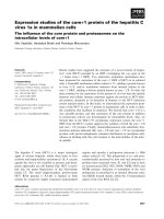
Figure 1: Organization of microtubules typically observed in tissue cultured
cells during M phase.
M phase consists of mitosis (nuclear division) and cytokinesis (cytoplasmic
division). Mitosis is divided into five stages: prophase, prometaphase, metaphase
and telophase. Cytokinesis usually starts at late anaphase or telophase. At
prometaphase, chromosomes attach to kinetochore microtubules. Till metaphase,
astral microtubules are relatively short. Upon anaphase, astral microtubules
elongate and a subset of astral microtubules reach equator. Antiparallel midzone
microtubules assembled between separated chromosomes.
4
1.2 Cytokinesis
Cytokinesis ensures the correct partitioning of the genetic material and cytoplasm
into two daughter cells and is thus crucial for cell proliferation. In animal cells,
cytokinesis can be split into four discrete steps; cleavage plane specification
(cleavage furrow positioning), cleavage furrow formation, ingression, and
abscission.
1.2.1 Cleavage plane determination
The mechanism for the determination of the position of the cleavage plane differs
among different organisms. In higher plants, the preprophase band (PPB), which
is mainly composed of microtubules and actin filaments, accumulates in a band on
the plasma membrane. The PPB becomes gradually thinner and marks the
position where the cell divides. Although the PPB is disassembled at metaphase,
the division sites remains marked by unknown mechanisms. During cytokinesis,
the new cell plate grows from the center of the cell and fused with the cell wall at
the zone formly occupied by PPB.
In budding yeast, the division site is formed at the interface between the mother
cell and daughter bud. Thus division site selection is a consequence of the
budding site selection. To mark the budding site, in G1 phase, a filamentous ring
of protein, septin, accumulates at the cortex near the previous budding site. Septin
ring directs formation of a new bud. As the bud grows, the ‘bud neck’ becomes
the future site of cytokinesis.
5
In fission yeast, a nucleus is positioned in the center of the cell by microtubules
during interphase. In interphase, a protein termed Mid1p localizes in the nucleus.
Upon mitosis, Mid1p is released from the nucleus and accumulates at the cortex
overlaying the nucleus, which recruits the other components to form the
contractile structure.
In animal cells, the position of the cleavage furrow is determined during anaphase.
It has been suggested that the anaphase spindle is responsible for the
determination of the position of the cleavage furrow (Rappaport, 1986; Hiramoto,
1981). Previous studies have attempted to understand the functions of the
different populations of microtubules in mitotic spindle in positioning of the
cleavage furrow.
1.2.1.1 Roles of microtubules in positioning of the cleavage furrow
Pioneering studies in marine invertebrates embryos have suggested that astral
microtubules are responsible for the positioning of the cleavage furrow. In the
Rappaport’s experiment (Rappaport, 1961), ectopic furrowing was observed
between neighboring spindle poles when a torus-shaped sand dollar embryo
generated using a glass ball entered into second mitosis, suggesting that astral
microtubules are sufficient for furrow formation (Figure 2A). When the nucleus
was removed from the heart urchin embryos, a significant number of these cells
were able to form the cleavage furrow between two asters (Lorch et al., 1953),
further suggesting that astral microtubules stimulate furrow formation.
6
There is equally compelling evidence that midzone microtubules are responsible
for the position of the cleavage furrow positioning. When a perforation was
created between the equatorial cortex and the mitotic spindle at one side of tissue
cultured cells at metaphase, no cleavage furrow was formed at the perforated side
(Figure 2B). In the cells with triple poles, the cleavage furrow was formed in the
region where midzone microtubules were bundled (Rieder et al., 1997).
Expression of the non-degradable mutant of cyclin B inhibited the formation of
midzone microtubules, leading to a failure of furrow formation (Wheatley and
Wang, 1996).
These differences in the role of astral versus midzone microtubules are not likely
due to differences in cell type as elegant micromanipulation experiments in
grasshopper spermatocytes indicate that both astral and midzone microtubules can
induce furrow formation. When asters were dissociated from the spindle at
metaphase/early anaphase and then sequestered into a membrane pocket by
microneedles, astral microtubules bundles were formed at late anaphase and
cortical ingression occurred at the cortex proximal to the bundles of microtubules
in the pocket, suggesting that astral microtubules are sufficient for furrow
formation (Alsop and Zhang, 2003). Interestingly, when both chromosomes and
asters were removed from late mitotic cells to produce the cells possessing only
midzone microtubules, the cortex adjacent to the bundled midzone microtubules
7
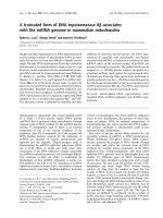
Figure2: Classic experiments demonstrate that astral microtubules position
the cleavage furrow in embryos whereas midzone microtubules stimulate
furrow formation in tissue cultured cells.
(A) In a “torus”-shapped sand dollar embryo, ectopic furrow formed between two
adjacent asters of two spindles. (B) In a tissue cultured cell, when a perforation is
made between the equatorial cortex and the spindle, only cortex at the non-
perforated site ingressed.
8
showed furrow ingression even after the array of microtubules was rotated to
avoid signals placed at an earlier phase (Bonaccorsi et al, 1998; Bucciarelli et al,
2003). Taken together, these experiments suggest that both astral and midzone
microtubules are able to stimulate furrow formation.
The other elegant experiments using PtK1 cells have suggested that the position of
the cleavage furrow is determined by the specialized microtubules of the anaphase
spindle. In cells with monopolar spindles generated by inhibiting centrosome
separation, chromosomes accumulate around the one side of undivided
centrosomes. When these cells were microinjected with an inhibitor for Mad2, a
key protein for the spindle checkpoint, they entered anaphase (although both sister
chromatids migrated toward the centrosomes) and, strikingly, the cleavage furrow
was formed on the side opposite to the centrosomes. In these cells, microtubules
emanating around the chromosomes elongated toward the cell cortex facing the
chromosomes and bound to the cortex (Canman et al., 2003). Kymographic
analyses revealed that microtubules in the furrow region were stable while those
in the polar region were dynamic (Canman et al., 2003). A similar difference in
the dynamics of microtubules was also observed in the normal cells with bipolar
spindle. Microtubules associated with the equatorial cortex were more stable than
those along the polar cortex. Taken together, these results suggest that stabilized
microtubules are able to stimulate furrow formation.
1.2.1.2 Molecular mechanisms for the determination of the position of the
cleavage furrow
9
How are microtubules associated with the equatorial region stabilized and how do
these microtubules determine the position of the cleavage furrow?
Proteins/protein complexes that interacted with microtubules such as PRC1, the
centraspindlin complex, and the chromosome passenger complex are involved in
the stabilization of microtubules at the equatorial region. In addition, the
centralspindlin complex and a member of the choromosome passenger complex,
aurora B kinase (see below) are also involved in the regulation of equatorial RhoA
activity (Miller, Bement, 2009), which aids in the the formation of the cleavage
furrow (see below). Stabilized microtubules might further contribute to the
positioning of the cleavage furrow by catalysing the activity of aurora B (Fuller et
al, 2008) or localization of the centralspindlin complex at the equator.
PRC1 is originally identified in HeLa cells as a Cdk substrate (Jiang et al., 1998).
Subsequently it was shown that PRC1 has the microtubule bundling activity and
forms oligomers in vivo. When it was overexpressed in mammalian cells,
extensive bundling of interphase microtubules was induced (Jiang et al., 1998;
Mollinari et al., 2002; Zhu et al., 2006). Phosphorylation of PRC1 by Cdc2-
cyclinB inhibits oligomerization and its association with microtubules (Zhu et al.,
2006). Upon anaphase onset, PRC1 is deposphorylated and translocates to the
spindle midzone by a kinesin motor protein KIF4 (Kurasawa et al., 2004; Zhu and
Jiang, 2005) where it bundles and stabilizes microtubules in the midzone
microtubules
The centralspindlin complex is comprised of a microtubule plus-end directed
motor protein MKLP1 (in vertebrates) /ZEN4 (in C. elegans)/ Pavarotti (in
10
Drosophila), and an upstream regulator of RhoA, a RhoGAP protein
MgcRacGAP (in vertebrates) / CYK-4 (in C. elegans)/ RacGAP50C (in
Drosophila). Unless otherwise stated, these components of the centralspinlin
complex are refered to as MKLP1 and MgcRacGAP, respectively. Biochemical
experiments demonstrated that MgcRacGAP and MKLP1 assembled into
homodimers seperately, prior to forming a heterotetrameric complex (Pavicic-
Kaltenbrunner, 2007). During anaphase, centraspindlin complex is localized to
spindle midzone and cross-linked midzone microtubule. In vitro experiments
demonstrated that the centraspindlin complex induces extensive bundling of
microtubules (Mishima et al., 2002). Its microtubule bundling activity requires
the intact complex (Mishima et al., 2002).
The chromosomal passenger complex has an important role in regulating the
organization of midzone microtubules. This complex includes a single enzymatic
subunit aurora B kinase, and three other regulatory subunits, INCENP (inner
centromere protein), survivin and borealin. INCENP functions as the scaffold for
the whole complex. Moreover, binding of aurora B to INCENP stimulates the
kinase activity of aurora B (Bishop and Schumacher, 2002; Sessa et al., 2005). It
remains controversial whether survivin and borealin regulates the kinase activity
of aurora B. However, ablating any subunit of the chromosome complex caused
impaired localization of the other subunits and disrupts mitosis and cytokinesis
(Ruchaud et al., 2007), suggesting that the entire complex is essential for both
mitosis and cytokinesis.
Until anaphase onset, the chromosomal passenger complex is mainly localized to
11
the centromeres of chromosomes. After chromosomes separatation, this complex
relocates from centromeres to the spindle midzone, where it regulates the
organization of midzone microtubules via several different mechanisms.
Both in vivo and in vitro experiments have demonstrated that aurora B can
phosphorylate both MKLP1 (Guse et al., 2005) and MgcRacGAP (Minoshima et
al., 2003). In C. elegan, the chromosome passenger complex is required for the
proper localization of MKLP1 at the spindle midzone. Non-phosphorylated
MKLP1 failed to localize to spindle midzone stably. Overexpression of non-
phosphorylated MKLP1 led to aberrant cytokinesis (Guse et al., 2005).
Aurora B is requried to maintain the stability of midzone microtubules through
phosphorylation of a microtubule destabilizing protein MCAK (M
itotic
C
entromere-Associated Kinesin) which belongs to a kinesin-13 family. Aurora B
phosphorylates MCAK and inhibits its microtubule depolymerizing activity (Ohi
et al., 2004; Lan et al., 2004). During anaphase, MCAK localizes in the cytoplasm
and at the spindle poles. A recent study showed that aurora B kinase generated an
intracellular phosphorylation gradient of MCAK, with the highest concentration of
phosphorylate MCAK at the spindle midzone, leading to the microtubule
depolymerase activity of MCAK at the lowest level at that region (Fuller et al.,
2008), which contributes to the stabilization of microtubules at equator.
Interestingly, equatorial microtubules are also important in the regulation of the
chromosome passenger complex. Aurora B kinase remained associated with
chromosomes in cells overexpressing non-degradable cyclin B mutants that failed