Cu(II) and Ni(II) complexes of n (2 hydroxybenzyl) amino acid ligands synthesis, structures, properties and catecholase activity 4
Bạn đang xem bản rút gọn của tài liệu. Xem và tải ngay bản đầy đủ của tài liệu tại đây (995.84 KB, 22 trang )
Chapter 4
Ni(II) Helical Staircase Coordination
Polymer Encapsulating Helical Water
Molecules
209
Chapter 4
4-1. Introduction
Currently, in view of the idea that the constrained microenvironment of organic and
metal-organic host lattices are excellent solid-state media to isolate and analyze
different hydrogen bonded water clusters,
1
there is a surge of interest in applying the
principles of supramolecular chemistry.
2
Consequently, supramolecular chemistry is
now at a phase of understanding of various hydrogen-bonded water clusters in the
form of tetramers,
3
hexamers,
4
octamers,
5
decamers
6
and dodecamers,
6c
and
(H
2
O)
15
(CH
3
OH)
3
clusters
7
in diverse environments of various crystal hosts. Zeolite-
like 3D network structures with chiral channels filled with highly ordered water
molecules are well known.
8
Recent reports by Infantes and Motherwell,
9
and Gillon
et al.
10
illustrated an extensive survey on the patterns of water clusters in several
varieties of hydrate structures obtained from CSD.
1D hydrogen bonded helical water chains
Among various assembly modes of water molecules, 1D hydrogen bonded water
chains have drawn a great deal of attention because of their intriguing hydrogen
bonding features among themselves as well as with the host molecules.
11
In this
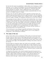
connection, particularly, the hydrogen bonded 1D helical water chains occupy a
special place due to their crucial role in the fundamental biological processes such as
transport of water, protons and ions (Figure 4-1). For example, the selective transport
of water across cell involves the hydrogen bonded assembly of single H-bonded
helical chains of water molecules in the constricted pore of the aquaporin-1.
12
These
1D water chains appear to be stabilized by strong H-bonding between neighboring
water molecules along the chain as well as H-bonding between water molecules and
donor-acceptor groups associated with channels.
210
Chapter 4
Figure 4-1. Schematic representation of water transport in aquaporin proteins.
11c
While such 1D helical water chains are prevalent in biological systems, it is highly
difficult to construct them in the synthetic hosts by design because the structural
constraints required in stabilizing the 1D water chains are yet to be fully understood.
Such water chains could model the biological systems for the transport of water or
ions across the membrane proteins with aquapores. However, some amount of success
has been achieved while generating helical 1D water chains in synthetic hosts.
Chakravarty et al. reported a hydrogen-bonded helical dicopper(II) complex as
supramolecular host anchored by hydrogen bonding to alternate water molecules
(Figure 4-2) that were assembled as a single-stranded, both right- and left-handed,
helical chain.
13
211
Chapter 4
Figure 4-2. 1D helical water chain constructed by alternate water molecules
anchoring the supramolecular Cu(II) complex.
13
Hong et al.
14
reported a left handed 1D helical water chains (Figure 4-3)
encapsulated in a chiral 3D hydrogen bonded supramolecular network structure in a
dicopper(II) complex of a Schiff base derived from L-histidine.
In a recent report, Nangia et al.
15
observed infinite 1D helical chain of water
molecules (Figure 4-4) in nanoporous channels of organic hexahosts, (Cl-
PHG.(H
2
O)
3
) and (Br-PHG.(H
2
O)
3
). It has been shown that the weak
halogen···halogen interactions directed the handedness of the water helices
surrounding the Cl-PHG and Br- PHG hosts.
212
Chapter 4
Figure 4-3. Left handed 1D helical water chain observed by Hong et al.
14
Figure 4-4. (left) O
w
–HO
w
hydrogen bonding in a water helix of Cl–
PHG.(H
2
O)
3
(disordered protons are shown). (right) along with the spiral assembly of
host molecules (green, blue) around the right-handed water helix in Br–PHG.(H
2
O)
3
.
15
4-2.
Aim of the current investigation
Inspired by the fascinating structural features of helices demonstrating the
cooperative self-assembly, recognition and their remarkable functions such as
chemical transport and screening activities of membrane channels in biological
213
Chapter 4
systems, the helicity has been successfully introduced into artificial systems by the
chemists in the field of metalla-supramolecular chemistry.
16-18
It is also explored
recently that the transport of water or protons across the cell involves highly mobile
hydrogen-bonded water molecules assembling into a single helical chain at the
positively charged constricted pore of the membrane channel protein aquaporin-1.
19
While 1D water chains are more predominant in biology to play crucial role in
stabilizing the native conformation of biopolymers, such helical water chains are
extremely rare in synthetic crystal hosts.
11, 13-15
It is well known that a chiral ligand can often lead to the formation of helical
structure.
16b
The presence of one or more non-chelating side arms in a chiral ligand
may provide the possibility for selective and complementary aggregation of the metal
complexes. Among various ligands designed and their Cu(II) and Ni(II) complexes
explored in Chapter 3, only the Ni(II) complex of the chiral ligand H
3
Sglu, has been
found to generate spiral coordination polymer.
H
3
Sglu
This chapter presents a interesting helical staircase coordination polymeric
architecture of a Ni(II) complex with a captivating feature of hosting 1D helical chain
of water molecules inside the chiral helical pores through hydrogen bonds.
214
Chapter 4
4-3. Results and Discussion
The ligand, H
3
Sglu has been prepared according to the same procedure as described
in Chapter 3. As the ligand is found to be freely soluble in water, aqueous solution of
H
3
Sglu has been employed for the complexation with Nickel. The Ni(II) complex,
[(H
2
O)
2
⊂{Ni(HSglu)(H
2
O)
2
}]⋅H
2
O IV-1 has been synthesized by the reaction of
aqueous H
3
Sglu with aqueous nickel nitrate hexahydrate in 1:1 stoichiometry. During
the slow diffusion of the reactants, greenish rod-like single crystals of IV-1 were
obtained after one week from the clear reaction mixture on slow evaporation.
4-3-1. Crystal Structure of [(H
2
O)
2
⊂{Ni(HSglu)(H
2
O)
2
}]⋅ H
2
O, IV-1
IV-1 crystallized with two independent molecules in the asymmetric unit as shown
in Figure 4-5. Each Ni(II)
unit has octahedral geometry with dianionic HSglu
2-
ligand
coordinated through phenolic oxygen atom (Ni(1)-O(1), 2.089(4) Å and Ni(2)-O(6),
2.101(3) Å) and secondary amine N atom (Ni(1)-N(1), 2.084(4) Å; Ni(2)-N(2),
2.082(4) Å) and the α-carboxylate oxygen atom (Ni(1)-O(2), 2.047(4) Å and Ni(2)-
O(7), 2.042(4) Å) in a facial manner, two aqua ligands and another carboxylate
oxygen from the neighboring molecule. Selected bond lengths and bond angles are
given in Table 4-1.
215
Chapter 4
Figure 4-5. A view of the asymmetric unit of IV-1.
Table 4-1. Selected bond lengths and bond angles in IV-1
Ni(1)-O(12) 2.043(4) Ni(1)-O(2) 2.047(4)
Ni(1)-O(11) 2.051(4) Ni(1)-N(1) 2.084(4)
Ni(1)-O(1) 2.089(4) Ni(2)-O(14) 2.039(4)
Ni(2)-O(7) 2.042(4) Ni(2)-O(5)
a
2.048(4)
Ni(2)-O(13) 2.070(3) Ni(2)-O(6) 2.101(3)
O(5)-Ni(2)
b
2.048(4)
O(14)-Ni(2)-O(5)
a
89.4(2) O(7)-Ni(2)-O(5)
a
92.9(2)
O(5)
a
-Ni(2)-O(13) 84.5(2) O(5)
a
-Ni(2)-N(2) 173.7(2)
O(5)
a
-Ni(2)-O(6) 88.7(2) C(12)-O(5)-Ni(2)
b
129.1(5)
C(12A)-O(5)-Ni(2)
b
125.0(6) O(12)-Ni(1)-N(1) 97.3(2)
O(2)-Ni(1)-N(1) 80.3(2) O(9)-Ni(1)-N(1) 173.6(2)
O(12)-Ni(1)-O(1) 88.1(1)
Symmetry transformations used to generate equivalent atoms: a: -x+1,y-1/2,-z+1; b: -
x+1,y+1/2,-z+1
The intermolecular connectivity via second carboxylate O atom generates a left-
handed helical staircase-like coordination polymeric architecture with a pesueo-4
1
screw axis. In this helical staircase, the aqua ligands trans to phenolic oxygen atoms
(namely O(11) and O(13)) are pointing inside the tube normal to the helical axis. The
216
Chapter 4
N-H and O-H protons are hydrogen-bonded to the carboxylate oxygen atoms
complementing along the surface of the helical staircase as shown in Figure 4-6. The
hydrogen-bond parameters are given in Table 4-2.
Figure 4-6. (Left) Display of helical water chain encapsulated in IV-1, (Right) Top
view of the staircase polymer without helical water chain.
In this square shaped cavity the dimensions are 7.65 and 7.53 Å (based on Ni···Ni
distances). Of the six lattice water molecules present in the asymmetric unit, four have
been found inside the helical pore and two outside. Of these, two water molecules
O(15) and O(16) are hydrogen-bonded to produce 1D helical polymer with a pseudo-
4
1
screw axis (Figure 4-7). This helical water chain, as a pole of the helical staircase,
also supports and stabilizes the orientation of helical staircase by maintaining the
hydrogen bonding with aqua ligands. The other two water molecules O(17) and
O(18) are found to propagate the hydrogen bonding both with the helical water chain
and aqua ligands and it appears that their hydrogen bonding tendency would have
217
Chapter 4
facilitated the positioning and orientation of water molecules forming the helical
chain.
Figure 4-7. (Left) Top view of IV-1 showing water filled helical channel (Right)
Hydrogen bonded helical water chain with space filling model.
218
Chapter 4
Table 4-2. Hydrogen bond lengths (Å) and bond angles (º) parameters in IV-1
D-H d(D-H) d(H···A) d(D···A)
∠D-H···A
A Symmetry
O1-H1* 0.93 2.14 2.484(5) 100 O10
N1-H1A* 0.91 2.08 2.953(6) 161 O3 x, y+1, z
N2-H2* 0.91 2.06 2.937(6) 161 O8 x, y+1, z
O6-H6* 0.93 1.98 2.453(9) 109 O4 x-1, y-1/2, z-1
O11-H11C 0.89(3) 1.84(3) 2.713(6) 165(3) O15
O11-H11D 0.89(2) 2.10(3) 2.801(6) 135(4) O17
O12-H12A 0.89(2) 1.87(2) 2.745(5) 167(3) O2 x, y+1, z
O12-H12B 0.90(3) 1.83(3) 2.695(9) 160(1) O19
O13-H13A 0.89(2) 2.03(3) 2.774(5) 141(4) O18 x, y+1, z
O13-H13B 0.89(2) 1.84(2) 2.724(6) 172(2) O16
O14-H14A 0.09(3) 2.35(5) 2.803(15) 111(3) O20B x-1, y-1/2, z-1
O14-H14B 0.90(3) 1.99(4) 2.773(6) 145(5) O7 x, y+1, z
O15-H15A 0.90(3) 1.92(4) 2.772(7) 158(4) O16 x-1, y+1/2, z-
1
O15-H15B 0.89(4) 1.86(4) 2.727(7) 163(5) O17 x, y-1, z
O16-H16A 0.90(4) 1.88(4) 2.767(7) 169(4) O15
O16-H16B 0.90(5) 1.93(5) 2.732(7) 148(5) O18
O17-H17A 0.89(4) 2.26(4) 3.120(6) 162(4) O5
O17-H17A 0.89(4) 2.35(3) 2.943(6) 124(3) O13 x-1, y+1/2, z-
1
O17-H17B 0.90(4) 1.99(4) 2.842(6) 157(4) O3 x, y+1, z
O18-H18A 0.90(3) 1.86(3) 2.729(6) 164(4) O8
O18-H18B 0.89(4) 2.11(4) 2.996(6) 161(3) O9 x, y-1, z
O18-H18B 0.89(4) 2.49(4) 3.011(5) 118(3) O11 x, y-1, z
O20B-H20C 0.90(4) 2.26(3) 3.035(15) 145(4) O4
*
The hydrogen atoms have been placed in the calculated positions.
The total potential solvent area in the lattice including the helical and the lattice
water molecules was found to be 405.1 Å
3
(22.7% of the unit cell.
20
All the tubular
coordination polymers are aligned in b-axis (Figure 4-8) and two more water
molecules (O(19) and disordered O(20) occupy the empty space in the lattice outside
the helical cavity.
219
Chapter 4
Figure 4-8. Packing of the staircase polymer IV-1 viewed along b axis showing chiral
channel. The water molecules in the channels are omitted for clarity.
As in the majority of the supramolecular syntheses, self-assembly of metal ions and
ligands resulted in the formation of single, double, triple and quadruply stranded
helical structures.
17
However, helical chain inside a helical structure is very rare.
Unlike a water helix inside a hydrogen-bonded helical supramolecular host,
13
the
structure of IV-1 has a hydrogen-bonded helix inside a helical 1D coordination
polymer. Highly ordered stream of helical water molecules inside another helical
polymer seems to be striking and has unique structural feature among those existing
porous helical structures
17c, 21-23
and other patterns of the water structures observed in
diverse environments of both inorganic
5-6
and organic
3-4, 11
hosts
and two dimensional
supramolecular (H
2
O)
12
rings.
6c
Whereas designing chiral materials from achiral
molecular compounds presents a promising theme in materials science, using simple
and available chiral precursor as an alternative remains another practical approach.
The structure of IV-1 exemplifies the feasibility of such an approach.
24
220
Chapter 4
At this point, it is important to highlight that the same HSglu
2-
anion has displayed
a completely different coordination environment and connectivity, when the metal ion
is changed from Ni(II) to Cu(II), resulting in 1D zigzag coordination polymeric
structure in [Cu(HSglu)(H
2
O)].H
2
O, III-2 as described in the previous chapter. This
variation from helical staircase coordination polymeric structure in six- coordinated
Ni(II) complex to 1D zigzag coordination polymeric structure in five- coordinated
Cu(II) complex displayed by the same HSglu
2-
anion demonstrates that the overall
topology depends on the nature of the metal ion and the coordination geometry at the
metal centers.
4-4. Physicochemical Studies
4-4-1. IR spectra
The X-ray crystal structure, IV-1 contains both aqua ligands and lattice water
molecules and the IR absorption bands observed between 3300 and 3450 cm
-1
also
suggest their presence
25a
which has been further supported by the weight loss
observed in TG analysis. The ν(N-H) band has been shifted from 2960 cm
-1
for the
free H
3
Sglu ligand to 2746 cm
-1
for the complex indicating the complexation. The
asymmetric ν
as
(COO
-
) and symmetric ν
s
(COO
-
) stretching vibrations of
carboxylate
in the free ligand have been observed at 1673 and 1388 cm
-1
respectively. For the
complex IV-1 the ν
as
(COO
-
) and ν
s
(COO
-
) stretching frequencies are observed at
1623 and 1348 cm
-1
respectively.
25b
The difference (Δν > 200) between ν
as
(COO
-
)
and ν
s
(COO
-
) indicates the terminal or monodentate coordination mode of carboxylate
group.
25c
The stretching frequencies characteristic of phenolic C-O in the ligand and
complexes are observed in the range of 1253 cm
-1
. The assigned IR stretching
221
Chapter 4
frequencies here are in agreement with the available literature for the related Ni(II)
complexes.
26
4-4-2. Electronic spectra
Electronic spectrum of IV-1 recorded as nujol mull transmittance displayed the
medium intensity d-d bands typical of octahedral Ni(II) at 642 and 730-737 nm while
the CT band corresponding to phenolate-to-Ni(II) transition was observed in the range
of 350-354 nm. The d-d bands at 642 nm can be assignable to the spin allowed
3
A
2g
(F) Æ
3
T
1g
transitions where as the shoulder at around 737 nm originates from the
spin forbidden
3
A
2g
Æ 1E
g
transitions frequently observed in Ni(II) octahedral
complexes.
26-27
4-4-3. Thermogravimetric studies
The TG analysis of IV-1 reveals that the weight loss occurs in the temperature
range 26-232 °C as shown in the Fig. 4-9. The total weight loss observed (21.6%)
agrees with the calculated value (22.5%) for the loss of five water molecules per
Ni(II) ion. The single crystal crumbles upon removal of water molecules or cooled to
-50°C. Our earlier attempts to collect X-ray data at low temperature failed due to this
phenomenon.
222
Chapter 4
Figure 4-9. TGA of IV-1
The effect of thermal dehydration on the single crystals of IV-1 is shown in Figure
4-10. The structure is not expected to be robust when dehydrated due to the fact that
these coordination polymers are not supported by strong non-covalent interactions.
(Figure 4-8).
Figure 4-10. (Left) Single crystals of IV-1 at RT before heating. Single crystals of
IV-1 after heating to 150 ºC (Right).
223
Chapter 4
4-5. Summary
The versatile role of water in many biological, chemical and physical processes has
stimulated intensive research efforts, but it is yet not a fully understood liquid owing
to the complexities and fluctuations in hydrogen bonding leading to the association.
Therefore, structural data of hydrogen bonded water clusters are essential to gain
deeper knowledge on the association of water molecules in different surroundings.
The structure of the left-handed helical coordination polymer IV-1 encapsulating
the hydrogen-bonded helical stream of water molecules illustrates another novel
cooperative assembly and recognition of water molecules in the inorganic crystal host.
These results may exemplify the maxim that the structural constraints operating on
orientation of water by its surrounding and vice versa can be very significant. This
captivating structural feature of IV-1 displaying the helical chain of water molecules
supporting the metal coordination helical staircase brings to light yet another
fascinating model for the water chains in membrane aquaporin proteins for the
transport of water or protons and it appears to be extremely rare among metal
coordination polymers until the present investigation.
28
4-6. Experimental
4-6-1. Synthesis of ligand
N-(2-hydroxybenzyl)-L-glutamic acid, H
3
Sglu
H
3
Sglu ligand has been synthesized according to the procedure described in
Chapter 3.
224
Chapter 4
4-6-2. Synthesis of complex
[(H
2
O)
2
⊂{Ni(HSglu)(H
2
O)
2
}]⋅ H
2
O, IV-1
A clear solution of H
3
Sglu (0.25 g, 1.0 mmol) in of water (2.5 mL) was allowed to
diffuse slowly into a clear aqueous solution (2.5 mL) of nickel(II) nitrate hexahydrate
(0.29 g, 1.0 mmol). The greenish rod-like crystals suitable for X-ray diffraction were
obtained after a week from the clear reaction mixture on slow evaporation. Yield:
0.28 g (70%). Anal. Calcd. for C
12
H
23
NO
10
Ni: C, 36.0; H, 5.8; N, 3.5; H
2
O, 22.5.
Found: C, 36.2; H, 5.6; N, 3.7; H
2
O, 21.6 (from TG).
4-6-3. X-ray crystallography
The solid state structure of IV-1 has been determined by single crystal X-ray
crystallographic technique. The details of crystal data and structure refinement
parameters are shown in Table 4-3.
225
Chapter 4
Table 4-3. Crystallographic data and structure refinement details
Complex IV-1
Formula C
12
H
23
NNiO
10
f.w
400.02
T/K 296(2)
λ/Å
0.71073
Crystal system Monoclinic
Space group P2
1
a/Å 17.135(1)
b/Å 6.194(4)
c/Å 17.160(1)
β/
o
101.220(2)
V/Å
3
1786.6(2)
Z
4
D(cald)/g.cm
-3
1.487
μ/mm
-1
1.134
Reflns col. 10561
Ind. reflns. 6001
R
int
0.0326
GooF 1.050
Flack parameter -0.004(16)
Final R[I>2σ], R
1
a
0.0500
wR
2
b
0.1191
a
R
1
= Σ||F
o
| - |F
c
||/Σ|F
o
|.
b
wR
2
= [Σw(F
o
2
- F
c
2
)
2
/Σw(F
o
2
)
2
]
1/2
226
Chapter 4
4-7. References
1. a) Ludwig, R. Angew. Chem. Int. Ed. Engl. 2001, 40, 1808; b) Ludwig, R.
ChemPhisChem. 2000, 1, 53.
2. Atwood, J. L.; Steed, J. W. (Eds.), Encyclopedia of Supra-molecular
Chemistry, Vols. 1-2, Marcel Dekker, New York, 2004.
3. a) Pal, S.; Sankaran, N. B.; Samanta, A. Angew. Chem. Int. Ed. Engl. 2003, 42,
1741; b) Ye, B. H.; Sun, A. P.; Wu, T. F.; Weng, Y. Q.; Chen, X. M. Eur. J.
Inorg. Chem. 2005, 1230.
4. a) Custelcean, R.; Afloroaei, C.; Vlassa, M.; Polverejan, M.; Angew. Chem.
Int. Ed. Engl. 2000, 39, 3094; b) Moorthy, J. N.; Natarajan, R.; Venugopalan,
P. Angew. Chem. Int. Ed. Engl. 2002, 41, 3417; c) Ghosh, S. K.; Bharadwaj,
P. K. Eur. J. Inorg. Chem. 2005, 4880; d) Garcia, R. L.; Damian-Murillo, B.
M.; Barba, V.; Hopfl, H.; Beltran, H. I.; Zamudio-Rivera, L. S. Chem.
Commun. 2005, 5527.
5. a) Blanton, W. B.; Gordon-Wylie, S.W.; Clark, G. R.; Jordon, K. K.; Wood, J.
T.; Geiser, U.; Collins, T. J. J. Am. Chem. Soc. 1999, 121, 3551; b)
Custelcean, R.; Afloroaei, C.; Vlassa, M.; Polverejan. M. Angew. Chem. Int.
Ed. Engl. 2000, 39, 3094; c) Atwood, J. T.; Barbour, L. J.; Ness, T. J.; Ratson,
C. L.; Ratson, P. L. J. Am. Chem. Soc. 2001, 123, 7192; d) Ghosh, S. K.;
Bharadwaj, P. K. Eur. J. Inorg. Chem. 2005, 4886.
6. a) Barbour, L. J.; Orr, W. G.; J. L. Atwood, Nature, 1998, 393, 671; b)
Barbour, L. J.; Orr, W. G.; Atwood, J. L. Chem. Commun. 2000, 859; c) Ma,
B. Q.; Sun, H. L.; Gao, S. Angew. Chem. Int. Ed. Engl. 2004, 43, 1374.
7. Raghuraman, K.; Katti, K. K.; Barbour, L. J.; Nagavarakishore, P.; Barnes, C.
L.; Katti, K. V. J. Am. Chem. Soc. 2003, 125, 6955.
227
Chapter 4
8. a) Zhao, B.; Cheng, P.; Chen, X.; Cheng, C.; Shi, W.; Liao, D.; Yan, S.; Jiang,
Z. J. Am. Chem. Soc. 2004, 126, 3012; Dong, Y. B.; Zhao, X.; Tang, B.;
Wang, H. Y.; Huang, R. Q.; Smith, M. D.; Loye, H. C. Z. Chem. Commun.
2004, 220; Runde, W.; Bean, A.C.; Scott, B. L. Chem. Commun. 2003, 1848.
9. a) Infantes, L.; Chisholm, J.; Motherwell, S. CrystEngComm. 2003, 5, 480; b)
Infantes, L.; Motherwell, S. CrystEngComm. 2002, 4, 454.
10. Gillon, A. L.; Feeder, N.; Davey, R. J.; Storey, R. Crystal Growth & Design,
2003, 3(5), 663
11. a) Cheruzel, L. E.; Pometun, M. S.; Cecil, M. R.; Mashuta, M. S.; Wittebort,
R. J.; Buchanan, R. M. Angew. Chem. Int. Ed. Engl. 2003, 42, 5452; b) Pal, S.;
Sankaran, N. B.; Samanta, A. Angew. Chem. Int. Ed. Engl. 2003, 42, 1741; c)
Ludwig, R. Angew. Chem. Int. Ed. Engl. 2003, 42, 258. d) Fei, Z.; Zhao, D.;
Geldbach, T. J.; Scopelliti, R.; Dyson, P. J. Antonijevic, S.; Bodenhausen, G.
Angew. Chem. Int. Ed. Engl. 2005, 44, 5720
12. a) Konozo, D.; Yasui, M.; King, L. S.; Agre, P. J. Clin. Invest. 2002, 109,
1395; b) Roux, R.; MacKinnon, R. Science. 1999, 285, 100.
13. Mukherjee, A.; Saha, M. K.; Nethaji, M.; Chakravarty, A. R. Chem. Commun.
2004, 716.
14. Lou, B.; Jiang, F.; Yuan, D.; Wu, B.; Hong, M. Eur. J. Inorg. Chem. 2005,
3214.
15. Saha, B. K.; Nangia, A. Chem. Commun. 2005, 3024.
16. (a) Lehn, J. M. Supramolecular Chemistry, VCH, Weinhein, 1995; b)
Albrecht, M. Chem. Rev. 2001, 101, 3457; c) Piguet, C.; Bernardinelli, G.;
Hopfgartner, G. Chem. Rev. 1997, 97, 2005; d) Hannon, M. J.; Childs, L. J.
Supramolecular Chemistry, 2004, 16, 7.
228
Chapter 4
17. a) Mamula, O.; von Zelewsky, A.; Bark, T.; Bernardinelli, G. Angew. Chem.
Int. Ed. Engl. 1999, 38, 2945; b) Kaes, C.; Hosseini, M. W.; Rickard, C. E. F.;
Skelton, B. W.; White, A. H. Angew. Chem. Int. Ed. Engl. 1998, 37, 920; c)
McMorran, D. A.; Steel, P. J. Angew. Chem. Int. Ed. Engl. 1998, 37, 3295; d)
Baum, Constable, G.; Fenske, E. C.; D.; Housecroft, C. E.; Kulke, T. Chem.
Eur. J. 1999, 5, 1862; e) Hannon, M. J.; Painting, C. L.; Alcock, N. W.; Chem.
Commun. 1999, 2023.
18. Ranford, J. D.; Vittal; J. J.; Wu, D; Yang, X. Angew. Chem. Int. Ed. Engl.
1999, 38, 3498.
19. Mitsuoka, K.; Murata, K.; Walz, T.; Hirai, T.; Agre, P. Heymann, J. B.; Engel,
A.; Fujiyoshi, Y. J. Struct. Biol. 1999, 128, 34.
20. Spek, A.L. Acta. Crystallogr. 1990, A46, C34.
21. Wu, C. D.; Lu, C. Z.; Lin, X.; Wu, D. M.; Lu, S. F.; Zhuang, H. H.; Huang, J.
S. Chem. Commun. 2003, 1284.
22. Wu, C. D.; Lu, C. Z.; Lu, S. F.; Zhuang, H. H.; Huang, J. S. J. Chem. Soc.,
Dalton Trans. 2003, 3192.
23. Pickering, A. L.; Seeber, G.; Long, D. L.; Cronin, L. Chem. Commun. 2004,
136.
24. Moulton, B.; Zaworotko, M. J. in Crystal Engineering: From Molecules and
Crystals to Materials, (Eds.: D. Braga, F. Grepioni, A. G. Orpen), Kluwer,
Dordrecht, 1999, pp 311.
25. a) Ferraro, J. R.; Low-frequency Vibrations of Inorganic and Coordination
Compounds, Plenum press, New York, 1971; b) Nakamato, K. Infrared and
Raman Spectra of Inorganic and Coordination Compounds, 4
th
edn., John
229
Chapter 4
Wiley & Sons, New York, 1986, pp. 191; c) Deacon, G. B.; Philips, R. Coord.
Chem. Rev. 1980, 33, 327.
26. (a) Chandra, S.; Kumar, U. Spectrochim. Acta Part A. 2005, 61, 219; b) Liu,
H.; Wang, H.; Niu, D; Lu, Z. Synth. React. Inorg. Met Org. Chem. 2005, 35,
233; c) Koga, T.; Farutachi, H.; Nakamura, T.; Fukita, N.; Ohba, M.;
Takahashi, K.; Okawa, H. Inorg. Chem. 1998, 989; d) Zakrzewski, G.;
Sacconi, L. Inorg. Chem. 1968, 7, 1034.
27. a) Lever, A. B. P. Inorganic Electronic Spectroscopy, 2
nd
Edn., Elsevier,
Amsterdam, 1984; b) Chow, S. T.; Johns, D. M.; McAuLiffe, C. A Inorg.
Chim. Acta 1977, 22, 1.
28. a) Schmuck, C. Angew. Chem. Int. Ed. Engl. 2003, 42, 2448; b) Rowan, A.E.
Nolte, R. J. M. Angew. Chem. Int. Ed. Engl. 1998, 37, 63.
230