An asthma allergen specific animal model for the study of responses to dust mite allergen induced asthma
Bạn đang xem bản rút gọn của tài liệu. Xem và tải ngay bản đầy đủ của tài liệu tại đây (18.7 MB, 254 trang )
AN ASTHMA ALLERGEN SPECIFIC ANIMAL MODEL FOR THE
STUDY OF RESPONSES TO MITE ALLERGEN INDUCED ASTHMA
KENNETH WONG HOK SUM
B.Sc. (Hons), NUS
A THESIS SUBMITTED FOR THE DEGREE OF DOCTOR OF
PHILOSOPHY
YONG LOO LIN SCHOOL OF MEDICINE
DEPARTMENT OF MICROBIOLOGY
NATIONAL UNIVERSITY OF SINGAPORE
2013
!
!"#$%&'()*+*$,
!
!
!
i
Acknowledgements
First and foremost, I would like to express my gratitude to Prof.
Kemeny for his patient guidance and support. When things did not work, and
that happened a lot, you were always a calming influence and helped get me
back on track. I also learnt so much more than just science from you and I
really appreciate the discussions we had.
To Dr Gijsbert Grotenbreg, thank you for taking me under your wing. I
learnt a lot from you and your focus and drive is always a source of inspiration.
Things certainly wouldn’t have gotten this far without your help on the project.
To Dr Paul MacAry, thank you for all your advice and guidance
throughout this project.
To the members of the DMK lab, past and present, I am very grateful
to have had the opportunity to work with you all. Most of you had been more
than just good colleagues. You are great friends. I could not have asked for a
better bunch of people to work with. Thank you especially to Yafang and
Sophie for guiding me with the asthma studies. Thank you as well to Nayana
for all the discussions and your help on those busy harvest days. So many
people, so little time to acknowledge them. A big thank you to Benson as well.
The lab would have been a mess without you handling the orders and the mice
colonies.
I am also very grateful to the people in the GMG lab. It had been great
to work with you guys and we had more than our fair share of laughs. This is
especially with Cynthia as we embarked on the big scary world of protein
expression together, both knowing absolutely nothing to start with and making
!
!"#$%&'()*+*$,
!
!
!
ii
every mistake in the book. To Joanna, Lionel and Michelle, I missed those
weekends in the lab with you guys. Who says working overtime is not fun!
And to Adrian Sim, Fatimah, Chien Tei, Michael, Lawrence and
everyone else, thank you for your help and for simply adding color to my life.
I also owe a debt of gratitude to my family in Malaysia. Thank you for
your unconditional support in everything I do, no matter how dumb.
Finally, to my long-suffering wife, the biggest thank you. You’ve
endured the past few years with this grumpy ogre and bore the brunt of it
when my experiments did not work. Hopefully I can make it up to you after
this! I wouldn’t have made it through this without you.
!
-/++!01.
!
!
!
iii
Summary
T cells play a central role in the pathogenesis of allergic asthma.
However, many studies into the roles of T cells in asthma had been performed
using ovalbumin as a surrogate allergen. This is mainly due to the greater
availability of research tools for use in the ovalbumin model. However, true
asthma allergens had been shown to behave very differently than ovalbumin.
In this study, we aim to expand the tools available for the study of mite
allergen-induced asthma.
Using a plasmid DNA immunization method, we induced T cell
responses against allergens from Blomia tropicalis and Dermatophagoides
pteronyssinus. We identified a number of epitopes recognized by allergen-
specific CD4 T cells, including several novel epitopes for Blo t 5. We next
demonstrated that the Blo t 5-specific CD4 T cells identified in this study were
recruited into the lungs following Blo t 5 inhalation. When administered
intradermally, the peptides induced a tolerogenic response and attenuated the
allergic airway inflammation induced by Blo t 5 sensitization and challenge.
The identification of CD4 T cell epitopes for Blo t 5 would allow for the study
of T cell responses to Blomia tropicalis, a major source of mite allergen in the
tropics that remained poorly studied to date.
Work done on the OVA model suggested a role for CD8 T cells in the
attenuation of the allergic airway responses to allergen. In this study, we
adoptively transferred Der p 1 specific T cells into mice sensitized and
challenged with house dust mite (HDM) extract. CD8 T cell responses were
tracked by class I MHC tetramers produced in-house. Our results showed that,
!
-/++!01.
!
!
!
iv
unlike in the ovalbumin model, the CD8 T cells were unable to attenuate the
allergic airway inflammatory responses to HDM. However, we showed that
the Th2 airway inflammation was reduced following the adoptive transfer of
Der p 1 specific CD8 T cells when mice were sensitized and challenged by
purified Der p 1 protein. We proceeded to demonstrate in vivo and in vitro that
exogenous HDM was a poor inducer of CD8 T cell responses. Finally, using
peptide-pulsed BMDCs to induce a Der p 1-specific immune response, we
observed that CD8 T cell responses exacerbated the allergic lung
inflammation response to HDM by increasing the number of infiltrating
immune cells and the production of IL-5 and IL-13. Therefore, our results
suggested that the induction of a CD8 T cell response by HDM was markedly
inefficient and it is unlikely that CD8 T cells could play a role in the acute
phase of asthma development. However, our results showed also that a CD8 T
cell response might actually be detrimental and exacerbate the inflammatory
responses in the lung.
Finally, we cloned and characterized the T cell receptor (TCR) gene
from a Der p 1 specific CD8 T cell. The TCR genes were cloned into
expression cassettes for the generation of TCR transgenic mice with CD8 T
cells specific for the HDM allergen, Der p 1. We believe that these mice could
be useful in the study of chronic asthma, where CD8 T cells had been shown
to play a role.
!
'2-,.%3.4/5'2"!,2%$
!
!
!
v
List of publications
1. Wong KL, Tang LF, Lew FC, Wong HS, Chua YL, MacAry PA,
Kemeny DM. CD44
high
memory CD8 T cells synergize with CpG
DNA to activate dendritic cell IL-12p70 production. J Immunol. 2009
Jul 1; 183(1):41-50
2. Tang Y, Guan SP, Chua BY, Zhou Q, Ho AW, Wong KH, Wong KL,
Wong WS, Kemeny DM. Antigen-specific effector CD8 T cells
regulate allergic responses via IFN-γ and dendritic cell function. J
Allergy Clin Immunol. 2012 Jun; 129(6):1611-20.e4
3. Ge MQ, Ho AW, Tang Y, Wong KH, Chua BY, Gasser S, Kemeny
DM. NK cells regulate CD8
+
T cell priming and dendritic cell
migration during influenza A infection by IFN-γ and perforin-
dependent mechanisms. J Immunol. 2012 Sep 1; 189(5):2099-109.
4. Prabhu N, Ho AW, Wong KH, Hutchinson PE, Chua YL, Kandasamy
M, Lee DC, Sivasankar B, Kemeny DM. Gamma interferon regulates
contraction of the influenza virus-specific CD8 T cell response and
limits the size of the memory population. J Virol. 2013 Dec;
87(23):12510-22
!
,!5'*.%3."%$,*$,
!
!
!
vi
Table of Contents
Chapter 1 Introduction 1.
1.1. Asthma 1.
1.1.1. Prevalence and cost of asthma 1.
1.1.2. Causes of asthma 2.
1.2. The immunology of allergic asthma 4.
1.2.1. The innate immune response in allergic asthma 7.
1.2.1.1. (6789:;:<.<6==>.?78.;@6:9.9A=6.:7.?==69B:<.?>;@C?.DDDDDDDDDDDDDDDDDDDDDDDDDDDDDDDDDDDDDDDDDDDDD.E.
1.2.1.2. *A>:7AF@:=>.DDDDDDDDDDDDDDDDDDDDDDDDDDDDDDDDDDDDDDDDDDDDDDDDDDDDDDDDDDDDDDDDDDDDDDDDDDDDDDDDDDDDDDDDDDDDDDDDDDDDDDDDDDDDDDDDDDD.GG.
1.2.1.3. +?>;.<6==>.DDDDDDDDDDDDDDDDDDDDDDDDDDDDDDDDDDDDDDDDDDDDDDDDDDDDDDDDDDDDDDDDDDDDDDDDDDDDDDDDDDDDDDDDDDDDDDDDDDDDDDDDDDDDDDDDDDDDDDD.GH.
1.2.1.4. 5?>AF@:=>.DDDDDDDDDDDDDDDDDDDDDDDDDDDDDDDDDDDDDDDDDDDDDDDDDDDDDDDDDDDDDDDDDDDDDDDDDDDDDDDDDDDDDDDDDDDDDDDDDDDDDDDDDDDDDDDDDDDDDDD.GI.
1.2.1.5. $6J;9AF@:=>.DDDDDDDDDDDDDDDDDDDDDDDDDDDDDDDDDDDDDDDDDDDDDDDDDDDDDDDDDDDDDDDDDDDDDDDDDDDDDDDDDDDDDDDDDDDDDDDDDDDDDDDDDDDDDDDDDDD.GK.
1.2.1.6. +?<9AF@?B6>.DDDDDDDDDDDDDDDDDDDDDDDDDDDDDDDDDDDDDDDDDDDDDDDDDDDDDDDDDDDDDDDDDDDDDDDDDDDDDDDDDDDDDDDDDDDDDDDDDDDDDDDDDDDDDDDD.GL.
1.2.1.7. 277?;6.=MCF@A:8.<6==>.DDDDDDDDDDDDDDDDDDDDDDDDDDDDDDDDDDDDDDDDDDDDDDDDDDDDDDDDDDDDDDDDDDDDDDDDDDDDDDDDDDDDDDDDDDDDDDD.GN.
1.2.1.8. $?;J9?=.O:==69.,.P$#,Q.<6==>.?78.γδ.,.<6==>.DDDDDDDDDDDDDDDDDDDDDDDDDDDDDDDDDDDDDDDDDDDDDDDDDDDDDDD.GN.
1.2.1.9. ,@6.?:9R?M.6F:;@6=:JC.DDDDDDDDDDDDDDDDDDDDDDDDDDDDDDDDDDDDDDDDDDDDDDDDDDDDDDDDDDDDDDDDDDDDDDDDDDDDDDDDDDDDDDDDDDDD.GE.
1.2.2. The adaptive immune system and allergic asthma 19.
1.2.2.1. "(S.,.<6==>.:7.?==69B:<.?>;@C?.DDDDDDDDDDDDDDDDDDDDDDDDDDDDDDDDDDDDDDDDDDDDDDDDDDDDDDDDDDDDDDDDDDDDDDDDDDDDDD.HT.
1.2.2.2. "(E.,.<6==>.:7.?==69B:<.?>;@C?.DDDDDDDDDDDDDDDDDDDDDDDDDDDDDDDDDDDDDDDDDDDDDDDDDDDDDDDDDDDDDDDDDDDDDDDDDDDDDD.HL.
1.2.2.3. ,@6.@JCA9?=.96>FA7>6.:7.?==69B:<.?>;@C?.DDDDDDDDDDDDDDDDDDDDDDDDDDDDDDDDDDDDDDDDDDDDDDDDDDDDDDD.HE.
1.3. Asthma allergens 29.
1.3.1. The mite allergens 30.
1.3.2. Mite allergens and the immune system 32.
1.4. Aims of the study 34.
Chapter 2 Materials and Methods 38.
2.1. Media and buffers 38.
2.1.1. PBS buffer 38.
2.1.2. MACS buffer 38.
2.1.3. FACS buffer 38.
2.1.4. Red Blood cell (RBC) lysis solution 39.
2.1.5. Complete RPMI for cell culture 39.
2.1.6. Buffers for ELISA 40.
2.1.7. LB broth 40.
2.1.8. LB agar 40.
2.1.9. CaCl
2
solution for preparation of competent cells 40.
2.1.10. SDS-PAGE gel electrophoresis buffers 40.
2.1.11. Buffers for protein purification from E.coli 41.
2.1.12. Buffers for protein refolding and purification 41.
2.2. List of antibodies used 42.
2.3. Mice 43.
2.4. Molecular biology protocols 43.
2.4.1. Preparation of chemically competent E.coli 43.
2.4.2. Transformation of E.coli 44.
2.4.3. General PCR protocol 45.
2.4.4. Agarose gel extraction 47.
2.4.5. Subcloning into TOPO vector by TA cloning 47.
2.4.6. Plasmid miniprep 49.
2.4.7. Restriction enzyme digest 49.
2.4.8. PCR purification 50.
!
,!5'*.%3."%$,*$,
!
!
!
vii
2.4.9. Ligation into plasmid vector 51.
2.4.10. RNA extraction and purification 51.
2.4.11. Reverse transcription of mRNA 52.
2.4.12. 5’ RACE reaction 52.
2.5. Protein expression and purification protocols 55.
2.5.1. SDS-PAGE gel electrophoresis 55.
2.5.2. Production of class I MHC tetramers 56.
2.5.3. Production of recombinant Blo t 5 60.
2.6. Cell isolation protocols 62.
2.6.1. Processing of splenic and lymph node cells 62.
2.6.2. Isolation of CD8 cells by magnetic separation 63.
2.7. Cell culture 64.
2.7.1. T cell line production and maintenance 64.
2.7.2. Bone-marrow derived Dendritic Cells 65.
2.8. Protocols for evaluation of cell functionality 66.
2.8.1. IFN-γ ELISPOT 66.
2.8.2. ELISA 68.
2.8.3. 3H-thymidine proliferation assay 68.
2.8.4. Chromium-51 release assay 69.
2.9. Flow cytometry 70.
2.9.1. Cell surface marker staining for flow cytometry 70.
2.9.2. Intracellular cytokine staining for flow cytometry 70.
2.9.3. Peptide exchange and class I MHC tetramer staining 71.
2.10. Intradermal immunization of mice with plasmid DNA by skin
tattoo 72.
2.11. Murine model of asthma 72.
2.11.1. Intranasal sensitization or challenge of mice 72.
2.11.2. Bronchoalveolar lavage analysis 72.
2.11.3. Analysis of lung cells 73.
2.11.4. Culture of lung-draining mediastinal lymph node (MLN) cells 74.
2.11.5. Lung histology 75.
Chapter 3 Expression and purification of recombinant Blo t 5 78.
3.1. Introduction 78.
3.2. The Blo t 5 gene 79.
3.3. Cloning of the Blo t 5 gene into pET 28 expression vector 81.
3.4. Expression of recombinant Blo t 5 in E. coli BL 21 83.
3.5. Purification of recombinant tBlo t 5 85.
Chapter 4 Production of class I MHC tetramers 90.
4.1 Introduction 90.
4.1. Expression and purification of class I MHC proteins 91.
4.2. Testing the functionality of UV cleavable class I MHC tetramers
96.
Chapter 5 Mapping of mite allergen T cell epitopes 99.
5.1. Introduction 99.
5.2. DNA immunization constructs 102.
5.3. DNA vaccination of mice 109.
5.4. Epitope mapping studies 112.
5.1.1. Mapping of Blo t 5 epitopes 116.
!
,!5'*.%3."%$,*$,
!
!
!
viii
5.1.2. Mapping of Der p 2 epitopes 124.
5.5. Further characterization of Blo t 5 epitopes 128.
5.6. Discussion 138.
Chapter 6 Der p 1 specific CD8 T cells and HDM-induced asthma.
145.
6.1. Introduction 145.
6.2. Immunization of mice 147.
6.3. Generation of a Der p 1 specific CD8 T-cell line. 153.
6.4. Characterization of T cell receptor gene 156.
6.5. Cloning of T cell receptor gene into cassette vectors 162.
6.6. Role of CD8 T cells in immune response to mite allergens 173.
6.7. Discussion 198.
Chapter 7 Final discussion 203.
7.1. Summary of findings 203.
7.2. Limitation of current study 210.
7.3. Future work 212.
T cells epitopes as tools for immunotherapy 216.
!
'2-,.%3.32)/0*
!
!
!
ix
List of Figures
Figure 1.1 Overview of the immune responses involved in allergic asthma 6.
Figure 1.2 Mite allergens and their biological activities 31.
Figure 3.1 DNA sequence of Blo t 5 and the amino acid translation of codon80.
Figure 3.2 SignalP 4.1 prediction of signal peptide cleavage in Blo t 5. . 80.
Figure 3.3 Blo t 5 and tBlo t 5 gene constructs in pET28 plasmid. 82.
Figure 3.4 Overview of the process for the expression of Blo t 5 84.
Figure 3.5 Ammonium sulfate purification of bacterial lysate containing
recombinant Blo t 5 protein 87.
Figure 3.6 Anion exchange chromatography and SDS-PAGE gel analysis 88.
Figure 3.7 Size exclusion chromatography and SDS-PAGE analysis of
fractions 89.
Figure 4.1 Steps involved in the production of class I MHC tetramers 93.
Figure 4.2 Large-scale expression of class I MHC subunits 94.
Figure 4.3 S75 gel filtration chromatography for purification of class I MHC
monomers 95.
Figure 4.4 UV-mediated peptide exchange for class I MHC tetramers 97.
Figure 4.5 Functinoality of refolded UV-cleavable class I MHC tetramers 98.
Figure 5.1 Murine optimized Blo t 5 gene with restriction sites and Kozak
initiation sequence 105.
Figure 5.2 Murine optimized Der p 2 gene with restriction sites and Kozak
initiation sequence 106.
Figure 5.3 Gene constructs produced for immunization studies and list of
primers used in the cloning process 107.
Figure 5.4 PCR cloning of Der p 2-SIINFEKL into pVAX1. 108.
Figure 5.5 Plasmid DNA immunization of mice by skin tattoo 111.
Figure 5.6 List of peptides used in mapping epitopes of the Bo t 5 protein. . 113.
Figure 5.7 List of peptides used in mapping epitopes of the house dust mite
protein, Der p 2. Peptides are 15 amino acids in length (except Dp2#27, a
16-mer peptide) and overlap by 10 amino acids. 114.
Figure 5.8 Overview of the epitope mapping process. 115.
Figure 5.9 Mapping of Blo t 5 epitopes in C57BL/6 mice 119.
Figure 5.10 Mapping of Blo t 5 epitopes in BALB/c mice 120.
Figure 5.11 Top 20 peptides predicted to bind H-2Db and H-2Kb. 122.
Figure 5.12 Tetramer guided epitope mapping. 123.
Figure 5.13 Mapping of Der p 2 epitopes in C57BL/6 mice. 126.
Figure 5.14 Mapping of Der p 2 epitopes in BALB/c mice. 127.
Figure 5.15 IFNγ producing T cells recognizing Blo t 5 epitopes in C57BL/6
mice following plasmid DNA immunization. 129.
Figure 5.16 T cells induced by intradermal plasmid DNA immunization were
able to induce lung inflammation following antigen exposure. 131.
Figure 5.17 Intracellular cytokine staining of draining lymph node cells. 135.
Figure 5.18 Peptide immunotherapy of Blo t 5 sensitized and challenged mice.
137.
Figure 5.19 Summary of T cell epitopes identified in this study 140.
Figure 5.20 Murine Der p 2 T cell epitopes identified in other studies. 140.
Figure 6.1 Gene construct for immunization of mice 150.
!
'2-,.%3.32)/0*
!
!
!
x
Figure 6.2 Immunization of mice with pVAX-TTFH-FGISNYCQI. Mice were
immunized intradermally by skin tattoo. Splenocytes of immunized mice
were screened for antigen specific cells by (a) ELISPOT or (b) class I
MHC tetramer staining. 152.
Figure 6.3 Establishment of a Der p 1 specific T cell line. 155.
Figure 6.4 Molecular characterization of TCR chains. 159.
Figure 6.5 Primers used in 5' RACE characterization of TCR chains. 160.
Figure 6.6 T cell receptor chains of the Der p 1 specific T cell line. 161.
Figure 6.7 List of primers used. 167.
Figure 6.8 Genomic DNA sequences of the TCR chains of the Der p 1 specific
CD8 T cell line 168.
Figure 6.9 Cloning of TCRα chain into pTα
cass
. 170.
Figure 6.10 Cloning of TCRβ chains into pTβ
cass
. 172.
Figure 6.11 Murine model for house dust mite-induced asthma 174.
Figure 6.12 Adoptive transfer of Der p 1 specific CD8 T cells into the murine
model of HDM-induced asthma 179.
Figure 6.13 DNA immunization with plasmid encoding epitope recognized by
Der p 1 specific CD8 T cells and the effect on lung responses to HDM.
182.
Figure 6.14 Role of Der p 1 specific CD8 T cells in lung responses to Der p 1.
188.
Figure 6.15 Sensitization by HDM-pulsed BMDCs followed by CD8 adoptive
transfer and HDM challenge 191.
Figure 6.16 Der p 1 CD8 T cell responses to house dust mite extract. 195.
Figure 6.17 Induction of pulmonary CD8 T cell response by transfer of
peptide-pulsed BMDCs and the effect on asthma development 197.
Figure 7.1 CD4 and CD8 T cell responses in HDM culture of MLN. 215.
!
'2-,.%3.!550*U2!,2%$
!
!
!
xi
List of abbreviations
AHR – Airway Hyperresponsiveness
AF488 – Alexa Fluor 488
AF647 – Alexa Fluor 647
APC - Allophycocyanin
APC – Antigen Presenting Cell
BMDC – Bone Marrow-derived Dendritic Cell
BV421 – Brilliant Violet 421
CD – Cluster of Differentiation
DC – Dendritic cell
ELISA – Enzyme-Linked ImmunoSorbent Assay
FACS – Fluorescense activated cell sorting
FBS – Fetal bovine serum
HDM – House Dust Mite
IFN – Interferon
IL – Interleukin
LB – Luria Bertani
MACS – Magnetic Activated Cell Sorting (Miltenyi, Singapore)
MHC – Major Histocompatibility Complex
PB – Pacific Blue
PBS – Phosphate Buffered Saline
PCR – Polymerase Chain Reaction
PE – Phycoerythrin
PE-Cy7 – Phycoerythrin-Cyanine 7
PerCP – Peridinin-Chlorophyll Protein
!
'2-,.%3.!550*U2!,2%$
!
!
!
xii
PerCP-Cy5.5 – Peridinin-Chlorophyll Protein – Cyanine 5.5
PFA – Paraformaldehyde
PRR – Pattern Recognition Receptor
RPMI – Roswell Park Memorial Institute
TCR – T cell receptor
TLR – Toll-like Receptor
TSLP – Thymic Stromal Lymphopoietin
WT – Wild type
!
"V!4,*0.GW.2$,0%(/",2%$.
!
!
!
1
Chapter 1. Introduction
1.1 Asthma
Asthma is a chronic airway disease characterized by chronic airway
inflammation, airway remodeling and hyperresponsiveness and presents with
symptoms of recurrent attacks of breathlessness, wheezing and coughing.
Clinically, the measurement of forced expiratory volume in 1 second (FEV
1
)
by spirometry is often used in the diagnosis and management of asthma.
Airway obstruction that is at least partially reversible following the
administration of a bronchodilator such as salbutamol strongly supports the
diagnosis of asthma. It is a highly heterogeneous disease, varying in severity
and frequency from patient to patient and with a number of different triggering
factors that affect different individuals in different ways.
1.1.1 Prevalence and cost of asthma
Asthma is not a recent disease. The term “asthma” was derived from the
Greek verb “aazein”, meaning to pant or to exhale with the mouth open. In the
Corpus Hippocraticum by the ancient Greek physician Hippocrates (460-360
BC), the word asthma was used for the first time as a medical term.
Worldwide, an estimated 300 million people suffer from asthma, leading
to approximately 250,000 annual deaths (1). The World Health Organization
projects that the number of people afflicted with asthma would increase by
over 100 million by 2025 (1). Developed, highly urbanized countries have a
particularly high prevalence of asthma, with the highest prevalence found in
the United Kingdom (greater than 15%), New Zealand (15.1%), Australia
!
"V!4,*0.GW.2$,0%(/",2%$.
!
!
!
2
(14.7%) and the United States (10.9%) (2, 3). In Singapore, asthma is
estimated to affect 5% of all adults and 20% of children (statistics from Health
Promotion Board, Singapore).
The Centre for Disease Control and Prevention (CDC), USA, estimated
that the Unites States spent approximately USD30 billion per year on
treatment and prevention of asthma. In Singapore, the total cost of asthma was
estimated to be USD33.93 million per annum in 1999 and is likely to be much
higher now (4). This includes the direct cost incurred by hospitalization and
other medical costs as well as indirect costs incurred due to the loss of
productivity. Generally, asthma is expected to account for approximately 1-
2% of the healthcare budgets of developed countries each year (2, 3).
Therefore, asthma is a disease with a profound health and economic
impact on the global population. The disease burden is expected to increase
significantly, with increasing industrialization and development in many
countries, particularly in the populous nations such as the People’s Republic
of China and India. As the treatment of asthma is generally restricted to
symptomatic treatments, there exists a need for greater understanding of the
mechanism of the disease to assist in the development of a cure or at least,
more effective therapies.
1.1.2 Causes of asthma
Asthma is a complex disease that can be induced by a number of
environmental factors, alone or in combination, and also potentially involves a
large number of susceptibility genes (5-8). This complexity is a part of the
problem of asthma treatment as the disease often varies from patient to patient
in severity and causative agent. Patients allergic to the same triggering factor
!
"V!4,*0.GW.2$,0%(/",2%$.
!
!
!
3
differ in the severity of their responses and many patients were responsive to
more than one triggering factor.
The most common form of asthma and the focus of most of the research
in the field is allergic asthma. Patients demonstrated aberrant pulmonary
responses to any number of allergens, commonly house dust mites (HDM),
pollen, ragweed, cockroach or mold. The lung is generally a tolerogenic
environment, as is the nature of most organs with direct exposure to the
external environment. However, in allergic asthma patients, inhalation of these
allergens triggered an immune response in the lung, leading to the
development of Th2 inflammation and the infiltration of immune cells such as
eosinophils, mast cells and the production of IgE antibodies. Studies have
shown that the nature of these allergens plays a big role in the induction of a
pulmonary immune response. This, particularly in the context of the house
dust mite, would be further discussed later in this chapter.
Asthma could also be induced by air pollutants such as ozone, cigarette
smoke and diesel exhaust particles (reviewed in (9)). Recent studies have
suggested that exposure to these factors may trigger epigenetic changes in the
patients. Cigarette smoke, for example, has been shown to inhibit the
expression of histone deacetylase (HDAC) in alveolar macrophages, leading
to the increase in transcription of genes encoding inflammatory cytokines (10).
Changes to DNA methylation and microRNA expression were also reported
(11, 12).
However, there also exists a genetic factor that could impact susceptibility
to developing asthma. While studies did not indicate a classical Mendellian
inheritance of asthma susceptibility, it was shown that certain asthma
!
"V!4,*0.GW.2$,0%(/",2%$.
!
!
!
4
phenotypes such as AHR or total serum IgE could have a heritable component
(7). The ADAM33 gene, first identified in A/J mice, had been implicated in
the susceptibility to develop AHR (13). Since then, many other susceptibility
genes have been identified (5-8).
1.2 The immunology of allergic asthma
The human immune system is a highly sophisticated mechanism that
protects us from external threats. The protection mechanism is layered and in
order of increasing specificity. Initial protection is offered by physical barriers
that prevent the entry of pathogenic organisms into the host. This includes the
barriers such as the skin or cilia in the respiratory tract that actively propel
mucus away from the lung, removing any particulate matter including
pathogens. Chemical means, such as β-defensins secreted by the skin and
respiratory tract and lysozyme in tears and saliva, also help prevent the entry
of infectious agents.
Should the physical barriers not suffice, other elements of the innate
immune system would then be recruited as the next line of defense. Innate
immune cells such as neutrophils and macrophages actively phagocytose and
kill the infectious agents. Another phagocytic cell type, the dendritic cells,
also phagocytose the infectious agents. Additionally, these cells then present
antigens from the phagocytosed invaders to T cells, triggering an adaptive
immune response. Hence, dendritic cells provide an important link between
the innate and the adaptive immune response. Basophils, eosinophils and mast
cells release immune mediators that can modulate the immune response.
Eosinophils can also degranulate to release an array of cytotoxic granule
proteins that can kill bacteria and parasites. Finally, natural killer (NK) cells
!
"V!4,*0.GW.2$,0%(/",2%$.
!
!
!
5
can recognize and kill cells infected with the foreign organism. Unlike the
adaptive immune response, the innate immune response is not specific and
does not develop a memory response. This means that the innate immune
response would not respond more effectively upon re-encounter with the same
pathogen.
Finally, the adaptive immune system comes into play. The adaptive
immune response is triggered more slowly than the innate response and
requires a period of time to become fully effective. T cells, specifically
recognizing the invader, come into play. T helper cells produce a number of
inflammatory cytokines that orchestrate the immune response to the invader,
while cytotoxic T cells actively kill infected host cells. B cells produce
antibodies specific to the invader, which help to neutralize the invading
organism. The adaptive immune response is generally long lasting and highly
specific. Memory T cells and plasma cells persist long after the infection had
abated and respond more rapidly and effectively upon re-encounter with the
same invader.
However, there exist occasions where an immune response occurs when
undesired. This could lead to the manifestation of diseases such as asthma and
autoimmunity. In allergic asthma, the exposure to innocuous antigens present
in the inhaled air induced an immune response similar to that against an
invading pathogen. Both the adaptive and innate immune systems were
directed against these allergenic antigens, leading to infiltration of immune
cells and pulmonary inflammation. The inflammatory responses in the lung
could also result in structural changes such as airway smooth muscles
hypertrophy, increased mucus production and increased airway resistance.
!
"V!4,*0.GW.2$,0%(/",2%$.
!
!
!
6
This results in long-term changes to lung function that characterizes chronic
asthma.
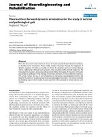
Figure 1.1 Overview of the immune responses involved in allergic asthma
The protease activity of the allergen allows the allergen to penetrate through
the epithelium. Activation of PRRs on the dendritic cells lead to the
maturation of the dendritic cells and antigen presentation to T cells. TSLP and
IL-33 produced by the epithelial cells induce the polarization of the immune
response to a Th2-type response. Th2 CD4 T cells secrete a variety of
cytokines that act on other immune cells. This leads to immunoglobulin class-
switching and IgE secretion from B cells. The cross-linking of FcεR1
receptors on mast cells by allergen binding to IgE result in mast cell
degranulation and release of various immune mediators. IL-5 produced from
Th2 T cells recruit eosinophils into the lungs. Also, IL-13 had been shown to
induce airway hyperresponsiveness and mucus production by goblet cells.
Regulatory T cells (Treg) have been shown to suppress the immune response
to allergens and are a key target in asthma therapy. Additionally, some studies
had suggested a role for CD8 T cells in controlling the Th2 inflammation.
!
"V!4,*0.GW.2$,0%(/",2%$.
!
!
!
7
1.2.1 The innate immune response in allergic asthma
The innate immune system is made up of a number of different cell types
that express pattern recognition receptors (PRRs) that can recognize pathogen-
associated molecular patterns (PAMPs) or danger-associated molecular
patterns (DAMPs). PAMPs are molecules found on pathogens that can be
recognized by receptors present on immune cells. This includes bacterial cell
wall components such as lipopolysaccharides (LPS), peptidoglycan, flagellin,
bacterial DNA or double stranded virus RNA. Unlike PAMPs, DAMPs do not
originate from pathogens but are found within the host cells. The presence of
DAMPs such as adenosine, ATP, heat shock proteins, uric acid and DNA are
indicative of cell damage or death and are capable of eliciting an immune
response. These are recognized by PRRs found on innate cells encompassing
the Toll-like receptors (TLRs), the nucleotide binding oligomerization domain
(NLRs), C-type lectin receptors (CLRs) and scavenger receptors (14). A
number of secreted PRRs such as complement proteins, mucins, ficolins,
pentraxins also play a role in protection from infections.
The role of the innate immune system of the lung is not only to protect the
host from respiratory pathogens but also to maintain homeostasis. The innate
immune system is the first component of the immune system to make contact
with the allergen and hence a key determinant in the nature of the response
mounted against the allergen. In many cases, encounter with the allergen
results in elimination of the allergen and maintenance of immune tolerance.
However, in allergic asthma, this may lead to the triggering of an immune
response to the allergen.
!
"V!4,*0.GW.2$,0%(/",2%$.
!
!
!
8
1.2.1.1 Dendritic cells and their role in allergic asthma
The first dendritic cells (DCs) were observed by Paul Langerhans and
were named “Langerhans cells”. This subset of innate immune cells were
coined as dendritic cells in 1973, in reference to their distinct morphology,
specifically the presence of numerous long branched projections (dendrites)
(15). DCs are specialized antigen-presenting cells (APCs) and are extremely
effective in initiating T cell responses compared to other APCs (16, 17).
Importantly, dendritic cells are highly effective in priming naïve T cells (18-
20). Immature DCs efficiently phagocytose and process antigens but are not
efficient antigen presenters. DC maturation can be induced by engagement of
pattern recognition receptors found on these cells such as the TLR 4 receptor
that recognizes bacterial lipopolysaccharide. Upon maturation, DCs down-
regulate their antigen uptake capability but upregulate the surface expression
of class I and class II MHC molecules as well as co-stimulatory molecules
such as CD40, CD80 and CD86 (21). Mature dendritic cells also upregulate
the expression of CCR7, a chemokine receptor that allows the dendritic cells
to home to lymphoid tissues, where the naïve T cells are located (22).
DCs are a heterogeneous population of cells that can be broadly
categorized into several subsets. Firstly, DCs can be divided into classical DCs
or plasmacytoid DCs (pDCs). Classical DCs possess the typical DC
morphology, with long dendrites, and express high levels of CD11c and
intermediate to high levels of class II MHC on the cell surface. pDCs,
however, lack dendrites but have a plasmacytoid shape and do not express
CD11c. pDCs express TLR 7 and 9 and respond to viral infections by
producing large quantities of IFNα/β. These cells express lower levels of class
!
"V!4,*0.GW.2$,0%(/",2%$.
!
!
!
9
II MHC and are very much less effective at presenting antigen to T cells
compared to classical DCs.
Classical DCs can arise from either lymphoid or myeloid progenitors.
These cells can be divided into steady state DCs which can be further divided
according to their tissue localization, for example, Langerhans cells in the
epidermis, dermal DCs in the skin dermis and lung DCs. There also exist a
subset of inflammatory DCs such as monocyte-derived DCs, which are not
found in steady state but induced to develop following infection or an
inflammatory response.
In the lung, two major subsets of DCs had been identified: the
CD11b
+
CD103
-
and the CD11b
-
CD103
+
DCs (23, 24). CD11b
+
CD103
-
DCs
effectively present antigen to CD4 T cells via the class II MHC molecule
while CD11b
-
CD103
+
DCs were shown to be more effective at cross-
presentation of exogenous antigens to CD8 T cells (25). Cross-presentation
refers to the phenomenon where exogenous antigens bound for class II MHC
presentation, were diverted to the class I MHC presentation pathway, normally
reserved for endogenous antigens. DCs can be found in the upper layers of the
epithelium and lamina propria of the airways. These DCs are at an immature
state. Therefore, at steady state, uptake and presentation of antigen by these
DCs would result in tolerance rather than induce an inflammatory response.
For example, the administration of ovalbumin (OVA) into the lung of mice
had been shown to result in immunologic tolerance rather than an allergic lung
response to the antigen (26, 27). Immature or partially mature DCs can induce
regulatory T cells that produce the immunosuppressive cytokines IL-10 and
TGF-β (28, 29). When the DCs encounter a danger signal in the form of a
!
"V!4,*0.GW.2$,0%(/",2%$.
!
!
!
10
PAMP or DAMP, they undergo maturation and migrate to the draining lymph
nodes. These DCs then prime naïve T cells specific for the antigen, eliciting an
immune response against the antigen. A study by Piggott el al, 2005,
demonstrated that the co-administration of OVA with a low level of TLR 4
agonists was sufficient to induce a Th2 response by inducing DC maturation
(30). Therefore, in order for the lung DCs to induce an immune response to
the allergen, both the antigen and a danger signal must be present to induce
DC maturation and antigen presentation. Strikingly, it has been demonstrated
that uric acid, a DAMP, was released following primary exposure to HDM in
the lungs of mice as well as upon allergen challenge in both human and mice.
The uric acid released was shown to be sufficient to induce a Th2 response
and the symptoms of allergic asthma (31). Similarly, extracellular ATP
released as a result of allergen challenge had also been shown to activate lung
dendritic cells and induce a Th2 inflammation in the lung (32).
Adoptive transfer of antigen-pulsed DCs into the lungs of mice showed
that DCs were able to induce Th2 allergic lung responses to the inhaled
allergen (33). Depletion of lung CD11c+ dendritic cells prior to allergen
challenge abolished the key features of asthma such as eosinophilic infiltration,
AHR and mucus secretion (34, 35). This shows that DCs are key to initiating
a Th2 lung response to the allergen. Finally, it was demonstrated that the
induction of a Th2 response is mediated by the FCεR1
+
inflammatory DCs
(generated in the presence of GM-CSF or IL-3) but not the conventional
steady state DCs (generated in the presence of Flt3L) (35). Furthermore, uric
acid released following allergen challenge efficiently recruited inflammatory
DCs into the lung, leading to the priming of a Th2 response (31).
!
"V!4,*0.GW.2$,0%(/",2%$.
!
!
!
11
1.2.1.2 Eosinophils
Eosinophils were first described in 1879 by Paul Ehrlich. These cells
were termed eosinophils as they were stained by eosin, an acidic dye.
Eosinophils develop from the pluripotent progenitor cells in the bone marrow
and can be found in small numbers in the peripheral blood. The large specific
granules of the eosinophils are stores for an array of effector molecules,
predominantly the peroxidase enzyme, major basic protein (MBP), the
eosinophil cationic protein (ECP) and the eosinophil-derived neurotoxin
(EDN) but also smaller quantities of cytokines, enzymes and growth factors
(36).
Eosinophils express the IL-5 receptor subunit α (IL-5Rα) and the CCR3
receptor (36). IL-5 plays a central role in the development and activation of
eosinophils (37). The presence of IL-5 has also been shown to prolong the
survival of eosinophils, which otherwise would only have a life-span of 18
hours (38). CCL11 (eotaxin) is the ligand for CCR3 and together with IL-5,
plays a key role in the recruitment of eosinophils (39). The sialic acid-binding
immunoglobulin-like lectin, Siglec-F (in mice) or Siglec-8 (in human) is also
expressed on eosinophils.
Eosinophilia is a common feature of allergic asthma. IL-5 produced by
Th2 helper T cells induced the development and recruitment of eosinophils
into the lung. This was augmented by the production of IL-4 and IL-13, which
upregulate CCL11 and promote eosinophil trafficking to the site of
inflammation (40). Although not strictly a “professional APC”, eosinophils do
express class II MHC molecules and the co-stimulatory molecules CD80 and
CD86 and are able to stimulate T cells in an antigen-specific manner (41).