Cytophysiologic effects and molecular inhibition of a functional actin specific ADP ribosyltransferase CDT from clostridium difficile 2
Bạn đang xem bản rút gọn của tài liệu. Xem và tải ngay bản đầy đủ của tài liệu tại đây (876.55 KB, 61 trang )
1
Chapter 1
Review of Literature
1.1. Clostridium difficile
Clostridium difficile is a spore-forming gram-positive bacilli. The organism was first
referred to as “the difficult Clostridium” in 1935 because of its fastidious and slow growth in
culture (Hall and O'Toole 1935). C. difficile produces an array of acid fermentation products
which can be detected by gas-liquid chromatography (Hill, Osterhaut et al. 1988). Its isolation
from stool specimen is significantly enhanced in selective agar medium supplemented with
cefoxitin, cycloserine, fructose and egg yolk called cycloserine-cefoxitin fructose agar (CCFA).
C. difficile produces the volatile fatty acid isocaproic acid, the tyrosine metabolic by-product p-
cresol and a yellow fluorescence on blood containing media (Bongaerts and Lyerly 1997).
The organism has been isolated from a variety of sources including humans especially in
the hospital setting, farmyard, domestic animals, soil sites, swimming pool and tap water samples
(Al Saif and Brazier 1996; Wilcox, Cunniffe et al. 1996). Subterminal spore formation which
makes this organism persistent and difficult to eradicate can be stimulated by sodium cholate and
sodium taurocholate for improved recovery from clinical specimen. Resistant spores survive in
adverse conditions, hence serving as the vector by which the infection is spread by food handlers
or healthcare personnel (Samore, Venkataraman et al. 1996). This explains why hospitalized
elderlies and neonates become colonized with this bacterium.
The pathogen can cause a spectrum of disease symptoms ranging from mild self-limited
diarrhea called C. difficile associated diarrhea (CDAD) to the formation of pseudomembrane
leading to severe colitis called pseudomembranous colitis (PMC) and toxic megacolon (Kelly,
Pothoulakis et al. 1994). PMC was first described in 1893 as diphtheritic colitis and it was not
until the 1970s that C. difficile was implicated as the etiologic agent (Tedesco, Stanley et al.
1974a; Larson, Parry et al. 1977; Larson, Price et al. 1978). PMC was earlier thought to be
caused by viral infection or mucosal ischemia until it was realized that stool specimen from
2
patients contained a toxigenic factor with cytopathic effect in tissue culture cells (Larson, Parry et
al. 1977). Afterwhich, C. difficile was identified as the source of the cytotoxin (Bartlett, Chang et
al. 1978; Larson, Price et al. 1978), followed by reports on effective therapy using vancomycin
(Keighley, Burdon et al. 1978; Tedesco, Markham et al. 1978).
1.2. Diagnosis
Although colonoscopy or sigmoidoscopy have been useful as diagnostic tools for colitis,
these techniques must be used judiciously due to the invasiveness of procedure (Hurley and
Nguyen 2002). The “gold standard” for diagnosis of C. difficile infection is the cytotoxicity assay
which detects toxin B. The test has 94% to 100% sensitivity and 97% specificity (Bond, Payne et
al. 1995; Bartlett 1998). However, it requires a tissue culture facility and 1-2 days to complete. It
should also be noted that toxin B is heat-labile, therefore, the shortest transport time and
refrigeration of specimen is imperative so as to minimize proteolytic degradation of the toxin.
Growth of C. difficile in egg yolk-enriched CCFA medium is a good adjunct but less specific than
the cytotoxin assay. Positive assay results have been confounded by significant proportion of
asymptomatic hospitalized patients who are colonized with C. difficile (George, Sutter et al.
1979).
Rapid toxin testing methods can generate results within few hours, however, sensitivity is
lower than the cytotoxicity assay at 85% with specificity at 100%. Enzyme-linked
immunosorbent assay (ELISA) is the most widely used method in clinical setting (Knoop, Owen
et al. 1993; Lyerly and Wilkins 1995; Brazier 1998). Another test which uses latex particle
agglunation (LPA) detects glutamate dehydrogenase produced by C. difficile (Lyerly, Barroso et
al. 1991). Caution should be observed however when interpreting LPA results because glutamate
dehydrogenase produced by other anaerobes including Clostridium sporogenes, certain types of
Clostridium botulinum and Peptostreptococcus anaerobius can cross react with antibody against
the enzyme.
3
Recently, Alfa et al. (2002), compared various diagnostic methods and found that cell
culture cytotoxin detection is most specific and the Triage C. difficile test (TCT) as a more
sensitive rapid screening test than ELISA. TCT detects toxin A and glutamate dehydrogenase
surface antigen directly from stool samples within the day of receipt. This is the reason why
cytotoxin test is recommended only for stools that required further testing. Another rapid
diagnostic test that is comparable to ELISA involves the use of polymerase chain reaction (PCR)
with 96% sensitivity and 100% specificity. Although primarily adopted in research settings, its
application in clinical diagnosis has become popular.
1.3. Epidemiology
Clostridium difficile is the most frequently implicated cause of antibiotic-associated
diarrhea (AAD) and colitis (Frost, Craun et al. 1998; Djuretic, Wall et al. 1999). In the U.S., C.
difficile infection is estimated at 3 million cases of diarrhea and colitis annually. Most cases are
nosocomially acquired with only about 20,000 diagnosed from outpatient setting (Kelly and
LaMont 1998). However, public incidence maybe underestimated as community-acquired
diarrhea is not routinely investigated for the presence of C. difficile or its toxin (Riley, Cooper et
al. 1995). As disease incidence has been increasingly reported especially in healthcare facilities
for the elders and large medical centers, C. difficile is now recognized as a major cause of
nosocomial diarrhea in industrialized countries (Lyerly 1993).
Approximately 15%-25% of hospitalized adults and debilitated patients carry the
organism particularly those receiving chemotheraphy prior to surgery (Thomas, Bennett et al.
1990; Privitera, Scarpellini et al. 1991; Zimmerman 1991; Yassin, Young-Fadok et al. 2001).
CDAD incidence in ambulatory adults has been estimated at 7 to 12 cases per 100,000 person-
years and up to 50% among infants and children (Bacon, Fekety et al. 1988; Hirschhorn, Trnka et
al. 1994; Levy, Stergachis et al. 2000). The resistance of infants to C. difficile infection may
provide some answers to the discovery of new treatment and information on pathogenesis of the
4
organism (Lyerly and Wilkins 1995). Toxigenic strain is carried by 50% of asymptomatic infants
with the protective mechanism involved still unclear (Lyerly and Wilkins 1995). Some argue that
infants usually carry the weakly toxigenic strains belonging to serogroup F. These strains have
toxin A-/B+ phenotype, produce lower levels of toxin and not typically associated with adult
disease. Another proposed explanation is the immaturity of toxin receptors on infant enterocyte
membrane (Eglow, Pothoulakis et al. 1992).
Some research findings suggest that immunity against C. difficile toxins may prove to be
an effective means of preventing infection. Kelly et al. (1992), reported that most adults secrete
anti-toxin A IgA antibody into the colonic lumen that could block toxin binding with intestinal
surface receptors. Furthermore, IgG antibody has been detected in about 60% of children and
adults in the U.S., implicating possible humoral immunity (Viscidi, Laughon et al. 1983). This is
supported by experimental results showing children with recurrent CDAD have lower serum
levels of anti-toxin A IgG than age-matched controls. Moreso, those with inadequate humoral
immune response were more predisposed to relapse (Leung, Kelly et al. 1991).
1.4. Pathogenesis
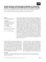
The development of CDAD is dependent on several factors (Fig. 1.1). The first factor is
acquisition of bacteria (Kelly and LaMont 1998). Since most patients become colonized during
hospital or nursing home stay and develop asymptomatic sequelae, colonization of the colon is
not sufficient for the development of disease. Instead, previous or concurrent antibiotic therapy is
the aggravating factor. The organism competes with normal intestinal flora when the latter is
disturbed by antibiotics which leads to overgrowth of C. difficile and elaboration of toxins. There
is a strong association between clindamycin use and development of CDAD (Tedesco, Barton et
al. 1974b) while broad-spectrum penicillins and cephalosporins are most commonly implicated
because of their widespread use (Nolan, Kelly et al. 1987). Studies involving molecular typing
reported that virulent C. difficile strains produce asymptomatic colonization more often than
5
Figure 1.1. Development and possible outcomes of C. difficile infection. Modified from
Abigail A. Salyers and Dixie D. Whitt, “Bacterial pathogenesis a molecular approach, 2
nd
edition (2002)”.
High number of non
-
spore forming
anaerobe over Normal Gut Flora
Microflora alteration
C. difficile proliferates
Symptoms
Abate
Vancomycin/
Metronidazole
CDAD
Ulceration
of Colon
Release of
Toxins A and B
Cessation of
treatment
Relapse
10-20%
Death
Antibiotic
Therapy
Asymptomatic
6
CDAD (Johnson, Clabots et al. 1990; Shim, Johnson et al. 1998), suggesting that extrinsic
bacterial factors like host immunity and timing and dosage of antimicrobial exposure must be
involved (Johnson and Gerding 1998).
As C. difficile grows, the toxins are released upon autolysis then enter the host cell via
receptor-mediated endocytosis and generalized pinocytosis. In rabbit, glycoprotein receptors for
toxin A on enterocyte membrane were found to be linked to nucleotide regulatory protein
(Pothoulakis, La Mont et al. 1991). Toxin A is an enterotoxin that causes excretion of fluid from
bowel whereas toxin B is primarily cytotoxic causing disruption in the signal transduction
pathway and disassembly of filamentous actin that leads to the collapse of cytoskeleton and cell
rounding (Hecht, Potoulakis et al. 1988). Bowel fluid released from damaged epithelial cells
containing polymorphonuclear neutrophil (PMN), lymphocyte, serum protein, erythrocyte and
mucus is inflammatory.
The toxins can also invoke inflammation through their ability to act as chemoattractant
for PMNs and stimulate release of mediators such as tumor necrosis factor alpha (TNF∝) and
interleukin 1 and 6 (Pothoulakis, Castagliuolo et al. 1993; Henderson, Wilson et al. 1999). The
infiltration and damage to the colonic mucosa result in the accumulation of fibrin, mucin, dead
cells and leukocytes forming yellowish patches of separate lesions on the mucosal surface. These
eventually coalesce into a sheetlike layer called pseudomembrane that distinguishes PMC from
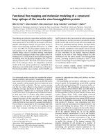
other types of colonic infection (Price and Davies 1977; Kelly and LaMont 1998) (Fig. 1.2A,B).
PMC is a potentially lethal gastrointestinal disease characterized by exudative plaques with
necrosis of the intestinal mucosal surface.
1.5. Disease management
Aside from diarrhea, abdominal pain, tenesmus and fever are other common symptoms of
C. difficile-mediated colitis (McClane and Mietzner 1999). The disease can be fatal as it can lead
to colonic perforation or systemic toxicity if left untreated. The treatment of choice involves
7
A
B
Figure 1.2. Morphology of pseudomembrane in the colon. A. Endoscopic image of PMC
with arrows pointing to pseudomembranes. B. Microscopic image of pesudomembrane
with a “volcanic eruption” appearance. Images were reprinted from Brian W. Hurley and
Cuong C. Nguyen, “The spectrum of pseudomembranous enterocolitis and antibiotic-
associated diarrhea (2002)”.
8
discontinuance of use of offending antibiotic and commencement of efficacious drugs against C.
difficile such as oral vancomycin or oral metronidazole (Briceland, Quintiliani et al. 1988;
Peterson and Gerding 1990). Clindamycin, lincomycin, ampicillin or the cephalosphorins were
involved in many cases of PMC and CDAD whereas aminoglycosides, trimethoprim-
sulfamethoxazole, erythromycin and the fluoroquinolones were less likely causes (Silva, Fekety
et al. 1984; Bingley and Harding 1987; McFarland 1998; Apisarnthanarak, Razavi et al. 2002;
Hurley and Nguyen 2002; Safdar and Maki 2002).
Once therapy is discontinued, relapses occur in 10 to 20% of cases due to failure to clear
the organism and restore the normal microbiota. In this case, various management approaches
have been recommended like improvement on handwashing and use of barrier precautions such
as isolation of symptomatic patients (Samore 1999), fluid and electrolyte replacement and
administration of agents that slows intestinal motility (i.e., Lomotil), slow and tapering
vancomycin therapy (Tedesco, Gordon et al. 1985), use of rifampin or cholestyramine (Tedesco
1982; Buggy, Fekety et al. 1987), bacteriotherapy with fecal enemas (Tvede and Rask-Madsen
1989), oral administration of nontoxigenic C. difficile (Seal, Borriello et al. 1987), and treatment
with the yeast Saccharomyces boulardii (Surawicz, Mc Farland et al. 1989).
1.6. Virulence Factors
1.6.1. Large clostridial cytotoxins (LCT)
The most studied diseases caused by C. difficile are those with symptoms caused by the
largest known single-molecule bacterial toxins, toxin A and toxin B (Dove, Wang et al. 1990).
These are well-studied amongst the clostridial exotoxins and cytotoxins and are encoded within a
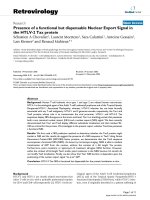
19.6 kb pathogenicity locus (PaLoc) of the C. difficile chromosome (Fig. 1.3). PaLoc contains a
putative positive regulator gene tcdD, LCT genes tcdA and tcdB, a putative holin gene tcdE and a
negative regulator gene tcdC. The toxins have no recognizable signal sequence and do not appear
9
Figure 1.3. The pathogenicity locus (PaLoc) of C. difficile VPI 10463 showing
conserved regions (GenBank accession nos. X51797, X53138, X92982, U25131,
U25132). Several gene portions encode for conserved structural features including the
glucosyltransferase or catalytic domains (striped block), nucleotide binding sites (solid
block), hydrophobic transmembrane domains (checkered block), repeating units (open
block) and binding domain for attachment to host cell receptor (speckled arrow). Solid
circles represent the DXD motif which is part of the catalytic domain responsible for
binding of Mn2+ whereas –SH symbolize conserved cysteines. The length of genes are
numerically indicated below the arrows (not drawn to scale) while arrowheads show the
transcriptional direction and line segments represent the size of monocistronic and
polycistronic transcripts. The figure is not drawn to scale for simplicity.
tcdE
tcdB
tcdC
tcdA
-
SH
tcdD
-SH
-
SH
-
SH
7098 bp
555bp
8133 bp
501bp
695 bp
10
to be proteolytically activated, as both toxins are released upon bacterial autolysis (Dove, Wang
et al. 1990).
They share around 49% of amino acid sequence identity with extensive structural
similarity in the C-terminal third consisting of small repeating subunits within larger units (Fig.
1.3). This portion is involved in receptor-binding specifically to galactose-rich residues and has
similarity to glucosyltransferases of Streptococcus mutans and Streptococcus sobrinus (GtfB,
GtfC and GtfI) which can bind to carbohydrates (von Eichel-Streiber, Laufenberg-Feldmann et al.
1992). Since monoclonal antibodies against the repeating subunits (amino acid residues starting
at 2097 and 2355) neutralize toxin A enterotoxic activity and inhibit its binding to carbohydrate
receptors, the repeating subunit portion appears to be immunodominant. Galα1-3Galβ1-
4GlcNAc on human intestinal cells has been identified as the toxin A receptor and it was
suggested to be the same receptor for toxin B (Lyerly and Wilkins 1995). Differences in receptor
composition and distribution may contribute to the level of toxicity among intestinal cells.
With about 50% homology, the N-terminal regions of LCTs are composed of a central
hydrophobic domain that represents a membrane spanning region involved in receptor-binding,
translocation of enzyme portion into the cell cytoplasm and intracellular processing. It is a
conserved domain with 4 cysteine residues and a putative nucleotide-binding domain that is
involved in the glucosylation of G-proteins (Fig. 1.3). Site-directed modification of toxin B
histidine residue of the nucleotide-binding site to glutamine resulted in 90% loss of toxic activity
(Aktories and Just 1995; Hofmann, Busch et al. 1997). The enzymatic activity of toxins A and B
was traced to a 63 kDa recombinant fragment located at around 516 to 542 residues of the N-
terminal region. These findings highlight the critical role of the N-terminal region in cytotoxicity
(Barroso, Moncrief et al. 1994).
Consistent to their considerable sequence homology, recent studies on both toxins
suggest similar molecular action (Jander, Rahme et al. 2000). Upon binding to membrane
receptors and internalization, the toxins act as monoglucosyltransferases capable of modifying
11
and consequently inhibiting members of the Rho family proteins including Rac, Cdc42 and Rho
subtypes RhoA, RhoB and RhoC. The toxins can cleave UDP-glucose and attach a glucose
moiety to residue Thr37 of Rho protein. This modifies Rho from an active (UDP-glucose + Rho)
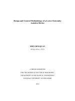
to an inhibited conformation (Glucose-Rho + UDP)(Fig. 1.4).
Rho proteins are members of a subfamily of small GTP-binding proteins (G-protein) that
act as “molecular switches” capable of regulating a number of essential functions in mammalian
cell including cell adhesion, microfilament organization, nuclear signaling, pseudopod formation
and re-shaping (intravasation) of phagocytes and many signal transduction pathways (Aktories
and Just 1995). The toxins preferentially glucosylate the GDP-bound form of G-protein because
its configuration exposes the threonine residue (Fig. 1.4). In contrast, the threonine gets buried in
the GTP-bound form that becomes inaccessible to toxin activity. As a consequence of Rho
glucosylation, GTPase activity of G-protein is reduced through intensified release of GTP which
disrupts signaling control and many cellular processes. This mechanism has been exploited by
molecular biologists as a useful tool the studies of cytoskeletal dynamics.
1.6.1.1. Toxin A
Toxin A is the largest bacterial toxin known having a molecular mass of 308 kDa
(Taylor, Thorne et al. 1981; Fiorentini and Thelestam 1991). The protein is stable at pH 4-10, has
a pl of 5.3 and possess many biological properties including enterotoxicity, cytotoxicity,
cytotonicity and red blood cell agglutinin (Taylor, Thorne et al. 1981; Donta, Sullivan et al. 1982;
Krivan, Clark et al. 1986). The toxin A receptor on rabbit erythrocytes and muscularis mucosae,
bovine thyroglobulin and rodent brush border membrane contains the trisaccharide sequence
Galα1-3Galβ1-4GlcNAc (Krivan, Clark et al. 1986; Clark, Krivan et al. 1987; Kamiya, Reed et al
al.1989; Percy, Burakoff et al. 1998).
12
Figure 1.4. Toxin A and toxin B action to G-protein. A, The normal cycling of G-
protein between the GDP- and GTP-bound form. The threonine residue (Thr) which will
be glucosylated by the toxin is shown. It is on the surface of the GDP-bound form of G-
protein but buried in the GTP-bound form. B, The GDP-bound form still converts to the
GTP bound form even though the Thr is glucosylated. However, the glucosylated GTP-
bound form has lower affinity and GTPase activity for its downstream effectors. These
events hinder the normal functions of G-protein causing changes in cell physiology.
Reprinted from Abigail A. Salyers and Dixie D. Whitt, “Bacterial pathogenesis, a
molecular approach, 2
nd
edition (2002)”.
Thr
GDP-bound
G-protein
GDP
PO
4
GTP
Thr
GTP-bound
G-protein
effect
Signal
Weak binding of GTP,
Low GTPase activity
B
. G-protein glucosylated by toxin
UDP-glucose
GDP
A
. Normal function
UDP
Thr
Glc
GTP
GDP
GDP
Glc
GTP
13
Human analogs of this core carbohydrate have been identified and designated as X antigen
(Galβ1-4[Fucα1-3]GlcNAcβ1-3Galβ1-4α[Glc]), Y antigen with the highest affinity to toxin has
an extra α1-2-linked fucose to X antigen and I antigen with 2 core structures (Galβ1-4GlcNAc)
per oligosaccharide (Tucker and Wilkins 1991). Carbohydrate moieties similar to X antigen are
expressed on secretory component of immunoglobulin and PMNs during inflammation. PMNs
initially attach to these receptor molecules on the surface of endothelial lining prior to
extravasation. Thus, a similar mechanism may explain the chemotactic and binding properties of
the toxin to PMN.
Other toxin A receptors on human epithelial cell may be involved since treatment with α-
galactosidase which does not affect the X,Y,I antigens (Lewis group) was found to reduce
binding (Smith, Cooke et al. 1997). The receptor was demonstrated to be a glycoprotein when
protease treatment likewise resulted in partial binding and later identified as a sucrase-isomaltase
glycoprotein on rabbit ileal brush border (Pothoulakis, La Mont et al. 1991; Pothoulakis, Gilbert
et al. 1996). Eventually other molecules such as substance P receptors were identified and shown
to be essential in causing enteritis (Pothoulakis, Castagliuolo et al. 1998). Through electron
microscopy, internalization of toxin A was observed via its localization on clathrin-coated pits.
Moreso, an acidic environment was required for its release from the endosome (Henriques, Florin
et al. 1987; Fiorentini and Thelestam 1991). Endosomal acidification leads to increased
hydrophobicity, revelation of toxin transmembrane structure, membrane insertion and
translocation (Qa'Dan, Spyres et al. 2000).
Toxin A acts as an enterotoxin when injected into ligated ileal loops inducing release of
bloody, viscous secretion upon mucosal damage (Triadafilopoulos, Pothoulakis et al. 1987).
Similar to toxin B, pertussis and diphtheria toxin however, toxin A is an intracellular-acting
cytotoxin which is internalized into target cell (Kushnaryov and Sedmak 1989). Various
mammalian cell lines were susceptible to toxin A including Chinese Hamster Ovary (CHO),
human cervix (HeLa), mouse adrenocortical (Y1), human larynx (Hep-2), human lung fibroblast
14
(MRC-5) and other cell lines (Donta, Sullivan et al. 1982; Henriques, Florin et al. 1987; Lima,
Lyerly et al. 1988; Kushnaryov and Sedmak 1989; Fiorentini, Malorni et al. 1990).
Many cellular effects of the toxin were observed. For example, cytoskeletal collapse
caused cell rounding and marginalization of nucleus in HeLa and CHO cells. After 3 h of toxin A
treatment, parallel filament bundles of 11 nm diameter were transiently visible in the nuclei with
extensive development of golgi apparatus, smooth endoplasmic reticulum and lysosomes
indicative of disturbance in synthetic and secretory functions of the cells (Kushnaryov and
Sedmak 1989; Fiorentini, Malorni et al. 1990). In IEC-6 cells, surface blebbing and nuclear
fragmentation ensued after early cytoskeletal disaggregation (Fiorentini, Donelli et al. 1993).
Intoxication of T8 cells showed increased tight junction permeability while induction of
interleukin 8 (IL-8), apoptosis and cell detachment were evident in human intestinal cells (Hecht,
Potoulakis et al. 1988; Mahida, Makh et al. 1996). Other observed biological effects were
induction of chloride secretion, actin condensation leading to membrane retraction and decline in
membrane potential, morphological changes of mitochondria and disaggregation, abrupt increase
in cellular ATP levels (Moore, Pothoulakis et al. 1990; He, Hagen et al. 2000). Overall, these
essential physiologic functions of cells were hindered directly through toxin enzymatic action or
indirectly as a result of Rho monoglucosylation or cytoskeletal disassembly.
The UDP-glucose hydrolase and glucosyltransferase activities of toxin A have been
attributed to the first 695 (Faust, Ye et al. 1998) or 562 amino acid residues of the N-terminus
(Ciesla and Bobak 1998). Similarly, glucosyltransferase activity of toxin B was detected within
the first 900 residues (Hofmann, Busch et al. 1997) owing to high sequence homology of LCTs.
The catalytic portion contains a conserved DXD motif flanked by hydrophobic regions (Wiggins
and Munro 1998). Amino acid residues composing this region are likely essential to function
since mutations in Asp286 and Asp288 of C. sordelii lethal toxin which is 90% homologous to
toxin B (Just, Selzer et al. 1996), inhibited both glycohydrolase and glucosyltransferase activities
and attachment of azido-UDP-glucose to the N-terminus of toxin (Busch, Hofmann et al. 1998).
15
The DXD motif was found to be important in Mn
2+
binding to allow the correct positioning of the
cosubstrate UDP-glucose which is subsequently cleaved and transferred and attached to Thr-37 of
Rho.
1.6.1.2. Toxin B
Toxin B is similarly large with its molecular weight at 270 kDa and pl at 4.1 (Sullivan,
Pellett et al. 1982). It is non-enterotoxic, non-cytotonic but a more potent cytotoxin than toxin A
that causes cell rounding but not fluid hypersecretion (Pothoulakis, Barone et al. 1986). Like
toxin A, toxin B is also composed of 3 major structural domains, has repetitive oligopeptides
suggestive of membrane binding via glycoprotein receptors and the presence of a flanked
hydrophobic region of 172 residues which was proposed to function for intracellular translocation
(von Eichel-Streiber, Laufenberg-Feldmann et al. 1992). A drop in intra-endosomal pH was also
observed, such that toxin B may assume a hydrophobic structural fold necessary for membrane
insertion and translocation, a process requiring the presence of Ca
2+
and calmodulin (Caspar,
Florin et al. 1987; Gilbert, Pothoulakis et al. 1995; Qa'Dan, Spyres et al. 2000). Therefore,
similarity in physical properties between toxins A and B is not surprising due to their conserved
nature. In addition, toxin B likewise functions in the UDP-glucosylation of small GTP-binding
proteins Rho, Rac and Cdc42 (Just, Selzer et al. 1995a) but not Rap which is a toxin A substrate
(Chaves-Olarte, Weidmann et al. 1997). The enzymatic domain lies within the N-terminus
(located at the first 546 or 467 amino acid residues) possessing a conserved residue Trp102 which
is crucial for UDP-glc binding (Busch, Hofmann et al. 1998).
Some of the more prominent effects of toxin B are directed towards immune cells.
Macrophages were demonstrated to release lipooxygenases including LTB4 and tumor necrosis
factor (TNFα) upon intoxication (Siffert, Baldacini et al. 1993; Souza, Melo-Filho et al. 1997).
Moreso, human monocytes released inflammatory mediators IL-1, IL-6 and TNFα which are
potent PMN chemoattractants. Therefore, it is evident that toxin B works through primary
16
induction of the release of proinflammatory cytokine by myeloid cells prior to biomolecular
disruption. Expectedly however, similar cellular effects of toxin A was observable on toxin B
treatment. For example, toxin B also induced calcium influx for actin disassembly (Gilbert,
Pothoulakis et al. 1995); nuclear fragmentation and chromatin condensation which are typical
features of apoptosis (Fiorentini, Fabbri et al. 1998); loss of anchorage due to actin, vinculin, talin
and vimentin reorganization (Ottlinger and Lin 1988; Ciesielski-Treska, Ulrich et al. 1989);
inhibition of protein synthesis (Pothoulakis, Barone et al. 1986); potocytotic surface blebbing
characterized by the presence of bleb matrix with ribosomes but devoid of organelles and
morphological alterations such as retraction of cytoplasmic projections and rounding (Wedel,
Toselli et al. 1983). However, unique effects of toxin B (100X more cytotoxic than toxin A) were
also reported (Riegler, Sedivy et al. 1995; Chaves-Olarte, Weidmann et al. 1997). Toxin B was
also observed to prevent proper signaling in human embryonic kidney cells via muscarinic
acetylcholine receptor and in rat basophilic leukemia cells via phospholipase D receptor (Ojio,
Banno et al. 1996; Schmidt, Rumenapp et al. 1996).
1.6.1.3. The Pathogenicity Locus (PaLoc)
1.6.1.3.1. Genetic profile of the PaLoc
In the late 1980s, a small fragment of toxin A gene (0.3 kb) was expressed from lambda
gt11 library with the unstable product found to be capable of causing CHO cell elongation
(Muldrow, Ibeanu et al. 1987). Thereafter, the use of a 4.7 kb PstI restriction fragment that
encodes a portion of tcdA as probe for chromosomal walking of VPI 10463 genome, led to the
discovery of both tcdA and tcdB sequences and their proximity (Barroso, Wang et al. 1990; Dove,
Wang et al. 1990). The tcdA gene is 8130 bp long of 26.9% GC content which encodes for 2710
amino acid residues (Sauerborn and von Eichel-Streiber 1990). Its fusion protein product showed
lethal cytotoxic and enterotoxic activities (Phelps, Lyerly et al. 1991). On the other hand, tcdB of
7098 nucleotides and 27.4% GC composition, encodes for 2366 amino acid residues whose
17
expressed protein exhibiting cytotoxicity and reactivity to toxin B antisera (Barroso, Wang et al.
1990). Likewise, von Eichel-Streiber et al. (1987; 1989; 1992) cloned the LCT genes which were
proposed to have arisen as a result of gene duplication due to their extensive sequence identity.
Recently, a toxinotyping scheme was proposed for the standardization of C. difficile
strain serogrouping (Rupnik, Avesani et al. 1998). Classification was further streamlined at the
first international C.difficile symposium where unified nomenclature for toxin genes and their
products were drafted, with relevant suggestions such as the use of tcd series in the designation of
PaLoc genes and renaming of tcdD as tcdR referring to the role of the protein product in
regulation (Rupnik, Dupuy et al. 2005).
Accordingly, toxin A and toxin B share common structural motifs (Fig. 1.3) like the N-
terminal third active site cleft with DXD motif and possessing enzymatic properties, a central
domain with 4 cysteines suggested to be critical for enzymatic function, a membrane-spanning
hydrophobic region which is necessary for toxin transport (von Eichel-Streiber, Meyer zu
Heringdorf et al. 1995) and the C-terminal domain which is comprised of repeating units of
aromatic amino acids tyrosine and phenylalanine that function for carbohydrate receptor binding
(von Eichel-Streiber and Sauerborn 1990).
The C-terminal portion was also observed to be
immunodominant portion of the toxin (von Eichel-Streiber, Harperath et al. 1987). While the
importance of the central hydrophobic region in toxin B in cytotoxicity and its function for
protein translocation in adenylate cyclase of Bordetella pertussis (Hanski 1989), alpha hemolysin
of E.coli and leukotoxin of Pasteurella haemolytica has been demonstrated (Strathdee and Lo
1987), the role of this membrane-spanning region in intracellular transport of LCTs is yet to be
determined.
Recent evidences have shown that bacterial virulence factors could be acquired through
horizontal transfer of genes often clustered into genetic blocks or modules called pathogenicity
island (Hammond and Johnson 1995; Groisman and Ochman 1996; Hacker, Blum-Oehler et al.
1997). Hammond et al. (1995), first reported the cluster of toxigenic elements in VPI 10463
18
chromosome composed of LCT genes and three smaller accessory orfs. In 1997, Hundsberger et
al. (1997), designated the block as the pathogenicity locus (PaLoc) and the genes as tcdA to tcdE
(Fig. 1.3) with tcdD, tcdB, tcdE and tcdA transcribed in the same direction in that order and tcdC
(adjacent to tcdA) in the opposite direction. The 3’ end of tcdB is 1350 bp upstream of the tcdA
translation start site (Fig. 1.3). tcdE which is composed of 501 bp and flanked by tcdB and tcdA,
is 122 bp downstream of tcdB stop codon. Located immediately upstream of tcdB is the 555 bp
tcdD gene that codes for TcdD protein with high lysine content of its C-terminus. In
nontoxigenic C. difficile strains, the PaLoc is replaced with either a 115 bp nucleotide segment
having transcriptional terminator features (Braun, Hundsberger et al. 1996) or a 127 bp segment
flanked by AT-rich inverted repeats characteristic of an insertion element (Hammond and
Johnson 1995) which are indicative of potential sites for genetic exchange.
1.6.1.3.2. Regulation of PaLoc genes
The divergent capabilities of C. difficile strains in their level of toxin production despite
identity of the PaLoc region, implicate heterogeneity in the regulatory process. One PaLoc gene
proposed to be involved in regulation is tcdD (txeR). TcdD displays a helix-turn-helix
configuration and shares significant identity with UviA, a promoter P1 DNA-binding bacterial
response regulator (Moncrief, Barroso et al. 1997) and positive regulators of C. botulinum
neurotoxin BotR (Hauser, Eklund et al. 1994) and C. tetani neurotoxin TetR (Marvaud, Eisel et
al. 1998). Indeed, it was observed that the expression of toxin A and toxin B promoter repeating
units (ARU) increased by 500 and 800-fold respectively, when tcdD was supplied in trans.
Furthermore, there are long intergenic distances between the transcriptional start sites (TSS) and
start codons of both toxin A (169 bases) and toxin B (239 bases) promoter regions typical among
clostridial sequences. These are strong indication of its role in the positive regulation of toxin
expression. However, when grown under conditions of limited nutrient, the level of toxin A and
toxin B was observed to increase (Yamakawa, Karasawa et al. 1996). Instead of acting as
19
transcriptional regulator therefore, TcdD may function as a sigma factor that promotes toxin
synthesis in response to stress. The PaLoc gene suggested to be involved in negative regulation is
tcdC (Hundsberger, Braun et al. 1997). It contains 695 bp located downstream of tcdA which
encodes for an acidic protein with various lengths of repetitive residues.
The proposed regulatory process starts with tcdD as being transcribed at high levels
during the early exponential growth phase which was immediately followed by increased
expression of tcdB, tcdE and tcdA at the stationary phase (Hundsberger, Braun et al. 1997).
Therefore, it was deduced that TcdD acts as a positive regulator of PaLoc gene expression. As
the bacteria enter the late stationary phase, TcdC expression becomes elevated to counter TcdD
action and antisense effect of transcripts from other genes. Toxin synthesis was most pronounced
during the early and mid-exponential growth phase, thus, the high toxin level at the stationary
phase is due to toxin accumulation. Finally, several studies have demonstrated the combined
monocistronic and polycistronic mode of PaLoc transcription through detection of individual and
readthrough mRNAs (Hammond, Lyerly et al. 1997; Hundsberger, Braun et al. 1997; Dupuy and
Sonenshein 1998). These connote inherent multiplicity in promoter sequences and discrepancy in
the expression level of different genes comprising the PaLoc.
1.6 2. CDT toxin
Some C. difficile strains possess accessory toxin genes cdtA (encodes for CDTa) and cdtB
(encodes for CDTb) that share 80 and 82% amino acid sequence identity
to C. perfringens Ia and
Ib, respectively (Popoff, Rubin et al. 1988b; Perelle, Gibert et al. 1997a) (Fig. 1.5). Around 6-
16% of C. difficile isolates from clinical setting contain both CDT components suggesting its
secondary role to LCTs (Stubbs, Rupnik et al. 2000; Geric, Johnson et al. 2003). The protein
components
were found interchangeable, thus catalytically functional chimeras from clostridial
bacteria have been generated (Popoff, Rubin et al. 1988b; Popoff, Milward et al. 1989; Perelle,
Scalzo et al. 1997b; Gulke, Pfeifer et al. 2001; Geric, Johnson et al. 2003). Accordingly, C.
20
difficile, C. perfringens, and C. spiroforme can all cause gastrointestinal diseases in humans as
well as animals (Borriello and Carman 1983; Braun, Herholz et al. 2000; Stoddart and Wilcox
2002), thus implying common evolutionary lineage.
1.6 2.1. ADP-ribosyltransferase
Aside from LCTs, a number of C. difficile strains simultaneously produce an ADP-
ribosyltransferase (ADPRT) designated as CDT toxin (Perelle, Gibert et al. 1997a). The enzyme
can mediate catalysis of nicotinamide adenine dinucleotide (NAD) and attachment of ADP-
ribosyl group to various protein substrates. Since several bacteria produce ADPRT (Table 1.1),
Figure 1.5. The cdt locus of C. difficile CD196 showing conserved regions (GenBank
accession no. L76081). Several gene portions encode for conserved structural features
including the catalytic domain (diagonally-striped block), nucleotide binding site (solid
block), hydrophobic transmembrane domains (checkered block), docking site (vertically-
striped block), oligomerization domain (horizontally-striped block) and binding domains
(speckled arrow). Arrows represent relative gene sizes (not drawn to scale) and point to
the direction of transcription. Relative gene sizes are indicated numerically below the
arrows.
they have been classified into four groups based on the type of substrate they act on. The first
group includes the diphtheria toxin (DT) of Corynebacterium diphtheriae (van Ness, Howard et
cdtA cdtB
2631 bp
1392 bp
21
al. 1980) and exotoxin A (ETA) of Pseudomonas aeruginosa (Wretlind, Bjorklind et al. 1987).
These toxins are involved in attaching the ADP-ribose to eukaryotic elongation factor 2 (EF-2)
which is active in protein synthesis. Members of the second group ADP ribosylates membrane
associated G-proteins. This group is comprised of cholera toxin (CT) produced by Vibrio
cholerae, pertussis toxin (PT) of Bordetella pertussis, Escherichia coli heat-labile enterotoxins
(LT1 and LT2) and Pseudomonas exoenzyme S (ExoS) (Moss and Vaughan 1988). Toxins that
act on low molecular weight GTP-binding protein Rho comprise the third group. These include
the C3 enzyme of Clostridium botulinum (Aktories, Rosener et al. 1988; Just, Mohr et al. 1992),
Clostridium limosum Sa and Sb exoenzymes (Just, Mohr et al. 1992) and epidermal cell
differentiation inhibitor (EDIN) of Staphylococcus aureus (Sugai, Hashimoto et al. 1992). The
fourth group are actin-specific ADPRTs which include the C2 toxin of Clostridium botulinum
types C and D (Aktories, Barmann et al. 1986b; Aktories, Weller et al. 1987),
iota toxin of
Clostridium perfringens type E (Simpson, Stiles et al. 1987), Clostridium spiroforme toxin (Stiles
and Wilkins 1986a; Simpson, Stiles et al. 1989), Clostridium difficile CDT toxin (Popoff, Rubin
et al. 1988b) and the Bacillus cereus vegetative insecticidal proteins, VIP1-VIP2 (Seungil, Craig
et al. 1999). These binary toxins which act independently as catalytic and membrane-binding
proteins are distinct from the classical A-B toxins like cholera toxin that needs to assemble to
form a functional complex with two different subunits (Madshus and Stenmark 1992).
1.6 2.2. Actin as substrate
Being one of the most abundant proteins in eukaryotic cells, actin plays a myriad of functions that
maintains homeostatic condition and normal physiology (Pollard 1986). Its propensity towards
reversible shift from the monomeric (G-actin) to filamentous (F-actin) form and vice versa makes
the accomplishment of its versatile roles possible. A number of its functions include organization
of organelles, cell division, protein translocation, support, mobility, secretion, phagocytosis and
more.
22
Table 1.1. Bacterial toxins produced as binary or preformed A-B structures
Bacteria Toxin (type
a
) Activity (substrate) Reference
C. difficile CDTa, CDTb (B) ADPRT
b
(G-actin) Popoff et al., 1988b
C. perfringens Ia,Ib (B) ADPRT (G-actin) Schering et al., 1988
C. botulinum C2I,C2II (B) ADPRT (G-actin) Ohishi & Tsuyama, 1986
C. spiroforme Sa,Sb (B) ADPRT (G-actin) Popoff & Boquet, 1988a
B. cereus VIP1,VIP2 (B) ADPRT (G-actin) Han et al., 1999
B. anthracis EF, LF (B)
adenylate cyclase and metalloprotease
(MAPKK) Leppla 1982, Vitale 1998
C. difficile Toxins A,B (P) glucosyltransferase (Rho proteins) Hippenstiel et al., 1997
C. novyi alpha (P) glucosyltransferase (Rho proteins) Selzer et al., 1996
C. botulinum C3 (P) ADPRT (Rho proteins) Evans et al., 2004
C. limosum exoenzyme (P) ADPRT (Rho proteins) Just et al., 1992
V. cholerae CT (P) ADPRT (G-protein) Moss & Vaughan, 1988
B. pertussis PT (P) ADPRT (G-protein) Moss & Vaughan, 1988
E. coli LT-1,LT-2 (P) ADPRT (G-protein) Moss & Vaughan, 1988
P. aeruginosa ExoS (P) ADPRT (G-protein) Sun et al., 2004
P. aeruginosa exotoxin A (P) ADPRT (eEF-2) Wretlind et al., 1987
a
(B) represents the binary type while (P) represents preformed type A-B toxin
b
ADPRT is ADP-ribosylating toxin
Actin is a single polypeptide comprised of 375 amino acid residues having a molecular
mass of 42 kDa. Crystallographic data showed that the molecule is made up of four subdomains
with one nucleotide binding cleft for ATP or ADP which attracts a divalent cation like Mg
2+
(Kabsch, Mannherz et al. 1990). Owing to its highly conserved nature, variability in amino acid
composition was reported at only 6% (Korn 1982). In mammalian cells, actin was classified into
6 major groups namely: the skeletal muscle α-actin, cardiac muscle α-actin, smooth muscle α-
and γ-actin, and cytoplasmic β- and γ-actin, which comprise the 3 isoforms α, β and γ,
distinguished by their isoelectric properties (Vandekerckhove and Weber 1979). Alpha-actin is
the most acidic while β-actin is partially acidic and the γ isoform is most basic. Iota toxin which
is a close homologue of CDT was shown to to act on all isoforms (Schering, Barmann et al. 1988)
whereas C2 only ADP-ribosylates the β and γ isoforms (Aktories, Ankenbauer et al. 1986a;
Ohishi and Tsuyama 1986).
23
The actin filament is a polar molecule having a fast-growing (barbed), positive end a
slow growing (pointed), negative end with the ATP-bound actin being more stable than ADP-
bound form in F-actin (Carlier 1991). The structural shift is regulated by several factors
including its interaction with actin-binding anchor proteins like α-actinin, profilin, gelsolin and β-
thymosin (Pollard and Cooper 1986). Therefore, the protein assumes a dynamic configuration as
simultaneous polymerization and depolymerization process takes place.
ADPRTs of the 4th group labels the monomeric actin form specifically at a conserved
acceptor amino acid Arg177 which was revealed through site-directed mutagenesis of the
enzymatic component (Aktories, Barmann et al. 1986b; Schering, Barmann et al. 1988). Since
the ADP-ribosylation site is located at the axis of the F-actin helix, the bulky ADP-ribose group
attached to Arg177 poses considerable steric hindrance to the polymerization process. The ADP-
ribose moiety behaves like a capping protein that blocks the positive end the filament.
Furthermore, ADP-ribosylation inhibits the actin ATPase activity. These disturbances may have
critical consequences such as breakdown of cytoskeletal network and deterioration of cellular
protein movement and function.
1.6.2.3. Biology of actin-specific ADPRT
CDT and other ADPRTs employ a binary mode for intoxication of eukaryotic cells. The
binary nature was first illustrated through the use of cross-reacting and neutralizing antibodies
against C. spiroforme toxin (Stiles and Wilkins 1986a; Stiles and Wilkins 1986b). The properties
of the binary components were later characterized based on electrophoretic mobility and found to
be nontoxic individually but potently cytotoxic in combination (Stiles and Wilkins 1986a).
Thereafter, iota Ia was discovered to be an ADP-ribosyltransferase whose substrate is the actin
monomer (Simpson, Stiles et al. 1987; Schering, Barmann et al. 1988) while Ib mediates
translocation of Ia into cells (Stiles, Hale et al. 2000; Blocker, Behlke et al. 2001; Richard,
Mainguy et al. 2002).
24
The functional toxin is composed of a precursor enzymatic or A protein component and a
translocation or B protein component forming an A-B complex (Table 1.1). This differs form
other bacterial binary toxins that engage target as a preformed holotoxin with an A-B structure
(Table 1.1). Intoxication by CDT and C2 initially occur through the binding of the B component
on target cell membrane, then oligomerization into heptamer on the cell surface. However, initial
processing of the B monomer by proteases (Ohishi and Miyake 1985; Simpson 1989) is required
before attachment to membrane receptors and formation of complexes. Thereafter, the B-receptor
complex functions as a docking site for the enzymatic A molecule. The aggregate is subsequently
translocated into the cytosol via an acidified endosomal vesicle (Simpson 1989).
that is optimal
for toxin to assume a conformation capable of membrane insertion and translocation.
Cytoplasmic toxin can then inhibit various normal cell physiologic functions primarily those
involving cytoskeletal structures.
1.6.2.4. Genetics of ADPRT
Among binary ADPRTs, the genes of components A and B have the same transcriptional
orientation with gene B located some 50 nucleotides downstream of gene A except for C2 which
is separated by 247 nucleotides (Perelle, Gibert et al. 1993; Fujii, Kubota et al. 1996; Kimura,
Kubota et al. 1998). And like most clostridial proteins, ADPRTs are encoded by genes of high
A+T content, specifically at about only 27 to 31% G+C content. Distinctive genetic features
among ADPRTs were also discovered including the chromosomal location of CDT and C2 toxin
versus the plasmid-encoded iota toxin. Furthermore, CDT chromosome contains sequences for a
signal peptide which was not present in C2. Whether C2 and other ADPRTs produce other
chaperone proteins for secretion like the PA83 protein from Bacillus species (Williams, Rees et
al. 2003) is still not clear.
25
PaLoc toxin genes tcdA and tcdB were shown to be transcribed both from gene-
specific promoters and from promoters of upstream genes with the gene-specific
transcripts represented the majority of toxin gene mRNAs and that their expression is
subject to catabolite repression by glucose (Dupuy and Sonenshein 1998). This glucose
effect was general to many toxinogenic strains having varying levels of toxin production.
Most contributory to PaLoc toxin synthesis is tcdR (formerly tcdD) which was found to
act as RNA polymerase sigma factor. In addition, external factors as temperature (Mani,
Lyras et al. 2002) and endogenous factors like cortocosteroids (Castagtliuolo, Karalis et
al. 2001), cysteine (Karlsson, Lindberg et al. 2000), intracellular calcium and NF-kappa
B (Jefferson, Smith et al. 1999) were reported to have considerable influence on toxin
expression.
Unlike the regulation on the expression of PaLoc genes, information on cdt regulation is
limited. However, ample reports have been published on the action of a plasmid pX01-encoded
B. anthracis atxA gene producing a 56 kDa protein (Dai, Sirard et al. 1995) as a positive regulator
for lethal toxins, capsule and other genes encoded on plasmids pX01 and pX02 (Guignot, Mock
et al. 1997; Hoffmaster and Koehler 1997). External factors such as pH and temperature also
affect the control of protein synthesis (Koehler 2002). As less virulent C. difficile strains were
observed to be devoid of atxA (Koehler 2002), it is plausible that C. difficile produce a similar
regulatory protein to enhance CDT secretion and pathogenicity.
1.6.2.5. Conserved ADPRT structure and function
Unlike the binding component of ADPRTs, the enzymatic component has been reported
to have substantial homology particularly at important catalytic regions of the toxin.
Accordingly, several protein structures necessary for NAD binding and hydrolysis were found
conserved across different families of ADPRT (Domenighini, Magagnoli et al. 1994). Based on