HCV functional genomic protein interactions with NS3 and their role in viral replication
Bạn đang xem bản rút gọn của tài liệu. Xem và tải ngay bản đầy đủ của tài liệu tại đây (2.33 MB, 105 trang )
I
HCV functional genomics:
Protein interactions with NS3 and
their role in viral replication
Khu Yee Ling
B. Sc. (Hons)
A THESIS SUBMITTED
FOR THE DEGREE OF DOCTOR OF PHILOSOPHY
INSTITUTE OF MOLECULAR AND CELL BIOLOGY
NATIONAL UNIVERSITY OF SINGAPORE
2004
I
Acknowledgements
I am indebted to my supervisor, Dr Goh Phuay Yee, for her patience and
guidance. Thanks are also due to my committee members, Drs Alan
Porter and Thomas Dick for their invaluable advices.
I am also grateful to Dr Goh Phuay Yee for the dimerization mutants
(Y267S, M288T and T266A) and Dr Tan Yee Joo for useful discussion and
help with using the FPLC machine.
The presence of wonderful lab members in the CAVR group, both past and
present, has made my stay in the institute a memorable experience. I
thank them for their friendship and gossip sessions, which were highly
useful for de-stressing. Besides the excellent sequencing services
provided by Dr Alice Tay, our prophet and guru of all things big and
small, I would also like to thank her for all the stimulating conversations
we have shared.
Closest to my heart, I would like to thank my parents, especially mum,
who encourages me, believes in me and been my greatest fan, always. My
husband, one of the most important men in my life, thanks for being there
whenever I needed you and Yong Teng, the other man in my life, who
brought out the patience in me I never knew I have.
II
Table of Contents
ACKNOWLEDGEMENTS I
TABLE OF CONTENTS II
LIST OF FIGURES IV
LIST OF TABLES VII
LIST OF PUBLICATIONS VIII
LIST OF ABBREVIATIONS IX
SUMMARY XI
1. INTRODUCTION 1
1.1 Medical Importance of HCV 1
1.2 Molecular biology of HCV 3
1.2.1 Structural Proteins 4
1.2.2 Non-structural Proteins 6
1.3 HCV protein-protein interaction 9
1.4 Aims and Objectives 11
2. MATERIALS AND METHODS 12
2.1 Construction of Plasmids 12
2.2 Yeast two-hybrid screens 12
2.2.1 NS3 NS3 interaction 12
2.2.2 NS3 Host interaction 13
2.3 Generation of mutations in NS3 helicase 14
2.4 NS3 helicase and helicase mutants expression, purification and analytical
gel filtration 16
2.5 Helicase activity assay 17
2.6 In vitro binding assay 18
2.7 FL-NS3, LMP7 expression and purification 18
III
2.8 In vitro protease activity assay 20
2.9 Proteasome activity assay 20
2.10 Immunoprecipitation(IP) 22
2.11 Western blot analysis 22
2.12 Tissue culture 23
3. RESULT 27
3.1 Characterization of NS3-NS3 interaction 27
3.1.1 Delineating the region of self-interaction in NS3 27
3.1.2 Expression and purification of recombinant NS3 helicase for gel filtration
analysis 32
3.1.3 Analytical gel filtration of dimerization mutants 37
3.1.4 Correlation between dimer formation and helicase activity 41
3.2 Characterization of NS3-LMP7 interaction 45
3.2.1 Screening for NS3 host interacting partner 45
3.2.2 Delineating the region of interaction between NS3 and LMP7. 47
3.2.3 Expression and purification of recombinant NS3 and LMP7 for in vitro
assays 50
3.2.4 Effect of LMP7 on NS3 protease activity 53
3.2.5 Effect of NS3 on proteasome activity 55
4. DISCUSSION 67
4.1 NS3 NS3 interaction 67
4.2 NS3 LMP7 interaction 70
5. CONCLUSION 76
6. REFERENCES 77
IV
List of Figures
FIGURE 1-2. HCV GENOME AND ENCODED VIRAL PROTEINS 5
FIGURE 2-1. SCHEME SHOWING THE GENERATION OF RANDOM MUTANTS THAT DISRUPT
HELICASE INTERACTION
15
FIGURE 3-1. A MINIMAL DOMAIN OF NS3 REQUIRED FOR INTERACTION DEFINED BY
YEAST
-TWO HYBRID ASSAY 28
FIGURE 3-2. IMMUNOPRECIPITATION BETWEEN FLAG-TAGGED NS3 AND MYC-TAGGED
NS3. 29
FIGURE 3-3. THE NS3 HELICASE INTERACTS IN AN N-TO-N ORIENTATION 30
FIGURE 3-4. MINIMAL REGION FOUND TO INTERACT WITH HELICASE DOMAIN AND
ITSELF
31
FIGURE 3-5. RECOMBIANT NS3 HELICASE EXPRESSION. 33
FIGURE 3-6. PURIFICATION OF NS3 HELICASE BY FPLC 33
FIGURE 3-7. GEL FILTRATION OF WILD-TYPE HELICASE. 34
FIGURE 3-8. GEL FILTRATION OF HELICASE MUTANTS 36
FIGURE 3-9. POSITIONS OF SOME OF THE MUTANTS THAT DISRUPTED INTERACTION
BETWEEN TWO MINIMAL REGIONS
38
FIGURE 3-10. GEL FILTRATION OF DIMERIZATION MUTANTS 40
FIGURE 3-11. HELICASE ASSAYS OF WILD-TYPE HELICASE, MUTANTS Y267 AND AAA. 41
FIGURE 3-12. DIMERIZATION MUTANTS SHOWS REDUCTION IN HELICASE ACTIVITY. 42
FIGURE 3-13. INHIBITION OF HELICASE ACTIVITIES BY THE ADDITIONS OF MUTANT
PROTEINS
. 44
FIGURE 3-14. RECOMBINANT GST-NS3 AND GST EXPRESSION. 46
FIGURE 3-15. NS3-LMP7 INTERACTION SHOWN BY IN VITRO BINDING ASSAY 46
V
FIGURE 3-16. LMP7 INTERACTS WITH THE PROTEASE DOMAIN OF NS3 48
FIGURE 3-17. NS3 INTERACTS WITH THE PROSEQUENCE OF LMP7. 49
FIGURE 3-18. PURIFICATION OF RECOMBINANT GST-NS3 51
FIGURE 3-19. PURIFICATION OF RECOMBINANT LMP7 52
FIGURE 3-20. IN VITRO BINDING OF PURIFIED LMP7 TO GST NS3 AND PROTEASE
ACTIVITY OF PURIFIED
NS3 54
FIGURE 3-21. NS3 BINDS TO THE IMMUNOPROTEASOME COMPLEX. 56
FIGURE 3-22. CHYMOTRYPSIN-LIKE ACTIVITY OPTIMIZATION IN HELA CELLS USING
SUBSTRATE
LLVY-AMC 59
FIGURE 3-23. NS3 DID NOT AFFECT IMMUNOPROTEASOME CHYMOTRYPSIN-LIKE
ACTIVITY IN
HELA CELLS 59
FIGURE 3-24. TRYPSIN-LIKE ACTIVITY OPTIMIZATION IN HELA CELLS USING SUBSTRATE
LRR-AMC 60
FIGURE 3-25. NS3 REDUCES IMMUNOPROTEASOME TRYPSIN-LIKE ACTIVITY IN HELA
CELLS
60
FIGURE 3-26. POST ACIDIC ACTIVITY OPTIMIZATION IN HELA CELLS USING SUBSTRATE
LLE-AMC 61
FIGURE 3-27. NS3 DID NOT AFFECT IMMUNOPROTEASOME POST ACIDIC ACTIVITY IN HELA
CELLS
61
FIGURE 3-28. CHYMOTRYPSIN-LIKE ACTIVITY OPTIMIZATION IN HUH-7 CELLS USING
SUBSTRATE
LLVY-AMC 62
FIGURE 3-29. NS3 DID NOT AFFECT IMMUNOPROTEASOME CHYMOTRYPSIN-LIKE
ACTIVITY IN
HUH-7 CELLS. 62
FIGURE 3-30. TRYPSIN-LIKE ACTIVITY OPTIMIZATION IN HUH-7 CELLS USING SUBSTRATE
LRR-AMC 63
VI
FIGURE 3-31. NS3 DID NOT AFFECT IMMUNOPROTEASOME TRYPSIN-LIKE ACTIVITY IN
HUH-7 CELLS. 63
FIGURE 3-32. POST ACIDIC ACTIVITY OPTIMIZATION IN HUH-7 CELLS USING SUBSTRATE
LLE-AMC 64
FIGURE 3-33. NS3 DID NOT AFFECT IMMUNOPROTEASOME POST ACIDIC ACTIVITY IN
HUH-7 CELLS. 64
FIGURE 3-34. EXPRESSION OF NS3-NS5B VIRAL PROTEINS REDUCES THE LMP7-
IMMUNOPROPTEASOME ACTIVITY. 66
VII
List of Tables
TABLE 2-1. VECTORS USED IN THIS STUDY 24
TABLE 2-2. PLASMIDS USED IN STUDYING NS3-NS3 INTERACTION 25
TABLE 2-3. PLASMIDS USED IN STUDYING NS3 LMP7 INTERACTION 26
VIII
List of Publications
Lim, S. P., Y. L. Khu, W. Hong, A. Tay, A. E. Ting, S. G. Lim, and Y. H. Tan. 2001.
Identification and molecular characterization of the complete genome of a Singapore
isolate of hepatitis C virus: sequence comparison with other strains and phylogenetic
analysis. Virus Genes. 23:89-95.
Khu, Y. L., E. Koh, S. P. Lim, Y. H. Tan, S. Brenner, S. G. Lim, W. Hong, and P. Y.
Goh. 2001. Mutations that affect dimer formation and helicase activity of the hepatitis
C virus helicase. J. Virol. 75:205-214.
Khu, Y. L., Y. J. Tan, S. G. Lim, W. Hong, and P. Y. Goh. 2004. Hepatitis C virus
nonstructural protein NS3 interacts with LMP7, a component of immunoproteasome,
and affects its proteasome activity. Biochem. J. (in press)
IX
List of Abbreviations
aa Amino acid
ABS Absorbance
ds Double stranded
DTT Dithiothreitol
ER Endoplasmic reticulum
FPLC Fast-performance liquid chromatography
GSH Glutathione
GST Glutathione S-transferase
HCV Hepatitis C virus
HVR Hypervariable region
IFN Interferon
IFN-
Interferon-gamma
IgG Immunoglobulin G
IPTG Isopropyl-1-thio-
-D-galactopyranoside
IRES Internal ribosomal entry site
ISDR Interferon sensitivity determining region
kd Kilo dalton
LMP Low molecular weight protein
LDLR Low-density lipoprotein receptor
NS Non-structural protein
nt Nucleotide
MHC Major histocompatibility complex
NTPase Nucleoside triphosphatase
ORF Open reading frame
X
PBS Phosphate buffered saline
PCR Random polymerase chain reaction
PEG Polyethylene glycol
PKR Double-stranded RNA-dependent protein kinase
PMSF Phenylmethylsufonyl fluoride
RFU Relative fluorescence unit
RdRp RNA dependent RNA polymerase
SDS-PAGE Sodium dodecyl sulfate-polyacrylamide gel electrophoresis
UTR Untranslated region
X-Gal 5-bromo-4-chloro-3-indolyl-
-D-galactopyranoside
YEPD Yeast extract-peptone-dextrose
-Gal
-Galactosidase
XI
Summary
Hepatitis C virus (HCV) is one of the major causes of liver diseases worldwide.
The non-structural protein 3 (NS3) of HCV, which is both a protease as well as a
helicase, plays important roles in the processing of the viral polyprotein and the
replication of viral RNA. This thesis attempts to answer several questions with regards
to viral and host interacting proteins of NS3, which may eventually assist in the
understanding of the mechanism of HCV replication and pathogenesis. Yeast two-
hybrid assays and co-immunoprecipitation experiments were employed to identify and
verify these interactions. The characterization and functional analysis of NS3
interacting partners are discussed essentially in two parts. The first part focuses on
NS3 NS3 self association while the second part describes in detail the interaction
between NS3 and a cellular protein, LMP7.
NS3 was found to bind strongly with itself and the minimal region required for
this interaction was mapped to a specific subdomain of 174 amino acids in the N
terminus of the helicase region. Random mutations in this minimal region were
generated by PCR, and mutants that failed to interact with a wild-type minimal
fragment were isolated using yeast two-hybrid assay as a screen. Three of these
mutations resulted in a reduction or a loss of interaction between helicases. Analytical
gel filtration showed that in the presence of an oligonucleotide, wild-type helicases
form dimers whereas the mutants remain mostly monomeric. All three mutants were
partially or almost inactive when assayed for helicase activity in vitro. Mixing a
dimerization mutant (Y267S) with wild-type helicase did not dramatically affect
helicase activity. These data indicate that dimerization of the helicase is important for
XII
helicase activity. The mutations that reduce self-association of the helicase may define
the key residues involved in NS3-NS3 dimerization (Khu et al., 2001).
Low molecular weight protein 7 (LMP7), an interferon-gamma (IFN-
)
inducible component of the proteasome isolated from a spleen cDNA library was also
found to bind NS3. The minimal domain of interaction was defined to be between the
prosequence region of LMP7 (a.a. 1-40), and the protease domain of NS3.
Recombinant LMP7 did not have any effect on NS3 protease activity in vitro. The
peptidase activities of the LMP7-immunoproteasomes, however, were markedly
reduced when tested in stable cell line containing a HCV subgenomic replicon
(Lohmann et al., 1999). The down regulation of viral antigens for presentation by
major histocompatibility complex (MHC) class I molecules may thus protect HCV
from host immune surveillance mechanisms to allow persistent infection by the virus.
1
1 Introduction
1.1 Medical Importance of HCV
In the past decades Hepatitis C has risen from obscurity as a disease to being
recognized today as a major heath problem worldwide. Hepatitis C was first
recognized by Prince and colleagues in 1974 as a distinct form of post-transfusion liver
disease caused by neither hepatitis A nor B virus (Prince et al., 1974). The search for
the etiological agent ended with the cloning of parts of the hepatitis C virus (HCV) in
1989 by Choo and coworkers through the use of random polymerase chain reaction
(PCR) assays in plasma of chimpanzees chronically infected with non-A non-B
hepatitis (Choo et al., 1989). Subsequently, a first generation HCV antibody
diagnostic kit was developed (Kuo et al., 1989) which helped in the screening of blood
products and serves as an important clinical diagnostic tool.
HCV infection is identified by World Health Organization (WHO) as one of
the leading public health problems with approximately 2.2 % of the world’s population
infected with the virus, which is nearly five times more than human immunodeficiency
virus (HIV) infected individuals (Tan et al., 2002, WHO 2004). HCV is primarily
transmitted through contaminated blood, blood products, and less effectively through
human body secretions such as saliva, and semen (Zanetti et al., 2003). Blood
transfusion was the most common mode of transmission in the early 1990s before
blood products were screened for HCV (Miyamura et al., 1990), more recently,
however, intravenous drug abuse has been the main route of transmission (Memon and
Memon, 2002).
2
The hallmark of HCV infection is the high frequency of viral persistence in the
host, and as much as 80 % of chronic HCV infections lead to chronic hepatitis. Most
infections are not diagnosed, as many patients can remain asymptomatic for decades.
As the disease progresses, a spectrum of liver conditions such as steatosis, cirrhosis
and hepatocellular carcinomas develop. Hepatitis C is the major indicator for liver
transplantation, making this pathogen a serious medical and socioeconomic problem
(WHO, 1998). At the moment, there is no protective vaccine against HCV and
therapeutic options are still limited. For more than a decade, interferon (IFN)-
was
used in the treatment of hepatitis C infection but the results have been disappointing as
most patients were unable to have sustained virologic response (Neumann et al., 1998).
Although recent therapies based on a combination of polyethylene glycol (PEG)
conjugated IFN-
and ribavirin, a synthetic guanosine analogue, were able to achieve
significant improvement in sustained response rates, HCV viremia is still not
eradicated in more than 50 % of patients treated and is accompanied by severe side
effects (Liang, 1998; Di Bisceglie and Hoofnagle, 2002).
Based on sequence analogy, HCV is divided into six major genotypes with more
than 20 subtypes and numerous quasispecies (Miyakawa et al., 1995; Simmonds et al.,
1999). The genotypes vary in their geographical distributions, response to therapy and
severity of the disease they cause. Subtypes 1a and 1b are common in United States
and Europe, while subtype 1b is most common in Asian countries (Dusheiko et al.,
1994; McOmish et al., 1994). Interestingly, patients infected with genotype 1b
respond poorly to IFN-
therapy as compared to those infected with genotypes 2 and 3
(Zein, 2000). The mechanism utilized by HCV to counteract IFN is still poorly
understood. Much work is needed in the formulation of new HCV therapies.
Unfortunately, the development of antiviral drugs has been hindered by the existence
3
of HCV quasispecies, the absence of small animal models and reliable cell culture
systems for robust propagation of the virus (Wyatt et al., 1998; Lohmann et al., 1999).
1.2 Molecular biology of HCV
HCV is a member of the family Flaviviridae classified under a separate genus
Hepacivirus. Other genera of this family include the Flaviviruses e.g. yellow fever
virus, Japanese encephalitis virus, and dengue virus, and the Pestiviruses e.g. classical
swine fever virus and bovine viral diarrhea virus (Robertson et al., 1998). Viruses of
the family Flaviviridae have in common a single sense strand RNA genome carrying a
long open reading frame (ORF) flanked at the 5’ and 3’ ends by untranslated regions
(UTR). The HCV genome is approximately 9600 nucleotides in length and encodes a
single polyprotein of about 3010 to 3033 amino acids (aa) depending on the genotype
(Miller and Purcell, 1990; Choo et al., 1991). A schematic depiction of the HCV
genome is shown in Figure 1-1. The 5’ UTR of HCV is typically 341 bases long and is
the most conserved portion of the HCV genome. This region is characterized by multi
stem-loop structures, which contribute to an internal ribosomal entry site (IRES),
mediating cap-independent translation of viral RNA (Friebe et al., 2001). The 3
’
UTR
of 200 to 300 nt contains a short variable sequence of approximately 40 nt followed by
a poly(U-U/C) region of variable length and a highly conserved 98 nt region
implicated to be important for minus strand synthesis (Friebe et al., 2002).
4
1.2.1 Structural Proteins
The HCV polyprotein is cleaved co- and post-translationally at several sites by
both viral encoded and host cellular proteases into mature viral proteins. About 10
distinct viral proteins have been identified which include at least three structural, Core,
E1 and E2 (and p7), six non-structural (NS) proteins, NS2, NS3, NS4A NS4B, NS5A
and NS5B (Hijikata et al., 1991) (see Figure 1-1). Cleavage of the structural proteins
by host signal peptidase in the lumen of the endoplasmic reticulum (ER) first releases
the core protein, followed by envelope proteins E1 and E2. These structural proteins
have in common hydrophobic domains in their C termini which are important for
membrane association and subsequent cleavage by the signal peptidases. The core
protein is strongly basic in nature and interacts with viral RNA to form the
nucleocapsid (Hussy et al., 1996a). This highly conserved protein is very
immunogenic and is used frequently for antibody detection in patient sera (Hosein et
al., 1991). Glycoproteins E1 and E2 are the viral envelope proteins (Hussy, 1996b).
These two proteins form heterodimers and dimerization is suggested to be important
for their correct folding during viral assembly (Michalak et al., 1997). The E2 protein
contains sequences at the N terminus that are the most variable within the HCV
genome, named hypervariable region (HVR) 1 and 2. The HVR regions seem to be the
only target for neutralizing antibodies (Weiner et al., 1992). The function of the small
p7 protein at the moment is still unclear but was recently shown to contain important
genotype specific sequences and is essential for the infectivity of HCV (Griffin et al.,
2003; Sakai et al., 2003).
5
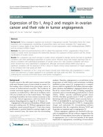
Figure 1-2. HCV Genome and encoded viral proteins.
Non-Structural
Proteins
Structural
Proteins
5’UTR
C
C
E
E
1
1
E
E
2
2
P
P
7
7
N
N
S
S
2
2
N
N
S
S
3
3
4
4
A
A
4
4
B
B
5
5
A
A
5
5
B
B
Envelope
glycoprotein
31-35 70
Serine
protease
NTPase/
Helicase
70
Metallo-
protease
23
?
7
Serine
protease
cofactor
8
?
27
?
58
RNA-
dependent
RNA
polymerase
68
MW
kDa
Putative
function
RNA
Binding
nucleocapsid
22-19
IRES
3’UTR
(U/UC)
Figure 1-1. HCV Genome and encoded viral proteins.
The HCV ORF is flanked by 5’ and 3’ UTR. The structural proteins (shaded) are located in the N terminus with the nonstructural proteins residing in the
remainder of the genome.
6
1.2.2 Non-structural Proteins
The NS proteins of HCV encode enzymes or regulatory factors that are
believed to catalyze and regulate the replication of the HCV RNA genome. The NS
polypeptide is processed by two viral proteases. The first protease, NS2/3, which
spans the C terminus of NS2 and N-terminus of NS3, is a zinc-dependent
metalloprotease that undergoes autocatalysis to generate NS2 and NS3 (Grakoui et al.,
1993). Once cleaved from NS3, NS2 is not essential for the HCV replication when
tested in subgenomic replicons (Lohmann et al., 1999; Blight et al., 2000).
The second protease activity of HCV is found in NS3, a multi-catalytic protein.
The N terminus one third of NS3 encodes a serine protease with three highly
conserved amino acid residues His-53, Asp-77, and Ser-138, which are catalytic triads
of the serine protease family (Miller et al., 1990). This protease cleaves at the
NS3/NS4A, NS4A/4B, NS4B/5A, and NS5A/5B junctions, releasing the mature NS3,
NS4A, NS4B, NS5A and NS5B. The cleavage between NS3/4A occurs in cis as a
spontaneous rapid autocatalytic event, while cleavage at the other sites can occur in
trans when exogenous NS3 is added (Bartenschlager et al., 1993; Tomei et al., 1993).
NS4A is a cofactor for NS3 protease activity, it is vital for cleavages at the NS3/4A
and NS4B/5A sites, and enhances processing of the NS4A/4B and NS5A/5B sites
(Tanji et al., 1995). The binding of NS4A to NS3 also helps anchor the NS3-NS4A
complex onto the ER where proteolytic processing takes place (Lin and Rice, 1995).
The remainder two thirds of NS3 encodes the viral nucleoside triphosphatase
(NTPase) and helicase. Viral helicase is thought to participate in viral replication and
transcription by unwinding the extensive RNA secondary structures in the HCV
genome for the synthesis of the complementary strand and the translation of viral
products. The intrinsic NTPase activity of the helicase provides the energy source for
7
unwinding by hydrolyzing nucleoside triphosphate (Suzich et al., 1993; Kim et al.,
1995). As revealed by sequence analysis and crystal structures of NS3 helicase, this
protein belongs to the DEXH box RNA helicase family with conserved sequences
G
207
SGKST, D
290
ECH, T
322
AT, and Q
460
RRGRTGRGRGG (Gorbalenya et al., 1988;
Cho et al., 1998). The G
207
SGKST sequence, also known as the Walker A sequence,
is found in most NTP hydrolyzing enzymes and is needed for binding NTP. Walker B
sequence D
290
ECH is involved in NTP hydrolysis (Walker et al., 1982). The T
322
AT
motif is important for unwinding RNA while the Q
460
RRGRTGRGRGG motif is
responsible for binding RNA (Pause and Sonenberg, 1992; Gross and Shuman, 1996).
The NS3 helicase can unwind double stranded (ds) RNA as well as dsDNA and RNA-
DNA heteroduplexes in a 3
’
to 5
’
direction. This activity also requires the presence of
divalent ions such as Mg
2+
or Mn
2+
and ATP (Tai et al., 1996; Wardell et al., 1999).
The NS3 protease activity is enhanced by NS4A (Bartenschlager et al., 1994; Failla et
al., 1994). NS4A was also shown to affect helicase activity (Gallinari et al., 1999;
Pang et al., 2002). The function of NS4B is poorly understood but is most likely to be
an integral part of the viral replication complex.
The role of NS5A, a highly phosphorylated protein, in HCV replication is
unclear. Sequence comparison of IFN-
sensitive and IFN-
resistant HCV isolates,
however, reveals a cluster of amino acid differences, termed interferon sensitivity
determining region (ISDR), which correlates with IFN response (Enomoto et al.,
1996). NS5B is the viral RNA dependent RNA polymerase (RdRp) with a GDD
motif, which is a hallmark of RNA polymerase of RNA viruses (Poch et al., 1989).
NS5B is the key enzyme involved in the generation of the complementary minus
strand RNA using the viral genome as template and the subsequent synthesis of the
progeny genomic plus strand RNA (Behrens et al., 1996). Similar to NS3, NS5B
8
activity is also dependent on the presence of divalent ions (Lohmann et al., 1998). The
high replication rate and low fidelity activity of NS5B were also associated with the
emergence of HCV quasispecies, a major obstacle to anti-viral therapy development
(Smith et al., 1997).
Although there has been much progress in the molecular biology of HCV,
studies on this virus are greatly impeded by the lack of small animal models and a
robust in vitro infectious cell culture system. Humans are the only known natural host
for HCV. There is no evidence for vector-mediated transmission. By far the
chimpanzees are the most reliable animal models for studying HCV infection.
Consequently, the mechanism of HCV replication is based primarily on experiences
drawn from closely related flavi- and pestiviruses. Recent years, however, have seen
some advancement in the HCV arena. Several groups have reported the use of the
Tupaia belangeri, a closely related primate of the chimpanzee (Xie et al., 1998; Zhao et
al., 2002), and chimeric mouse models, in which human hepatocytes are transplanted
on immunocompromised mice, as potential models for studying HCV infection
(Mercer et al., 2001). The generation of HCV-replicon systems, where the expression
of the HCV NS proteins drives the self-replication of subgenomic HCV RNAs
(Lohamnn et al., 1999; Blight et al., 2000), will also undoubtedly help accelerate our
understanding of HCV replication and propagation.
9
1.3 HCV protein-protein interaction
Viral proteins are known to interact with one another in the formation of the
viral replication complex. The HCV replicase complex is believed to be ER
membrane associated, comprising of at least NS3 and NS5B as well as several host
cofactors, similar to several plus strand RNA viruses, such as poliovirus and
flaviviruses (Bolten et al., 1998; Westaway et al., 1997). The HCV NS5B was
reported to complex with NS3 and NS4A (Ishido et al., 1998), reminiscent of the
association between NS5 and NS3 of dengue virus type 2 and Japanese encephalitis
virus (Kapoor et al., 1995; Chen et al., 1997). NS4A, NS4B and NS5A have also been
found to form a complex (Lin et al., 1997), so do NS5A and NS5B (Shirota et al.,
2002). All the HCV NS proteins interact with each other either directly or indirectly,
supporting the hypothesis of functional multi-subunit replicase complex.
Viruses are dependent on protein synthesis machineries in the host for viral
protein translation, and other cellular components for their replication. HCV proteins
were reported to associate with several host proteins. E2 binds the putative cellular
receptors, CD81 and the low-density lipoprotein receptor (LDLR) (Pileri et al., 1998;
Agnello et al., 1999; Wunschmann et al., 2000), which may act as receptors for HCV
entry into target cells. A cellular chaperon, HSP90, was reported to bind NS2/3 and is
needed for successful cleavage at the NS2/3 site by helping in the proper folding of
newly synthesized NS2/3 (Waxman et al., 2001). A human eukaryotic initiation factor
4AII with RNA dependent ATPase/helicase activity was found to bind NS5B and
facilitates viral translation by unwinding the secondary structures of the 5’ UTR
(Kyono et al., 2002). p68, a cellular helicase, was also reported to assist in HCV
replication by binding to NS5B (Goh et al., 2004).
10
The direct pathogenic effect of HCV replication, however, is not clear but
studies on transgenic mice models expressing either the structural proteins alone or
both structural and nonstructural proteins presented similar phenotypes, including
steatosis, oxidative stress, and hepatic tumors. The interaction between core and lipid
vesicles was implicated in HCV-related steatosis reflecting abnormal lipid metabolism
(Moriya et al., 1997). The core protein alone was also shown to be capable of causing
oxidative stress as well as tumor formation (Moriya et al., 1998; Okuda et al., 2002)
but another group showed that the full-length HCV polyprotein is needed to induce
tumors (Lerat et al., 2002). As to how, the virus can remain undetected by the host
immune system for decades remain controversial. One of the strategies suggested was
the binding of NS5A to double-stranded RNA-dependent protein kinase (PKR) at its
ISDR motif thereby avoiding the anti-viral effect of IFN (Gale et al., 1997). Besides
NS5A, E2 glycoprotein was also reported to bind and inhibit the activity of PKR
(Taylor et al., 1999).
Alignments of amino acid sequences have revealed that the serine protease and
NTPase/helicase motifs of NS3 are highly conserved in the Flaviviridae family and
among different HCV genotypes (Miller and Purcell, 1990). Productive replication
was also abrogated in vivo when NS3 is mutated at the active sites, making this protein
an attractive target for drug discovery (Kolykhalov et al., 2000). Besides the obvious
role of NS3 in viral replication, this protein may also play a role in regulating cell
proliferation through its interaction with p53 tumor suppressor (Ishido and Hotta,
1998). NS3 was also reported to transform NIH 3T3 cells (Sakamuro et al., 1995) as
well as rat 3T3 cells (Zemel et al., 2001), implicating its involvement in oncogenesis.
Taken together, most of the viral proteins interact and may affect the activities of host
proteins in the long-term. The understanding of the interplay between viral and host
11
proteins will shed light on the mechanism involved in HCV pathogenesis and help in
the formulation of more efficient treatments for HCV.
1.4 Aims and Objectives
The mechanisms by which HCV replicates and the tactics employed by the
pathogen to remain undetected for years are ill defined. Studies on the functions of
viral - viral as well as viral - host interactions will provide valuable information on
understanding these mechanisms of evasion from host immune surveillance, and will
be useful for the development of anti-HCV therapies. This thesis aims to identify both
viral and host interacting partners to NS3, a pivotal player in HCV replication, in the
hope of providing new insights into understanding function and effect of these
interactions. Yeast-two hybrid screens were set up to identify HCV proteins as well as
host proteins that interact with NS3, using a spleen cDNA library. The functions of
these interactions were investigated and discussed in two parts. The first section
describes the characterization of NS3 self-interaction, while the second part covers the
interaction between NS3 and host proteins, in particular, between NS3 and LMP7.
12
2. Materials and Methods
2.1 Construction of Plasmids
The NS3 coding sequence was amplified from HCV RNA extracted from
HCV-positive serum (Lim et al., 2001) by RT-PCR. For the yeast two-hybrid screen,
NS3 clones were fused in frame with the Gal4 DNA binding domain in pAS2-1 vector
and Gal4 DNA activating domain in pACT2 (Clontech). For mammalian expression
of flag- or myc-tagged proteins, DNA fragments were cloned into pXJ40-flag, pXJ40-
myc or pXJ100-myc (Manser et al., 1997) respectively. For expression of glutathione
S-transferase (GST) tagged proteins in bacteria, constructs were made in pGEX-2TK
vector (Pharmacia). LMP7 coding region was amplified by PCR from the spleen
cDNA library with primers OLG144
(CGCGGATCCATGGCGCTACTAGATGTATGC) which contained a BamHI site,
and OLG 145 (CCGCTCGAGTTATTGATTGGCTTCCCGGTA) which contained a
XhoI site. Vectors, plasmids used in studying NS3-NS3 interaction and NS3-LMP7
interaction are summarized in Tables 2-1, 2-2 and 2-3 respectively.
2.2 Yeast two-hybrid screens
2.2.1 NS3 NS3 interaction
Yeast two hybrid screens were performed as described in the Matchmaker
user’s manual (Clontech). Interaction between NS3 fragments were indicated by the
activation of the reporter genes HIS3 and ADE2, which would allow yeast cells to
grow on –His and –Ade media respectively, and LacZ, a
-galactosidase (
-Gal) that
produce a blue colour when the substrate X-Gal (5-bromo-4-chloro-3-indolyl-
-D-