Báo cáo khoa học: Krit 1 interactions with microtubules and membranes are regulated by Rap1 and integrin cytoplasmic domain associated protein-1 doc
Bạn đang xem bản rút gọn của tài liệu. Xem và tải ngay bản đầy đủ của tài liệu tại đây (992.54 KB, 15 trang )
Krit 1 interactions with microtubules and membranes are
regulated by Rap1 and integrin cytoplasmic domain
associated protein-1
Sophie Be
´
raud-Dufour
1
, Romain Gautier
1
, Corinne Albiges-Rizo
2
, Pierre Chardin
1
and Eva Faurobert
1
1 UMR 6097 CNRS-UNSA, Institut de Pharmacologie Mole
´
culaire et Cellulaire, Vabonne, France
2 CRI U823 Universite
´
Joseph Fourier, Institut Albert Bonniot e
´
quipe 1 DYSAD, Grenoble, France
The small G protein Rap1 (Krev-1), a member of the
Ras superfamily, has been brought to the forefront
subsequent to the discovery that it regulates diverse
cellular processes such as integrin activation and cell
adhesion, cell spreading, cell polarity and cell–cell
junction formation [1–3]. To gain more insight into
these pathways, a variety of effector proteins that
interact with the active Rap1GTP-bound form has
been identified. Among them, RAPL, which is enriched
in lymphoid tissues, activates the integrin aLb2,
most likely by interacting with aL integrin [4] and
RIAM, which binds to different actin regulators, and
Keywords
CCM1; FERM domain; Krit1; microtubules;
PIP
2
Correspondence
E. Faurobert, CRI U823 Universite
´
Joseph
Fourier, Institut Albert Bonniot e
´
quipe 1
DYSAD, Site Sante
´
La Tronche BP170,
38042 Grenoble, Cedex 9, France
Fax: +33 476 54 94 25
Tel: +33 476 54 94 74
E-mail:
(Received 15 May 2007, revised 13 July
2007, accepted 24 August 2007)
doi:10.1111/j.1742-4658.2007.06068.x
The small G protein Rap1 regulates diverse cellular processes such as inte-
grin activation, cell adhesion, cell–cell junction formation and cell polarity.
It is crucial to identify Rap1 effectors to better understand the signalling
pathways controlling these processes. Krev interaction trapped 1 (Krit1), a
protein with FERM (band four-point-one ⁄ ezrin ⁄ radixin ⁄ moesin) domain,
was identified as a Rap1 partner in a yeast two-hybrid screen, but this
interaction was not confirmed in subsequent studies. As the evidence sug-
gests a role for Krit1 in Rap1-dependent pathways, we readdressed this
question. In the present study, we demonstrate by biochemical assays that
Krit1 interacts with Rap1A, preferentially its GTP-bound form. We show
that, like other FERM proteins, Krit1 adopts two conformations: a closed
conformation in which its N-terminal NPAY motif interacts with its C-ter-
minus and an opened conformation bound to integrin cytoplasmic domain
associated protein (ICAP)-1, a negative regulator of focal adhesion assem-
bly. We show that a ternary complex can form in vitro between Krit1,
Rap1 and ICAP-1 and that Rap1 binds the Krit1 FERM domain in both
closed and opened conformations. Unlike ICAP-1, Rap1 does not open
Krit1. Using sedimentation assays, we show that Krit1 binds in vitro to
microtubules through its N- and C-termini and that Rap1 and ICAP-1
inhibit Krit1 binding to microtubules. Consistently, YFP-Krit1 localizes on
cyan fluorescent protein-labelled microtubules in baby hamster kidney cells
and is delocalized from microtubules upon coexpression with activated
Rap1V12. Finally, we show that Krit1 binds to phosphatidylinositol 4,5-
P
2
-containing liposomes and that Rap1 enhances this binding. Based on
these results, we propose a model in which Krit1 would be delivered by
microtubules to the plasma membrane where it would be captured by
Rap1 and ICAP-1.
Abbreviations
BHK, baby hamster kidney; CFP, cyan fluorescent protein; FERM, four-point-one protein ⁄ ezrin ⁄ radixin ⁄ moesin; ICAP, integrin cytoplasmic
domain associated protein; Krit1, Krev interaction trapped gene; MT, microtubules; PTB, phosphotyrosine-binding domain.
5518 FEBS Journal 274 (2007) 5518–5532 Journal compilation ª 2007 FEBS. No claim to original French government works
participates in an integrin activation complex that
binds to and activates b integrins [5]. VAV1 and
TIAM1 are localized by Rap1GTP to sites of cell
spreading and serve as exchange factors for Rac [6].
ARAP3 is a GTPase-activating protein for RhoA and
Arf6 that affects PDGF-induced lamellipodia forma-
tion [7]. In dictyostelium, Phg2 promotes myosin II
disassembly at the front of chemotaxing cell facilitating
filamentous-actin mediated leading edge protrusion [8].
Afadin ⁄ AF6 participates in the maturation of cell–cell
junctions [9].
Krev interaction trapped 1 (Krit1) was identified in
1997 as a Rap1 partner in a yeast two-hybrid screen
[10], but subsequent studies did not confirm their
interaction [11], leading to the conclusion that Krit1
is not a Rap1 partner [12]. However, several pieces
of evidence concerning a potential role of Krit1 in
Rap1-regulated cellular processes prompted us to
reconsider this question. First, it was demonstrated
recently that Rap1-dependent activation of integrins
requires talin binding to the cytoplasmic tail of
b integrin [13]. Talin is an essential integrin-activating
protein that connects the cytoplasmic tail of b inte-
grins to the actin cytoskeleton [14]. Integrin cytoplas-
mic domain associated protein (ICAP)-1 is a partner
of the cytoplasmic tail of b1 integrin and has been
shown to compete with talin for binding to b1 inte-
grin [15]. Consistently, on ICAP-1-null osteoblasts
and fibroblasts, fibronectin receptors are in an active
conformation and b1-dependent cell adhesion is
enhanced compared to that of wild-type cells [16,17].
By contrast, overexpression of ICAP-1 reduces cell
spreading and disorganizes focal adhesions [15].
These results suggest that, at resting state, b1 inte-
grin is kept inactive through binding of ICAP-1 to
its cytoplasmic tail. Interestingly, Krit1 is a partner
of ICAP-1 [11,18]. Yeast two-hydrid studies have
shown that Krit1 competes with b1 integrin for bind-
ing to ICAP-1 [11], suggesting that Krit1 could
relieve the inhibitory effect of ICAP-1 on b1 integrin
activation. The second piece of evidence concerns the
existence of a human genetic disease linked to muta-
tions in Krit1. Cerebral cavernous malformation 1
(CCM1) corresponds to brain capillary malforma-
tions characterized by clusters of dilated thin-walled
blood vessels [19]. These lesions usually hemorrhage,
resulting in seizures, focal neurological deficits or
stroke. Ultrastructural studies show that tight junc-
tions are absent between the endothelial cells in these
lesions and that the surrounding basal lamina is
hypertrophied [20].
Very little is known about the Krit1 protein and its
subcellular localization. First, the Krit1 C-terminus
amino acid sequence bears homologies with FERM
(band four-point-one ⁄ ezrin ⁄ radixin ⁄ moesin) domains.
FERM domains localize proteins to the plasma mem-
brane, where they can interact with phosphoinositides
and membrane proteins [21]. The FERM domain of
talin interacts with phosphatidylinositol 4,5-P
2
(PIP
2
)
and with the cytoplasmic tail of b integrins [14]. More-
over, proteins with FERM domains usually exist in
two conformational states: a closed ‘inactive’ confor-
mation where the FERM domain is masked by
another part of the protein and an open ‘active’ con-
formation where the FERM domain is unmasked.
Cleavage, phosphorylation or PIP
2
binding are activa-
tion signals [21]. Second, it has been reported that
Krit1 interacts with tubulin in bovine aortic endothe-
lial cells and decorates microtubules all along their
length [22]. However, this interaction has been ques-
tioned because the antibody used recognized a protein
of lower size on western blot. Another antibody has
subsequently identified a protein of the predicted size,
but the primary location of Krit1 awaits elucidation
[23]. Because microtubules are known to regulate the
dynamics of focal adhesion assembly [24,25], we read-
dressed this important issue.
In the present study, we investigated the interaction
of Krit1 with Rap1, microtubules and membranes. We
show that Krit1, like many proteins with FERM
domains, adopts a closed conformation. This closed
conformation is opened by ICAP-1. Importantly, we
confirm that Krit1 interacts with Rap1 and preferen-
tially with active Rap1 (GTP-bound form), ending the
debate about this point. We show that Rap1 binds to
the FERM domain of Krit1 both in its closed and
opened conformations and that, unlike ICAP-1, Rap1
does not open Krit1. Moreover, we demonstrate that
Krit1, Rap1 and ICAP-1 can form a ternary complex
in vitro. We show that Krit1 interacts with in vitro poly-
merized microtubules, and with PIP
2
on artificial mem-
branes. Remarkably, we demonstrate that Rap1 and
ICAP-1 inhibit in vitro Krit1 binding to microtubules
and that Rap1 stimulates Krit1 binding to membranes.
Consistently, YFP-Krit1 localizes on cyan fluorescent
protein (CFP)-labelled microtubules in baby hamster
kidney (BHK) cells and is delocalized from microtubules
upon coexpression with activated Rap1.
Results
Krit1 contains a putative FERM domain and binds
to PIP
2
The tri-dimensional structures of the FERM domain
of the archetypal ERM proteins, ezrin, radixin and
S. Be
´
raud-Dufour et al. Rap1 regulates Krit1 microtubule and lipid binding
FEBS Journal 274 (2007) 5518–5532 Journal compilation ª 2007 FEBS. No claim to original French government works 5519
moesin, have been resolved by crystallography [26–28].
They are composed of three subdomains, F1 to F3,
arranged in a clover-shaped fashion (Fig. 1A). The
F3 subdomain resembles a phosphotyrosine-binding
domain (PTB) domain, a conserved structural fold that
binds to protein NPxY motifs [29]. Analysis of Krit1
sequence using blast shows that the last 300 amino
acid residues of Krit1 bears approximately 20% iden-
tity with the F1, F2 and F3 subdomains of the typical
ERM proteins (ezrin, radixin, moesin, talin). Even
though this is the lower limit for comparative model-
ling, we were able to generate models for the structure
of Krit1 F1 and F2 to F3 subdomains based on
homology with radixin F1 in complex with IP3 and
with talin F2 to F3 (Fig. 1B). These models displayed
very stable secondary structures and energies with
respect to molecular dynamics simulations, strongly
supporting the idea that the Krit1 C-terminus folds as
a FERM domain.
A functional feature of FERM domains is their
capacity to interact with PIP
2
on plasma membrane.
In the radixin–IP3 cocrystal, IP3 binds to a basic cleft
located between the F1 and F3 subdomains and folded
around a tryptophan present at the hydrophobic base
of the cleft [27] (Fig. 1A). Interestingly, in our model
of the Krit1 FERM domain, basic residues are also
found in the cleft between subdomain F1 and F3 and
the tryptophan at the base of the pocket is conserved
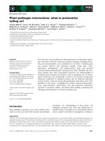
Fig. 1. Krit1contains a putative FERM domain and binds to PIP
2
. (A) Radixin–IP3 cocrystal structure. IP3 is shown in yellow; basic residues
of F1 (K53, K60, K63, K64) and F3 (R273, R275, R279) domains are shown in red. The tryptophan W58 is shown in green. (B) Homology
models of F1, F2 and F3 domains of Krit1. The three independently modelled subdomains have been manually arranged as on the radixin
structure. Basic residues of F1 (K475, K479, R485) and F3 (K713, K720, K724) domains are shown in red. The Tryptophan W487 is shown in
green. (C) Krit1 (1 l
M) was incubated with plasma membrane mix liposomes (0.75 mM) containing, or not, 2% PIP
2
at various NaCl concen-
trations. After centrifugation, proteins present in the supernatant (S) and the pellet (P) were analyzed by SDS ⁄ PAGE and quantified by fluo-
rometry. Protein precipitation in the absence of liposomes has been substracted. Bands below Krit1 band are E. coli contaminants that also
bind to PIP
2
.
Rap1 regulates Krit1 microtubule and lipid binding S. Be
´
raud-Dufour et al.
5520 FEBS Journal 274 (2007) 5518–5532 Journal compilation ª 2007 FEBS. No claim to original French government works
(Fig. 1B), implying that Krit1 would have the struc-
tural features required for binding to phospho-inosi-
tides on membranes. We therefore tested the ability of
Krit1 to bind to PIP
2
-containing membranes. Krit1
was incubated with artificial liposomes supplemented,
or not, with 2% PIP
2
at increasing NaCl concentra-
tions. At 100 mm NaCl, 70% of Krit1 bound to PIP
2
-
containing liposomes, whereas less than 10% bound to
liposomes without PIP
2
, showing that Krit1 interacts
mainly with PIP
2
on these liposomes (Fig. 1C). More-
over, increasing salt concentrations decreased Krit1
binding, highlighting the electrostatic status of this
interaction (Fig. 1C).
Krit1 exists in a closed conformation opened by
ICAP-1
Intriguingly, in addition to its C-terminal FERM
domain, Krit1 has a NPAY motif on its N-terminus
that binds to ICAP-1 [11,18]. This peculiarity of Krit1
led us to consider whether this motif could interact
with Krit1 F3 PTB-like subdomain. To test this
hypothesis, we separately expressed the His-tagged
N-terminus (amino acids 1–207) and GST-C-terminus
(207–end), which we called Krit1-NTer and GST-Hypo-
Krit1, respectively (Fig. 2A). GST pull-down assays
were performed by mixing these two fragments at a
concentration of 100 nm each. Remarkably, Krit1-
NTer bound specifically to GST-HypoKrit1 and to
GST-ICAP-1 used as a positive control, but it did not
bind to GST alone (Fig. 2B). To test whether the
NPAY motif is involved in this interaction, we
mutated Asn192 and Tyr195 into alanines in Krit1-
NTer (Fig. 2A). Krit1-NTer APAA mutant did not
interact with GST-HypoKrit1 and interacted only very
weakly with GST-ICAP-1 as previously reported [11]
(Fig. 2B). In another assay, ICAP-1 totally prevented
the binding of Krit1-NTer (5 lm) to GST-HypoKrit1
(3.5 lm) when added at an equimolar concentration
with Krit1-Nter (Fig. 2C), which correlates well with
the observation that ICAP-1 had a better affinity for
Krit1-Nter than HypoKrit1 (Fig. 2B).
These results show that Krit1 C-terminal FERM
domain interacts with Krit1 N-terminus in vitro, and
that this interaction requires the ICAP-1 binding motif
NPAY. They suggest that full-length Krit1 exists in a
closed conformation with the N-terminus folded on
the C-terminus and that ICAP-1 binding to the N-ter-
minal part disrupts Krit1 N- and C-termini interac-
tion.
Fig. 2. Krit1 N- and C-terminal parts
associate together via the NPAY motif
and ICAP-1 disrupts this association.
(A) Schematic representation of Krit1
fragments used. ANK, Ankyrin repeats.
(B) GST pull-down of 100 n
M Krit1-NTer WT
or mutant on 100 n
M GST-HypoKrit1 or GST
alone following the experimental procedure
described in the Experimental Prcedures.
(C). GST pull-down of 5 l
M Krit1-NTer on
3.5 l
M GST-HypoKrit1 in the presence of
5 l
M ICAP-1. GST fusions in (A) were
immunoblotted with GST antibodies. GST
fusions in (B) and (C) were stained with
Ponceau Red. Krit1-NTer and ICAP-1 were
immunoblotted with His-tag and ICAP-1
antibodies, respectively. Inputs correspond
to 5% of total proteins. These results are
representative of three independent
experiments.
S. Be
´
raud-Dufour et al. Rap1 regulates Krit1 microtubule and lipid binding
FEBS Journal 274 (2007) 5518–5532 Journal compilation ª 2007 FEBS. No claim to original French government works 5521
Krit1 interacts with Rap1 preferentially in its
GTPcS bound form
To answer the question of whether Krit1 is a Rap1
effector (i.e. whether Krit1 interacts preferentially with
Rap1 in its GTP-bound form), we performed GST
pull-down assays. Because we could not purify GST
full-length Krit1 due to its insolubility in Escherichia
coli, we studied the binding of Rap1 to GST-HypoK-
rit1. We compared the binding of 1 lm of Rap1 loaded
with GDP or GTPcSto10lm of GST-HypoKrit1 by
GST pull-down experiments. Unless specified, all the
experiments were performed with Rap1A isoform.
Rap1 binding to HypoKrit1 was specific because no
binding was observed to GST. Rap1GTPcS bound
two-fold more strongly to HypoKrit1 than did
Rap1GDP, reflecting a higher affinity of active Rap1
for HypoKrit1 (Fig. 3A). In the same assay conditions,
H-RasGTPcS did not bind to HypoKrit1 (Fig. 3A).
This experiment therefore shows that Krit1 interacts
with Rap1 and preferentially with Rap1GTP.
Rap1 binds to the C-terminus of Krit1
Serebriiskii et al. [10] mapped a binding site for Rap1
in the 50 last amino acid residues of the N-truncated
form of Krit1 that they originally identified. To verify
this result, this site was deleted in GST-HypoKrit1
(Fig. 2A). The resulting GST-HypoKrit1DC mutant
has a truncation of half of its F3 subdomain. We used
the same experimental conditions as in Fig. 3A. Dele-
tion of the C-terminus of Krit1 abolished the binding
of Rap1GTPcS to GST-HypoKrit1 (Fig. 3B), confirm-
ing that Rap1 binds to Krit1 C-terminal FERM
domain.
Krit1, Rap1 and ICAP-1 form a ternary complex
in vitro
Since Krit1 can interact independently with ICAP-1
and with Rap1, we tested whether a ternary complex
can form between the three proteins. We purified
His
-
tagged full-length Krit1 from E. coli and perform-
ed GST pull-downs by mixing full-length Krit1 with
Rap1GTPcS and GST-ICAP-1 at a final concentration
of 5 lm each (Fig. 4). Krit1 interacted specifically with
GST-ICAP-1 and not with GST alone. This interaction
was strong enough to be revealed by staining of the
proteins with Sypro Orange. As expected, Rap1GTPc S
did not interact with GST-ICAP-1. Interestingly,
when Krit1 and Rap1GTPcS were added together,
Rap1GTPcS was pulled-down with Krit1 on GST-
ICAP-1 beads. Immunoblotting of Rap1 was necessary
to reveal Rap1 binding, indicative of a weaker interac-
tion of Krit1 with Rap1 than with ICAP-1. Therefore,
this experiment shows that the three proteins form a
ternary complex in vitro.
Rap1 binds equally to Krit1 opened and closed
conformations and does not open Krit1
Next, we compared the binding of Rap1 to Krit1
closed and opened conformations. To do so, we
measured by GST pull-down the binding of 10 lm of
Fig. 3. Rap1 binds to Krit1 C-terminus
preferentially in its Rap1GTP form. (A) GST
pull-down of 1 l
M Rap1GDP, Rap1GTPcSor
H-RasGTPcSon10l
M GST-HypoKrit1 or
GST alone. Inputs correspond to 4% of total
proteins. GST fusions were immunoblotted
with GST antibodies. Rap1, ICAP-1 and
H-Ras were immunoblotted with His-tag,
ICAP-1 and H-Ras antibodies, respectively.
(B) GST pull-down of 1 l
M RapGTPcS
on 10 l
M GST-HypoKrit1 or 10 lM
GST-HypoKrit1DC. Input corresponds
to 25% of total Rap1. Rap1 was
immunoblotted with His-tag antibody. The
data are representative of four independent
experiments.
Rap1 regulates Krit1 microtubule and lipid binding S. Be
´
raud-Dufour et al.
5522 FEBS Journal 274 (2007) 5518–5532 Journal compilation ª 2007 FEBS. No claim to original French government works
Rap1 GTPcSon3lm of GST-HypoKrit1 beads in
absence or presence of 10 lm of Krit1-NTer. Impor-
tantly, Krit-NTer binding to GST-HypoKrit1 did not
affect the interaction of Rap1GTPcS to GST-Hypo-
Krit1 (Fig. 5A). Conversely, Rap1 did not modify
the binding of Krit1-NTer to HypoKrit1, suggesting
that Rap1 binds to Krit1 closed conformation and
does not open it. Moreover, the binding of
Rap1GTPcS to GST-HypoKrit1 was not modified
either when the complex between HypoKrit1 and
Krit1-NTer was disrupted by the addition of 10 lm
of ICAP-1, implying that the opening of Krit1 by
ICAP-1 does not affect Rap1 binding (Fig. 5A). This
result was confirmed on full-length Krit1 by a
FLAG pull-down experiment using FLAG-tagged
Krit1. Addition of ICAP-1 together with Rap1 did
not change Rap1 binding to Krit1 (Fig. 5B). There-
fore, Rap1 and ICAP-1 can bind independently to
Krit1. Taken together, our results suggest that,
unlike ICAP-1, Rap1 binding to the C-terminal
FERM domain does not induce the opening of Krit1
and that Rap1 binds as well on Krit1 closed and
opened conformations (Fig. 5C).
Fig. 5. Rap1 binding to HypoKrit1 is not modified by Krit-Nter or ICAP-1. (A) Comparaison by GST pull-down of the binding of 10 lM
Rap1GTPcSto3lM GST-HypoKrit1 in the absence or presence of 10 lM Krit1-Nter and 10 lM ICAP-1. Rap1 and Krit1-Nter were detected
by immunoblotting using anti-His serum. ICAP-1 was revealed by anti-ICAP-1 serumy. (B) FLAG pull-down of 3 l
M Rap1GTPcSor7lM
GST-ICAP-1 to FLAG-Krit1 beads. Control corresponds to beads incubated with nontransfected BHK cells and processed as FLAG-Krit beads.
Input corresponds to 2.5% of total proteins. Proteins were stained by Sypro Orange.These results are representative of three independent
experiments. (C) Model of the conformation of Krit1 in a binary complex with Rap1 or in a ternary complex with Rap1 and ICAP-1. Rap1
binds to the C-terminus of Krit1 closed and opened conformations. ICAP-1 binding opens Krit1 without perturbing Rap1 binding. For clarity
of presentation, only intramolecular folding is represented. A head to tail intermolecular folding between two molecules of Krit1 is however,
not excluded.
Fig. 4. Krit1, Rap1-GTPcS and ICAP-1 form a ternary complex
in vitro. GST pull-down of 5 l
M Krit1 and ⁄ or 5 l M Rap1GTPcSon
5 l
M GST-ICAP-1 fusion or GST alone. Krit1 binding to GST-ICAP-1
was visualized by Sypro Orange staining of the gel. Rap1 binding
was visualized by western blot using His-tag antibody. These
results were replicated three times.
S. Be
´
raud-Dufour et al. Rap1 regulates Krit1 microtubule and lipid binding
FEBS Journal 274 (2007) 5518–5532 Journal compilation ª 2007 FEBS. No claim to original French government works 5523
Krit1 interacts in vitro with microtubules via two
sites present, respectively, on its N- and
C-termini
It has previously been shown that Krit1 colocalizes
with microtubules in bovine aortic endothelial cells
[22]. However, the antibody used in these studies rec-
ognizes a 58 kDa protein on western blot that could
correspond either to a shorter splice variant of Krit1
or to another protein. To address this question, we
studied, by sedimentation on sucrose cushion, the
binding of purified His-tagged full-length Krit1 to
in vitro polymerized microtubules (MT). When
40 pmol of full-length Krit1 were incubated with MT
polymerized from 200 pmol of purified tubulin, 60%
of the protein cosedimented with MT (Fig. 6A), dem-
onstrating a direct interaction of Krit1 with MT.
Remarkably, the contaminant proteins contained in
Krit1 preparations, even those present at the same
concentration as Krit1, did not cosediment with MT
(Fig. 6A), highlighting the specificity of Krit1 interac-
tion with MT. To map the domain responsible for this
interaction, we studied the binding of different frag-
ments of Krit1 in the same conditions. Krit1-NTer
fragment bound strongly to MT (45%). Krit1 bears a
basic stretch of six lysines and arginine on its N-termi-
nus that could interact with MT. Mutations of amino
acids 47KKRK50 in four alanines almost completely
Fig. 6. Krit1 interacts directly with
microtubules in vitro via two sites in its
N- and C-termini and Rap1 and ICAP-1
inhibits this interaction. In each experiment,
40 pmol of Krit1 were incubated in the
absence or presence of taxol-stabilized MT
polymerized in vitro from 150 pmol of
purified tubulin and centrifuged on sucrose
gradient. Supernatant (S) and pellet (P) were
analyzed by SDS ⁄ PAGE and the percentage
of Krit1 bound to MT quantified by
fluorometry. Krit1 precipitation in the
absence of MT has been substracted.
(A) Identification of MT binding sites on
Krit1 using different Krit1 mutants.
Arrowheads indicate Krit1 WT or mutants
(T, tubulin). Each experiment was repeated
two to four times. (B) Inhibition of Krit1
binding to MT by Rap1 and ICAP-1.
200 pmol of Rap1 or ICAP-1 were added to
40 pmol of Krit1 and to polymerized MT.
Each experiment was repeated three times.
Rap1 regulates Krit1 microtubule and lipid binding S. Be
´
raud-Dufour et al.
5524 FEBS Journal 274 (2007) 5518–5532 Journal compilation ª 2007 FEBS. No claim to original French government works
abolished Krit1 binding to MT (Fig. 6A) whereas this
mutant was still able to interact with ICAP-1 (data not
shown). GST-HypoKrit1 also interacted with MT. By
contrast to full-length Krit1 and Krit1-NTer, only
20% of GST-HypoKrit1 bound to MT (Fig. 6A). In a
control, GST alone did not bind to MT (Fig. 6B). The
truncation in the F3 subdomain of GST-HypoKrit1
almost completely abolished the cosedimentation of
GST-hypoKrit1DC with MT (Fig. 6A). Thus two sites
are responsible for the interaction of Krit1 with micro-
tubules: a basic stretch of residues located in the N-ter-
minal part of the protein centered on residues 46–50
with high affinity for MT and a second site located on
the F3 subdomain with low affinity for MT.
Rap 1 and ICAP-1 inhibit in vitro Krit1 binding to
microtubules
Having shown that MT bind to the N- and C-termini
of Krit1, we considered whether ICAP-1 or Rap1,
which bind, respectively, to Krit1 N- and C-termini,
could modulate Krit1 interaction with MT. Thus, we
measured the binding of 40 pmol of Krit1 to MT in
the presence of 200 pmol of Rap1GTPcS or GST-
ICAP-1. Rap1GTPcS alone was not recruited to MT
and less than 10% of GST-ICAP-1 bound to MT
(Fig. 6B). Remarkably, both Rap1GTPcS and GST-
ICAP-1 inhibited Krit1 binding to MT whereas GST
alone had no effect (Fig. 6B). Similar inhibition was
obtained using Rap1B isoform (data not shown).
However, ICAP-1 was a more potent inhibitor than
Rap1GTPcS at these concentrations (80% inhibition
versus 50%). It is unlikely that the effect of ICAP-1
would be due to a competition with Krit1 for binding
to MT because only 20 pmol of GST-ICAP-1 bound
to the 150 pmol of tubulin, polymerized in MT, leav-
ing approximately 130 pmol of tubulin available for
interaction with Krit1.
YFP-Krit1 colocalizes with CFP-labelled
microtubules in transfected BHK cells and is
delocalized from microtubules by activated Rap1
To confirm our results obtained in vitro in a cellular
context, we coexpressed Krit1 fused to YFP together
with CFP-tubulin in BHK cells. Although the YFP-
ARNO signal, used as a negative control, was diffuse
in the cytosol, the YFP-Krit1 signal was superimpos-
able on the CFP-labelled microtubules signal (Fig. 7A),
indicating that YFP-Krit1 was localized along microtu-
bules from MTOC to the periphery. Coexpression of
the activated mutant of Rap1, HA-Rap1V12, flattened
the cells which became spread and displayed numerous
membrane spikes, a phenotype also observed with
HA-Rap1V12 alone (data not shown). Remarkably,
YFP-Krit1 was no longer colocalized with CFP-
labelled microtubules in Rap1V12 expressing BHK
cells (Fig. 7B). Thus, these experiments support our
in vitro observations indicating that Krit1 binds to
microtubules and that this binding is inhibited by
Rap1GTP.
Rap1 enhances HypoKrit1 binding to asolectin
vesicles
Another feature of Krit1 is its capacity to bind to
phospholipids. We considered whether Rap1, which
binds to the Krit1 FERM domain, could modulate
Krit1 association with membranes. Remarkably, when
3 lm of Rap1GTPcS were added to 0.5 lm GST-Hyp-
oKrit1, we observed a stimulation of GST-HypoKrit1
binding to asolectin vesicles (Fig. 8A). A fraction of
Rap1GTPcS also sedimented with the vesicles. Because
the recombinant unmodified Rap1 that we used did
not bind to lipids by itself (Fig. 8A), this fraction
corresponds to Rap1 complexed with HypoKrit1
(Fig. 8A). Moreover, the stimulation of GST-Hypo-
Krit1 binding to asolectin vesicles by Rap1GTPcS was
dose-dependent, up to six-fold (Fig. 8B).
Discussion
In the present study, we confirm that Krit1 interacts
in vitro with Rap1 and preferentially with active Rap1
(GTP-bound form), a criteria for Krit1 being an effec-
tor of Rap1. We show, for the first time, that Krit1
exists in a closed conformation in which its N-terminus
interacts with its C-terminus. ICAP-1 binding to the
N-terminal NPAY motif disrupts this interaction,
whereas Rap1 binding to the C-terminal FERM
domain does not. Moreover, we show that Krit1,
Rap1 and ICAP-1 can form a ternary complex in vitro.
Krit1 binds in vitro to microtubules via two sites on
its N- and C-termini. Remarkably, Rap1 and ICAP-1
inhibit in vitro Krit1 binding to microtubules. In
transfected BHK cells, YFP-Krit1 localizes along
CFP-labelled microtubules and is delocalized from
microtubules by coexpression of activated Rap1V12.
Finally, we show that Krit1 binds to phospholipids on
membranes and that Rap1 enhances this binding.
Krit1 is a Rap1 partner
Our detailed biochemical study demonstrates that Krit
interacts with Rap1 ending the debate about this ques-
tion. Indeed, both N-truncated Krit1, which we called
S. Be
´
raud-Dufour et al. Rap1 regulates Krit1 microtubule and lipid binding
FEBS Journal 274 (2007) 5518–5532 Journal compilation ª 2007 FEBS. No claim to original French government works 5525
HypoKrit1 (corresponding to the yeast two-hybrid
Rap1 partner originally identified) and the full-length
protein interact with Rap1, as shown by pull-down
and asolectin vesicle sedimentation experiments. More-
over, HypoKrit1 interacts with a higher affinity with
Rap1GTPcS than with Rap1GDP, indicating that
Krit1 could be a downstream effector of Rap1. Similar
results were obtained with FLAG-tagged full-length
Krit1 (data not shown). Our results differ substantially
from those of Zhang et al. [11] who failed to observe
an interaction between the two proteins. This discrep-
ancy might be related to the methods applied. These
authors used in vitro translation of Rap1 and Krit1.
The amount of proteins produced might not be suffi-
cient for coimmunoprecipitation because micromolar
concentrations of the two proteins were necessary in
our hands to observe a complex. Consistently, an
interaction of low affinity (K
d
¼ 4.7 lm) was found
between Krit1 and Rap1B, a very close homolog of
Rap 1A [30]. We did not detect any interaction of
H-Ras with Krit1, as previously reported [10,30,31],
suggesting that Krit1 is involved in a Ras-independent,
Fig. 7. YFP-Krit1 colocalizes with CFP-labelled microtubules in transfected BHK cells and is delocalized by activated Rap1. BHK cells were
transfected with plasmids encoding YFP-Krit1 or YFP-ARNO and CFP-tubulin (A) together with pMT2HA-Rap1V12. (B) HA-Rap1V12 was
detected with anti-HA 3F10 serum. Scale bars ¼ 15 lm.
Rap1 regulates Krit1 microtubule and lipid binding S. Be
´
raud-Dufour et al.
5526 FEBS Journal 274 (2007) 5518–5532 Journal compilation ª 2007 FEBS. No claim to original French government works
Rap1-dependent pathway. Even though it has been
proposed that Rap1 may bind to the Krit1 F1 subdo-
main because this subdomain bears homology with a
computerized model of Ras binding domain [32], the
absence of interaction between HypoKrit1DC and
Rap1 suggests that Rap1 interacts with the PTB-like
F3 subdomain. Similarly, talin and radixin FERM
domains interact via their F3 subdomain with integrin
and ICAM-2 cytoplasmic tails, respectively, as shown
by cocrystal structures [33,34]. Further mutagenesis
studies will be necessary to map precisely the site for
Rap1 interaction on the Krit1 FERM domain.
Krit1, like other FERM proteins, adopts closed
and opened conformations and binds to PIP
2
The molecular modelling of the last 300 amino acid
residues of Krit1 that we generated corroborates the
existence of a FERM domain at the C-terminus of the
protein. FERM domains are involved in localizing
proteins to the plasma membrane. It has been shown,
by sedimentation experiments and crystallographic
studies, that they bind to PIP
2
[e.g. radixin [27] and
talin [35]) via the interaction of the polar head of the
lipid with a basic cleft present between the F1 and F3
subdomains. Consistently, we show that Krit1 binds to
PIP
2
. Interestingly, several basic residues are exposed
to the F1 to F3 cleft on the Krit1 FERM model that
we generated. Mutagenesis analyses of these residues
will help to determine whether the binding site of
the polar head of PIP
2
on Krit1 is similar to that of
radixin.
Many members of the FERM family undergo intra
and ⁄ or inter molecular folding of their C-terminus
onto their N-terminal FERM domain [21]. Consis-
tently, we demonstrate the interaction of the N-termi-
nus of Krit1 with its C-terminus, with both parts being
produced separately. Cis or trans interaction between
these two domains are equally possible and would lead
to either an intramolecular folding in a closed confor-
mation or an intermolecular folding in an antiparallel
homodimer like talin. Furthermore, we have shown
that the N-terminal NPAY motif is involved in the
interaction, which suggests that the PTB-like F3 sub-
domain of the FERM is the C-terminal counterpart.
As such, in the dormant form of moesin, the F3 sub-
domain is masked by the N-terminal extended actin
binding tail domain [28]. Moreover, we show that
ICAP-1 binding disrupts Krit1 N- and C-termini inter-
action. This interaction, as well as its disruption by
ICAP-1, could be verified on the full-length protein by
fluorescence resonance energy transfer of Krit1 fused
to YFP and CFP at its extremities. This could repre-
sent a crucial mechanism of regulation of Krit1 activ-
ity. Indeed, both the NPAY motif and PTB-like F3
subdomain are binding sites for other partners, such as
ICAP-1, phospholipids and yet unknown partners,
most likely trans-membrane or peri-membrane pro-
teins. Masking of these two sites may prevent Krit1
interacting with other proteins until it is delivered to
its target(s). It has been shown that Krit1 interacts with
the CCM2 gene product malcavernin, a PTB domain
protein, in the context of a Rac ⁄ MEKK3 ⁄ MKK3
signalling complex that activates p38 mitogen-activated
protein kinase kinase [23]. Krit1–CCM2 interaction
does not involve the NPAY motif and a ternary
complex can form between Krit1, CCM2 and ICAP-1.
It is possible that Krit1, through its interaction with
different partners on the plasma membrane, partici-
pates in several linked signalling pathways involved in
angiogenesis [23].
Rap1 and ICAP-1 regulate Krit1 localization to
microtubules and membranes
Among the FERM family members, ERM proteins
and talin provide a regulated linkage between mem-
brane proteins and the cortical actin cytoskeleton.
Fig. 8. Rap1 stimulates Krit1 binding to
membranes. (A) GST-HypoKrit1 (0.5 l
M)
was incubated with asolectin vesicles
(1 mgÆmL
)1
) in the absence or presence of
Rap1GTPcS(3l
M). After centrifugation, the
supernatant (S) and the pellet (P) were
analyzed by SDS ⁄ PAGE and quantified by
fluorometry. Protein precipitation in absence
of liposomes has been substracted.
(B) Dose–response of stimulation of Krit1
binding to asolectin vesicles by increasing
concentrations of Rap1GTPcS. Each
experiment was repeated three times.
S. Be
´
raud-Dufour et al. Rap1 regulates Krit1 microtubule and lipid binding
FEBS Journal 274 (2007) 5518–5532 Journal compilation ª 2007 FEBS. No claim to original French government works 5527
No actin-binding site has been reported on Krit1.
Consistently, purified Krit1 does not interact with
in vitro polymerized actin (Eric Macia, personal com-
munication), nor does YFP-Krit1 localize in cells to
phalloidin-labelled F-actin (data not shown). Instead,
we confirm the previous report that Krit1 is a microtu-
bule-associated protein [22] by showing that it colocal-
izes with microtubules in cells and that it interacts
directly with in vitro polymerized microtubules. More-
over, our results show that Krit1 binds to MT through
two sites: one basic site in the N-terminal part of the
protein located on residues 46–51 with high affinity for
MT and a second on the C-terminus in the F3 subdo-
main with low affinity for MT. The N-terminal basic
motif has also been described as a functional nuclear
localization sequence [23], suggesting that it could be
involved in the shuttling of Krit1 between the micro-
tubules and the nucleus. Remarkably, the contribution
of these two sites to the binding to MT is additive
(Fig. 6A). Because we show that the N-terminus of
Krit1 interacts with the C-terminus, it is most likely
that these two MT binding sites actually form a con-
tinuous surface on the closed protein. We show that
ICAP-1 and Rap1 both inhibit the binding of Krit1 to
MT. Two hypotheses can be proposed concerning the
mechanism of this inhibition. The first one is that they
directly mask the surface that binds to MT. Indeed,
our results show that MT and Rap1 both bind to the
last 50 residues of Krit1. It is not so clear for the basic
stretch, which lies between residues 46–51. ICAP-1
binds further on the primary sequence on the
191NPAY194 motif. It is possible, however, that these
two sites are actually close in the folded protein. The
second hypothesis is that ICAP-1 binding provokes
structural changes of the basic site that lead to a
decrease of its affinity for MT. Nevertheless, the fact
that ICAP-1 is a more potent inhibitor than Rap1 cor-
relates well with, for one part, its stronger binding to
Krit1 at the micromolar concentrations used in these
experiments and, for the second part, with the higher
contribution to the binding to MT of the basic stretch
than the C-terminal site.
At the cell periphery, Rap1 shuttles between recy-
cling endosomes and the plasma membrane, with its
GTP form being localized at the plasma membrane
[36]. Interestingly, we show that Rap1GTPcS stimu-
lates HypoKrit1 binding to artificial membranes. Hyp-
oKrit1 is not recruited to membranes through Rap1
because the unmodified Rap1 that we used does not
bind to membranes. Instead, Rap1 binding to Krit1
FERM domain most likely induces a conformational
change of the PIP
2
binding pocket that gives Hypo-
Krit1 a better affinity for PIP
2
. Our attempts to
observe Rap1 stimulation of full-length Krit1 binding
to asolectin vesicles were not successful even in the
presence of ICAP-1, whose binding should unmask the
Krit1 FERM domain (data not shown). Several experi-
mental caveats may be responsible for this negative
result. In the absence of its C-terminal lipid anchor,
Rap1 positioning toward Krit1 and the membrane
may not be optimized and therefore the complex is not
fully stabilized. Moreover, ICAP-1 may not be suffi-
cient to stabilize Krit1 in its opened conformation.
Interaction with another partner may be required for
the protein to be stably opened.
Krit1 localization on microtubules and its delocaliza-
tion by activated Rap1 is very similar to reported
results obtained for RAPL, another Rap1 effector.
Indeed, RAPL also localizes on MT in endothelial cells
and directly binds to in vitro polymerized MT [38]. In
wound healing assays, RAPL localizes on MT oriented
toward the leading edge, an area where Rap1 is highly
activated and its interaction with Rap1 is required for
directional migration [38]. Moreover, coexpression of
Rap1V12 also induces dissociation of RAPL from MT
[37]. In lymphocytes, RAPL moves dynamically from
the perinuclear region to the leading edge upon chemo-
kine stimulation and activated Rap1 triggers the
association of RAPL with aLb2, leading to the redis-
tribution of this integrin to the immunological synapse
[4]. In addition to integrin activation, Rap1 induces cell
polarization and facilitates cell migration. Expression
of activated Rap1 polarizes lymphocytes, generating a
leading edge at the front and a uropod at the back. It
also stimulates lymphocyte endothelium transmigration
[38] and directional migration of endothelial cells [37].
Therefore, delivery of Rap1 effectors to focal adhesions
by the MT network might participate in the polarized
activation of integrins. In agreement with this, target-
ing of MT in close vicinity to adhesion foci on the
plasma membrane has been reported [39], as well as
the regulation of the turnover of focal adhesions by
MT [24,25]. Based on our results, we propose a model
of regulation of Krit1 binding to MT and plasma
membrane; in the cell, Krit1 would be transported in
its closed conformation along MT toward the plasma
membrane. On reaching the membrane, Krit1 would
detach from MT and be captured by activated Rap1
and ICAP-1 on the plasma membrane (Fig. 9). In the
absence of ICAP-1, Rap1GTP would bind to Krit1,
favouring its detachment from MT and its binding to
the plasma membrane, but without opening it. This
complex could serve as a stock for a rapid delivery of
Krit1 to its target sites on the plasma membrane.
An important breakthrough in unravelling the
Rap1-dependent pathway in integrin activation was
Rap1 regulates Krit1 microtubule and lipid binding S. Be
´
raud-Dufour et al.
5528 FEBS Journal 274 (2007) 5518–5532 Journal compilation ª 2007 FEBS. No claim to original French government works
achieved recently by Han et al. [13]. Their work dem-
onstrates that Rap1 promotes talin binding to the
cytoplasmic tail of b3 and b1 integrins. They show that
Rap1 induces the formation of an integrin activation
complex that contains talin and RIAM, a Rap1 effec-
tor. RIAM is known to bind to profilin and Ena ⁄
VASP [5], suggesting that it could participate in actin
network formation at the level of focal adhesion [13].
Concerning the activation of b1 integrin per se (i.e.
its switch to high affinity conformation), our present
data together with that of Han et al. [13] lead us to
hypothesize that this Rap1-dependent activation com-
plex could contain Krit1. Its binding to ICAP-1 would
displace ICAP-1 from its inhibitory site on b1 integrin,
therefore allowing talin binding and subsequent b1
conformational changes. Further work is in progress
to determine whether Krit1 is targeted by the micro-
tubule network to focal adhesions and whether Rap1,
RIAM and ICAP-1 regulate this localization. Further-
more, functional studies will aim to investigate the role
of Krit1 on cell adhesion and to determine whether
Krit1 is involved in the Rap1-dependent integrin acti-
vation pathway.
Experimental procedures
Plasmid constructions
pcDNA4 ⁄ V5-HISA-FLAG-Krit1 and pCDNA4 ⁄ V5-HISA-
FLAG-ICAP-1 were kindly provided by D. A. Marchuk
(Duke University Medical Center, Durham, NC, USA).
Krit1 was subcloned in pET21d (Novagen, Fontenay-sous-
Bois, France) and pEYFP-N1 (Clontech, Saint-Germain-
en-Laye, France), HypoKrit1 (208–736) in pGEX-2T
(Roche Diagnostics, Meylan, France). Krit1-Nter (1–207)
was subcloned in pQE 30 (Qiagen, Courtaboeuf, France)
and pETGEX-CT (National Institute of Genetics, Japan).
Asn192Ala-Tyr195Ala substitutions, alanine substitutions
of 47KRKK50 and the introduction of a stop codon at
amino acid 686 were produced using the QuikChange
mutagenesis kit (Stratagene, Amsterdam, the Netherlands).
pMT2-HA-Rap1A and pMT2-HA-Rap1AV12 were kindly
provided by J. L. Bos (University Medical Center, Utrecht,
the Netherlands). C-terminal-deleted (1–167) Rap1A was
subcloned into pET16b (Novagen). ICAP-1 was subcloned
in pGEX-2T.
Purification of recombinant proteins
The expression of Krit1 and Krit1 KRKK ⁄ AAAA in
E. coli BL21(DE3)codon+ cells (Stratagene) was induced
at an attenuance of 0.8 at D
600 nm
with 0.2 mm isopropyl
thio-b-d-galactopyranoside and grown for an additional
16 h at 18 °C. His-tagged Rap1, His-tagged Krit1-Nter WT
and APAA, GST-HypoKrit1, GST-HypoKrit1DC, Krit1-
Nter-GST, and GST-ICAP-1 were produced in E. coli Bl21
gold (Stratagene) induced with 0.2 mm isopropyl thio-b-d-
galactoside at an attenuance of 0.8 for 3 h at 28 °C.
Purifications were performed according to manufacturer
instructions using the nickel-charged resin Ni-NTA (Qia-
gen) for His-Tagged proteins or glutathione sepharose 4B
(GE Healthcare Europe GmbH, Orsay, France) for GST
fusions. Purified His-tagged Krit1 WT and KRKK ⁄ AAAA
were dialyzed and concentrated against 50 mm phosphate
buffer pH 8.0, 120 mm NaCl, 10% glycerol, dithiothreitol
1mm. All other proteins were dialyzed and concentrated in
20 mm Tris ⁄ HCl pH 7.5, 120 mm NaCl, 1 mm MgCl
2
, and
10% glycerol and supplemented with 20 lm GDP for His-
tagged Rap1. A fraction of purified GST-ICAP-1 was
cleaved by thrombin according to the manufacturer’s
instructions (GE Healthcare). Protein concentration was
determined by SDS ⁄ PAGE and fluorometric analysis of
Sypro Orange (Amresco, Interchim, Montluc¸ on, France)
stained gel using a Fuji LAS3000 fluorescence imaging sys-
tem (Fuji Film, Tokyo, Japan).
Preloading of Rap1 with GDP or GTPcS
Purified Rap1 was incubated for 45 min at 30 °Cin50mm
Tris ⁄ HCl pH 7.5, 120 mm NaCl, 1 mm MgCl
2
,2mm
EDTA, 10% glycerol and 2 mm GTPcS or GDP (Roche
Diagnostics). The loaded nucleotide was stabilized in the
nucleotide site by addition of 5 mm MgCl
2
.
GST pull-down
Purified GST fusions bound to 20 lL of glutathione beads
(GE Healthcare) were incubated under agitation with puri-
fied His-tagged proteins as indicated for 2 h at 4 °CinPD
Fig. 9. Model of regulation of Krit1 binding to microtubules and
plasma membrane. In the cell, Krit1 would be transported in its
closed conformation along the microtubules toward the plasma
membrane. When reaching the membrane, Krit1 would detach
from MT and be captured by activated Rap1 and ICAP-1 on the
plasma membrane.
S. Be
´
raud-Dufour et al. Rap1 regulates Krit1 microtubule and lipid binding
FEBS Journal 274 (2007) 5518–5532 Journal compilation ª 2007 FEBS. No claim to original French government works 5529
buffer (50 mm Tris ⁄ HCl pH 7.5, 120 mm NaCl, 1 mm
MgCl
2
, 10% glycerol, 1% NP-40, 2 mm dithiothreitol, pro-
tease inhibitors) in a final volume of 50 lL. Beads were
washed three times in 1 mL of PD buffer and eluted with
30 lL of Laemmli sample buffer. Input and bound proteins
were analyzed by Sypro Orange (Amresco) staining of the
gel or by western blotting.
Cell culture and transfection
BHK cells were grown in BHK-21 medium (Invitrogen BV,
Leek, the Netherlands) containing 5% FBS, 10% tryptose
phosphate broth, 100 unitsÆmL
)1
penicillin, 100 lgÆmL
)1
streptomycin and 2 mml-glutamine. The cells were trans-
fected with Fugene6 transfection reagent (Roche Diagnos-
tics) according to the manufacturer’s instructions.
FLAG pull-down
BHK cells were transfected with pcDNA4 ⁄ V5-HISA-
FLAG-Krit1. Forty-eight hours after transfection, cells
were washed with NaCl ⁄ Pi and scrapped in PD buffer.
After a 15 min of incubation at 4 °C, cells were centrifuged
at 14 000 g for 30 min at 4 °C using an Eppendorf centri-
fuge with 5415R rotor. The supernatant was incubated with
mouse anti-FLAG M2 coupled to agarose beads (Sigma
Aldrich, L’Isle d’Abeau Chesnes, France) for 2 h at 4 °C.
FLAG-Krit1 trapped on beads was incubated with 3 lm of
Rap1GTPcS, 7 lm of ICAP-1, or both, for 2 h at 4 °C
under agitation. Beads were then washed three times with
PD buffer and bound proteins were eluted with
200 lgÆmL
)1
of FLAG peptide in PD buffer. Input and
eluted proteins were analyzed by SDS ⁄ PAGE and Sypro
Orange staining of the gel.
Microtubule sedimentation experiments
Tubulin purification and polymer preparation
Purified lamb brain tubulin was polymerized for 15 min at
37 °C in BRB50 buffer (50 mm Pipes pH 6.8, 1 mm EGTA,
1mm MgCl
2
) containing 1 mm GTP, 5 mm MgCl
2
and
10% glycerol [40]. Then, every 10 min, 8, 80, and 800 lm
taxol were added at 37 °C. The mixture was layered onto
28 lL of 50% glycerol in BRB50 buffer containing 1 mm
GTP and 80 lm taxol and centrifuged at 16 000 g for
10 min at 25 °C using an Eppendorf centrifuge with 5415R
rotor. The pellet was resuspended in the same buffer.
Sedimentation experiments
Polymerized microtubules corresponding to approximatively
150 pmol of tubulin were incubated with the indicated pro-
teins in BRB50 buffer pH 7.5, in 20 lL for 30 min at 25 °C.
The reaction mixture was layered onto 20 lL of 30% glycerol
in BRB50 buffer pH 7.5 and centrifuged at 16 000 g for
10 min at 25 °C using an Eppendorf centrifuge with 5415R
rotor. The pellets were resuspended in 40 lL of 15% glycerol
in BRB50 buffer pH 7.5. The percentage of proteins cosedi-
mented with microtubules was determined by SDS ⁄ PAGE
and fluorometric analysis of the Sypro Orange stained gel
using fluorescence imaging or by western blotting.
Immunofluorescence and cell microscopy
BHK cells were cotransfected with pEYFP-Krit1 and
pECFP-tubulin with or without pMT2-HA-Rap1V12.
Twenty-four hours after transfection, cells were fixed in 4%
paraformaldehyde for 20 min and processed for immunoflu-
orescence analysis as already described [41]. HA-Rap1V12
was labelled with 3F10 anti-HA monoclonal serum (Roche
Diagnostics). Confocal microscopy analysis was carried out
using a Leica TCS-SP5 microscope (Leica Microsystemes
SAS, Rueil-Malmaison, France).
Asolectin vesicles and liposome sedimentation
experiments
Asolectin vesicles and liposomes preparation
Asolectin (Sigma Aldrich) vesicles were prepared by the
reverse phase method [42] and sucrose-loaded liposomes
[plasma membrane mix: 40% phosphatidylethanolamine,
15% phosphatidylserine, 20% cholesterol, 25% phosphatidyl-
serine, 0.2% N-(7-nitrobenz-2-oxa-1,3-diazol-4-yl)-1,2-dihexa-
decanoyl-sn-glycero-3-phosphoethanolamine (NBD-PE);
2mm lipids] were prepared by the extrusion method [43].
Lipids in chloroform were purchased from Avanti Polar
Lipids except for egg phosphatidylethanolamine (Sigma
Aldrich) and NBD-PE (Molecular Probes Europe, Leiden,
the Netherlands).
Sedimentation experiments
Proteins were incubated for 20 min at 25 °C with sucrose-
loaded liposomes or asolectin vesicles in 20 mm Tris ⁄ HCl
pH 8.0, 120 mm NaCl, 1 mm MgCl
2
, 10% glycerol, 1 mm
dithiothreitol and were centrifuged for 20 min at 400 000 g
at 20 °C using a TL100 Beckman centrifuge and TLA100.3
rotor. The percentage of proteins in the supernatant and
pellet was determined by fluorometric analysis of the Sypro
Orange stained gel using fluorescence imaging.
Modelling of Krit1 FERM domain
The psi-blast [44] web tools were used to obtain align-
ments of Krit1 last 300 residues with different template
sequences. The structure of radixin (protein databank code:
1GC6) was used as template for F1 FERM domain and
that of talin (protein databank code: 1Y19) for the F2 and
Rap1 regulates Krit1 microtubule and lipid binding S. Be
´
raud-Dufour et al.
5530 FEBS Journal 274 (2007) 5518–5532 Journal compilation ª 2007 FEBS. No claim to original French government works
F3 FERM domains. The alignments were improved manu-
ally, taking into account the predicted secondary structure
of Krit1 and the secondary structures of talin and radixin.
One hundred models of F1, F2 and F3 domains were con-
structed separately by comparative modelling with the mod-
eller 7 [45]. The best model was retained for each FERM
domain and the side chains were repositioned in optimized
conformations, using scwrl [46] and the SCit web server
[47]. The models were finally subjected to energy minimiza-
tion. The stability of these models was assayed by molecu-
lar dynamics simulations using the Gromos 96 force field
implemented in gromacs, version 3.2.1 [48]. Because there
is a good conservation of the spatial arrangement of F1–
F2–F3 among the crystallized FERM domains, the three
modellized Krit1 subdomains were manually positioned
according to the arrangement of radixin FERM domain.
Structures were visualized with pymol (Delano Scientific
LLC, Palo Alto, CA, USA).
Acknowledgements
We thank Fre
´
de
´
ric Brau for excellent technical support
with the confocal microscopy. We thank Daniel Bou-
vard, Julie Milanini, Michel Franco and Eric Macia
for useful discussions and critical reading. We thank
D. A. Marchuk for providing us with Krit1 and
ICAP-1 cDNAs, and J. L. Bos for Rap1 cDNA. We
thank Alfred Wittinghofer for providing H-Ras. This
work was supported by the Association pour la
Recherche sur le Cancer.
References
1 Bos JL (2005) Linking Rap to cell adhesion. Curr Opin
Cell Biol 17, 123–128.
2 Caron E (2003) Cellular functions of the Rap1 GTP-bind-
ing protein: a pattern emerges. J Cell Sci 116, 435–440.
3 Kooistra MR, Dube N & Bos JL (2007) Rap1: a key regula-
tor in cell–cell junction formation. J Cell Sci 120, 17–22.
4 Katagiri K, Maeda A, Shimonaka M & Kinashi T
(2003) RAPL, a Rap1-binding molecule that mediates
Rap1-induced adhesion through spatial regulation of
LFA-1. Nat Immunol 6,6.
5 Lafuente EM, van Puijenbroek AA, Krause M, Carman
CV, Freeman GJ, Berezovskaya A, Constantine E,
Springer TA, Gertler FB & Boussiotis VA (2004)
RIAM, an Ena ⁄ VASP and Profilin ligand, interacts
with Rap1-GTP and mediates Rap1-induced adhesion.
Dev Cell 7, 585–595.
6 Arthur WT, Quilliam LA & Cooper JA (2004) Rap1
promotes cell spreading by localizing Rac guanine
nucleotide exchange factors. J Cell Biol 167, 111–122.
7 Krugmann S, Andrews S, Stephens L & Hawkins PT
(2006) ARAP3 is essential for formation of lamellipo-
dia after growth factor stimulation. J Cell Sci 119,
425–432.
8 Jeon TJ, Lee DJ, Merlot S, Weeks G & Firtel RA (2007)
Rap1 controls cell adhesion and cell motility through the
regulation of myosin II. J Cell Biol 176, 1021–1033.
9 Zhang Z, Rehmann H, Price LS, Riedl J & Bos JL
(2005) AF6 negatively regulates Rap1-induced cell adhe-
sion. J Biol Chem 280, 33200–33205.
10 Serebriiskii I, Estojak J, Sonoda G, Testa JR & Golemis
EA (1997) Association of Krev-1⁄ rap1a with Krit1, a
novel ankyrin repeat-containing protein encoded by a
gene mapping to 7q21–22. Oncogene 15, 1043–1049.
11 Zhang J, Clatterbuck RE, Rigamonti D, Chang DD &
Dietz HC (2001) Interaction between krit1 and icap1-
alpha infers perturbation of integrin beta1-mediated
angiogenesis in the pathogenesis of cerebral cavernous
malformation. Hum Mol Genet 10, 2953–2960.
12 Revencu N & Vikkula M (2006) Cerebral cavernous
malformation: new molecular and clinical insights.
J Med Genet 43, 716–721.
13 Han J, Lim CJ, Watanabe N, Soriani A, Ratnikov B,
Calderwood DA, Puzon-McLaughlin W, Lafuente EM,
Boussiotis VA, Shattil SJ et al. (2006) Reconstructing
and deconstructing agonist-induced activation of inte-
grin alphaIIbbeta3. Curr Biol 16, 1796–1806.
14 Calderwood DA (2004) Integrin activation. J Cell Sci
117, 657–666.
15 Bouvard D, Vignoud L, Dupe-Manet S, Abed N, Four-
nier H-N, Vincent-Monegat C, Retta SF, Fassler R &
Block MR (2003) Disruption of focal adhesions by inte-
grin cytoplasmic domain-associated protein-1alpha.
J Biol Chem 278, 6567–6574.
16 Bouvard D, Aszodi A, Kostka G, Block MR, Albiges-
Rizo C & Fassler R (2007) Defective osteoblast function
in ICAP-1-deficient mice. Development 134, 2615–2625.
17 Bouvard D, Millon-Fremillon A, Dupe-Manet S, Block
MR & Albiges-Rizo C (2006) Unraveling ICAP-1 func-
tion: toward a new direction? Eur J Cell Biol 85, 275–282.
18 Zawistowski JSSI, Lee MF, Golemis EA & Marchuk
DA (2002) KRIT1 association with the integrin-binding
protein ICAP-1: a new direction in the elucidation of
cerebral cavernous malformations (CCM1) pathogene-
sis. Hum Mol Genet
11, 389–396.
19 Marchuk DA, Srinivasan S, Squire TL & Zawistowski
JS (2003) Vascular morphogenesis: tales of two syn-
dromes. Hum Mol Genet 12, R97–R112.
20 Clatterbuck RE, Eberhart CG, Crain BJ & Rigamonti
D (2001) Ultrastructural and immunocytochemical evi-
dence that an incompetent blood–brain barrier is related
to the pathophysiology of cavernous malformations.
J Neurol Neurosurg Psychiatry 71, 188–192.
21 Bretscher A, Edwards K & Fehon RG (2002) ERM
proteins and merlin: integrators at the cell cortex. Nat
Rev Mol Cell Biol 3, 586–599.
S. Be
´
raud-Dufour et al. Rap1 regulates Krit1 microtubule and lipid binding
FEBS Journal 274 (2007) 5518–5532 Journal compilation ª 2007 FEBS. No claim to original French government works 5531
22 Gunel M, Laurans MSH, Shin D, DiLuna ML, Voorh-
ees J, Choate K, Nelson-Williams C & Lifton RP (2002)
KRIT1, a gene mutated in cerebral cavernous malfor-
mation, encodes a microtubule-associated protein.
PNAS 99, 10677–10682.
23 Zawistowski JS, Stalheim L, Uhlik MT, Abell AN, Anc-
rile BB, Johnson GL & Marchuk DA (2005) CCM1
and CCM2 protein interactions in cell signaling: impli-
cations for cerebral cavernous malformations pathogen-
esis. Hum Mol Genet 14, 2521–2531.
24 Kaverina I, Rottner K & Small JV (1998) Targeting,
capture, and stabilization of microtubules at early focal
adhesions. J Cell Biol 142, 181–190.
25 Small JV & Kaverina I (2003) Microtubules meet sub-
strate adhesions to arrange cell polarity. Curr Opin Cell
Biol 15, 40–47.
26 Smith WJ & Cerione RA (2002) Crystallization and pre-
liminary crystallographic analysis of the ezrin FERM
domain. Acta Crystallogr D Biol Crystallogr 58, 1359–
1361.
27 Hamada K, Shimizu T, Matsui T, Tsukita S & Hako-
shima T (2000) Structural basis of the membrane-target-
ing and unmasking mechanisms of the radixin FERM
domain. EMBO J 19, 4449–4462.
28 Pearson MA, Reczek D, Bretscher A & Karplus PA
(2000) Structure of the ERM protein moesin reveals the
FERM domain fold masked by an extended actin bind-
ing tail domain. Cell 101, 259–270.
29 Balla T (2005) Inositol-lipid binding motifs: signal inte-
grators through protein–lipid and protein–protein inter-
actions. J Cell Sci 118, 2093–2104.
30 Wohlgemuth S, Kiel C, Kramer A, Serrano L, Witting-
hofer F & Herrmann C (2005) Recognizing and defining
true Ras binding domains I: biochemical analysis.
J Mol Biol 348, 741–758.
31 Serebriiskii I, Khazak V & Golemis EA (1999) A two-
hybrid dual bait system to discriminate specificity of
protein interactions. J Biol Chem 274, 17080–17087.
32 Kiel C, Wohlgemuth S, Rousseau F, Schymkowitz J,
Ferkinghoff-Borg J, Wittinghofer F & Serrano L (2005)
Recognizing and defining true Ras binding domains II:
in silico prediction based on homology modelling and
energy calculations. J Mol Biol 348, 759–775.
33 Garcia-Alvarez B, de Pereda JM, Calderwood DA,
Ulmer TS, Critchley D, Campbell ID, Ginsberg MH
& Liddington RC (2003) Structural determinants of
integrin recognition by talin. Mol Cell 11, 49–58.
34 Hamada K, Shimizu T, Yonemura S, Tsukita S, Tsukita
S & Hakoshima T (2003) Structural basis of adhesion-
molecule recognition by ERM proteins revealed by the
crystal structure of the radixin-ICAM-2 complex.
EMBO J 22, 502–514.
35 Martel V, Racaud-Sultan C, Dupe S, Marie C, Paulhe
F, Galmiche A, Block MR & Albiges-Rizo C (2001)
Conformation, localization, and integrin binding of talin
depend on its interaction with phosphoinositides. J Biol
Chem 276, 21217–21227.
36 Bivona TG, Wiener HH, Ahearn IM, Silletti J, Chiu
VK & Philips MR (2004) Rap1 up-regulation and acti-
vation on plasma membrane regulates T cell adhesion.
J Cell Biol 164, 461–470.
37 Fujita H, Fukuhara S, Sakurai A, Yamagishi A, Kami-
oka Y, Nakaoka Y, Masuda M & Mochizuki N (2005)
Local activation of Rap1 contributes to directional vascu-
lar endothelial cell migration accompanied by extension
of microtubules on which RAPL, a Rap1-associating
molecule, localizes. J Biol Chem 280, 5022–5031.
38 Shimonaka M, Katagiri K, Nakayama T, Fujita N,
Tsuruo T, Yoshie O & Kinashi T (2003) Rap1 translates
chemokine signals to integrin activation, cell polariza-
tion, and motility across vascular endothelium under
flow. J Cell Biol 161, 417–427.
39 Krylyshkina O, Anderson KI, Kaverina I, Upmann I,
Manstein DJ, Small JV & Toomre DK (2003) Nanome-
ter targeting of microtubules to focal adhesions. J Cell
Biol 161, 853–859.
40 Lee YC, Samson FE Jr, Houston LL & Himes RH
(1974) The in vitro polymerization of tubulin from beef
brain. J Neurobiol 5, 317–330.
41 Franco M, Boretto J, Robineau S, Monier S, Goud B,
Chardin P & Chavrier P (1998) ARNO3, a Sec7-domain
guanine nucleotide exchange factor for ADP ribosylation
factor 1, is involved in the control of Golgi structure
and function. Proc Natl Acad Sci USA 95, 9926–9931.
42 Franco M, Chardin P, Chabre M & Paris S (1995) Myr-
istoylation of ADP-ribosylation factor 1 facilitates
nucleotide exchange at physiological Mg
2+
levels. J Biol
Chem 270, 1337–1341.
43 Mayer LD, Hope MJ & Cullis PR (1986) Vesicles of
variable sizes produced by a rapid extrusion procedure.
Biochim Biophys Acta 858, 161–168.
44 Altschul SF, Madden TL, Schaffer AA, Zhang J, Zhang
Z, Miller W & Lipman DJ (1997) Gapped BLAST and
PSI-BLAST: a new generation of protein database
search programs. Nucleic Acids Res 25, 3389–3402.
45 Marti-Renom MA, Stuart AC, Fiser A, Sanchez R,
Melo F & Sali A (2000) Comparative protein structure
modeling of genes and genomes. Annu Rev Biophys
Biomol Struct 29, 291–325.
46 Canutescu AA, Shelenkov AA & Dunbrack RL Jr
(2003) A graph-theory algorithm for rapid protein side-
chain prediction. Protein Sci 12, 2001–2014.
47 Gautier R, Camproux AC & Tuffery P (2004) SCit: web
tools for protein side chain conformation analysis.
Nucleic Acids Res 32, W508–W511.
48 Van Der Spoel D, Lindahl E, Hess B, Groenhof G,
Mark AE & Berendsen HJ (2005) GROMACS: fast,
flexible, and free. J Comput Chem 26, 1701–1718.
Rap1 regulates Krit1 microtubule and lipid binding S. Be
´
raud-Dufour et al.
5532 FEBS Journal 274 (2007) 5518–5532 Journal compilation ª 2007 FEBS. No claim to original French government works