The role of BLNK, DOK 3 DIP in BCR signaling 2
Bạn đang xem bản rút gọn của tài liệu. Xem và tải ngay bản đầy đủ của tài liệu tại đây (284.63 KB, 8 trang )
International Immunology, Vol. 12, No. 3, pp. 397–404 © 2000 The Japanese Society for Immunology
B cell development and activation defects
resulting in xid-like immunodeficiency in
BLNK/SLP-65-deficient mice
Shengli Xu, Joy En-Lin Tan, Esther Poh-Ying Wong, Arunkumar Manickam,
Sathivel Ponniah and Kong-Peng Lam
Institute of Molecular and Cell Biology, National University of Singapore, 30 Medical Drive, Singapore
117609, Republic of Singapore
Keywords: adaptor protein, B cell antigen receptor, CD5
ϩ
B cells, signal transduction, gene targeting
Abstract
Engagement of the B cell receptor (BCR) leads to the activation of tyrosine kinases and other
signaling molecules that ultimately determine the type and magnitude of the B lymphocyte’s
cellular response. The adaptor protein BLNK/SLP-65 plays a pivotal role in BCR signal transduction
by coupling Syk activation to downstream elements such as Grb2, phospholipase C-γ, Vav and
Nck. We have generated BLNK
–/–
mice to determine the physiological role of this protein in B cell
development and activation. BLNK
–/–
mice exhibit an incomplete block in B cell development with a
severe inhibition of pro-B to pre-B cell differentiation. BLNK
–/–
sIgM
⍣
cells can develop, seed the
peripheral lymphoid tissues and accumulate in numbers overtime. However, these mutant B cells
failed to mature and are non-responsive to BCR cross-linking in terms of proliferation and up-
regulation of activation markers such as CD69 and CD86 (B7-2). In addition, the CD5
⍣
subset of
B cells is absent. The immune response to T cell-independent antigen but not T cell-dependent
antigen is also impaired. Overall, the phenotype of BLNK
–/–
mice bears a striking resemblance to
that of
xid mice which is the murine model of human XLA that has a mutation in Bruton’s tyrosine
kinase. This raises the interesting possibility that mutation in BLNK/SLP-65 may be responsible for
certain human immunodeficiencies.
Introduction
The pre-B cell receptor (pre-BCR) and the BCR play pivotal Signal transduction events have been studied extensively in
B lymphocytes. Engagement of the BCR activates cytoplasmicroles in the development of B lymphocytes. The pre-BCR
comprising the Ig heavy chain and surrogate light chains, and protein tyrosine kinases such as Syk, Lyn, Blk and Bruton’s
tyrosine kinase (Btk) (6), and can lead to a multitude of cellularthe BCR that is composed of the surface Ig, are complexed to
the signal transducing subunits Igα and Igβ (1). Studies responses, such as proliferation, activation, differentiation or
cell death. The current challenge in the field of B cell signalingwith the µMT mouse that has a targeted deletion of the
transmembrane exon of the Ig heavy chain (2) or the λ5T is to identify specific signaling pathways that associate with
a particular cellular response. Recently, it has been demon-mouse that lacks surrogate light chain (3) indicated that the
inability of these mutant mice to express a pre-BCR can lead strated that adaptor proteins play a major role in interfacing
tyrosine kinase activation by lymphocyte antigen receptorsto the arrest of B cell development at a very early stage. In
addition, mice with a compromised BCR resulting from the with selective downstream signaling molecules. One such
adaptor molecule termed BLNK (7), SLP-65 (8) or BASH (9)truncation of the cytoplasmic tail of Igα (4) do not accumulate
mature B cells in the periphery. Finally, the induced ablation has been identified in B cells and is specifically involved in
BCR signaling. BLNK can associate with Btk (10) and alsoof BCR on mature peripheral B cells leads to their rapid cell
death (5). Taken together, these studies implied that signals couple Syk activation to Grb2, phospholipase C (PLC)-γ,Vav
and Nck (7), and is intimately associated with intracellularfrom the pre-BCR and BCR are required for the progression
of B lymphopoiesis and the maintenance of B cell survival. Ca
2ϩ
mobilization which is essential for cell activation (11).
Correspondence to: K P. Lam
Transmitting editor: D. Tarlinton Received 6 December 1999, accepted 3 January 2000
398 Immunodeficiency in BLNK/SLP-65 knockout mice
BLNK contains a C-terminus SH2 domain, several SH3 rubber-stopper from a 5 ml syringe. Peritoneal cavity and
bone marrow cells were obtained by injecting staining mediumdomains and a series of YXXP motifs in the N-terminus (7–9).
It bears striking homology to another adaptor protein SLP-76 (PBS containing 3% FCS and 0.1% NaN
3
) into the peritoneal
cavity and femur and tibia respectively using a 10 and 1 mlthat is expressed in T cells (12) and intimately involved in
TCR signaling. In general, the order of the signaling events syringe with a 26-gauge needle. All cells were treated with
red blood cell lysing solution (0.15 M NH
4
Cl,1mMKHCO
3
from the TCR and BCR is quite similar, with the engagement
of the antigen receptors triggering the activation of similar and 0.1 mM Na
2
EDTA) to eliminate erythrocytes. For FACS
analyses, cells were stained with optimal amounts of FITC-,classes of intracellular cytoplasmic kinases. In analogy to
BLNK in B cells, SLP-76-coupled TCR induced ZAP-70 (the phycoerythrin (PE)- and biotin-conjugated mAb for 10 min
on ice, and washed 3 times with staining medium. Biotin-equivalent of Syk) activation to Ca
2ϩ
mobilization in T cells
(13). In addition, SLP-76 is essential for T cell development conjugated mAb were revealed with streptavidin–CyChrome.
Flow cytometry analyses were performed on a FACScanas its inactivation in the mouse germline leads to a profound
block in thymocyte maturation at a very early stage (14,15). (Becton Dickinson, Mountain View, CA).
Thus, given the central role of BLNK in BCR signaling and its
In vitro stimulation and proliferation assays
similarity to SLP-76 in T cells, we have inactivated BLNK in
the mouse to study its physiological role in B cell development
Splenic B cells were obtained from wild-type and mutant mice
and activation.
by negative selection using MACS (Miltenyi Biotech) with anti-
CD43 mAb-coupled magnetic beads that bind T cells and
macrophages. The purity of B cells obtained was Ͼ90% as
Methods
assessed by anti-B220 and anti-IgM mAb staining in FACS
analysis. For the
in vitro stimulation assay, 10
6
purified B cells
Generation of BLNK/SLP-65-deficient mice
were seeded into 48-well tissue culture plate and incubated
The cDNA for BLNK/SLP-65 was obtained by RT-PCR of RNA
with 10 µg/ml goat anti-mouse IgM F(ab)Ј
2
fragment overnight
isolated from mouse spleens using primers 5Ј-AGTG-
in RPMI medium supplemented with 10% FCS. Cells were
GCTTGAGTTCTTGAGGC-3Ј and 5Ј-AGAAAAGCTCGTGTG-
harvested and stained for the expression of activation markers.
AACGCC-3Ј, and used to screen a mouse 129 genomic DNA
A colorimetric MTT assay (Roche, Singapore) was used
library. Restriction enzyme digestion, Southern blotting and
according to the manufacturer’s instructions to measure cell
DNA sequencing were used to map a phage clone containing
proliferation
in vitro. Briefly, 5ϫ10
5
purified B cells were
some 5Ј exons of BLNK. Subsequently, a targeting vector
stimulated with varying concentrations of goat anti-mouse
was constructed to replace the exon containing the starting
IgM F(ab)Ј
2
fragment in a 96-well tissue culture plate. After
ATG and a further 4.5 kb of DNA upstream with a
neo
r
gene.
48 h, the cells were incubated with the MTT labeling reagent
A5kb
BamHI–ClaI fragment 5Ј anda2kbNheI–BamHI
for a further 4 h followed by the addition of solubilization
fragment 3Ј of the deleted exon were used as the long arm
solution overnight. Cell proliferation was quantified using an
and short arm of homology respectively. To inactivate BLNK
ELISA reader at 570 nm wavelength.
in the germline, 10
7
E14.1 embryonic stem (ES) cells were
electroporated with 10 µgof
NotI-linearized targeting vector
Immunizations of BLNK/SLP-65-deficient mice
and selected with 300 µg/ml G418 (Gibco, Hong Kong, ROC)
The ability of BLNK
–/–
mice to mount a humoral immune
and 2 µM gancyclovir. Double-drug-resistant ES cell clones
response was assessed by immunizing the animals with the
were screened by Southern blotting for homologous recombin-
hapten 4-hydroxy-3-nitrophenyl acetyl (NP). Wild-type and
ants using probe A as shown in Fig. 1. The frequency of
mutant mice were immunized i.p. with 10 µgNP
25
-Ficoll in PBS
targeting was 1:100. Two ES cell clones with the correct
to examine their immune responses to a T cell-independent
configuration of the targeted locus were injected into C57BL/
antigen. For the immune response to a T cell-dependent
6 blastocysts to generate chimeric mice for germline transmis-
antigen, mice were immunized i.p. with 100 µg alum-precipit-
sion of the mutant allele.
ated NP
17
-chicken globulin (CG). Sera were obtained from
mice at day 0 and 8 of immunizations to detect the presence
Antibodies
of NP-specific antibodies in an ELISA. To detect NP-specific
The following mAb used in the flow cytometry analyses were
antibodies, the ELISA plates were coated with 50 µlof5µg/
purchased from PharMingen (San Diego, CA): anti-B220
ml NP-BSA and blocked with 3% BSA. Pre-immune and
(RA3-6B2); anti-IgM (331.12), anti-IgD (1.3-5), anti-CD43 (S7),
immune sera were added at various dilutions to the wells of
anti-CD5 (53-7), anti-CD11b (M1/70), anti-CD23, anti-CD69,
the ELISA plates. Specific antibodies of class IgM and IgG3
anti-CD86 (B7-2), anti-µ
a
(DS-1) and anti-µ
b
(AF6-78.25). The
were quantified for the T-independent, and IgM and IgG1 for
goat anti-mouse IgM F(ab)Ј
2
fragment used in the in vitro
the T-dependent immune responses respectively.
stimulation assays was obtained from Chemicon (Temecula,
CA).
Results
FACS analyses
Generation of BLNK/SLP-65-deficient mice
Tissues and cell preparations for flow cytometric analyses
were prepared as previously described (16). In brief, single- BLNK/SLP-65-deficient mice were generated by deleting the
exon containing the starting codon ATG and a further 4.5 kbcell suspensions were obtained from spleen and lymph nodes
by dissociation of these tissues with a plastic mesh and a of DNA upstream in mouse ES cells (Fig. 1). The deletion of
Immunodeficiency in BLNK/SLP-65 knockout mice 399
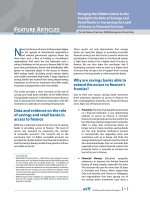
Fig. 1. (A) Inactivation of BLNK/SLP-65 in the mouse germline. Partial restriction endonuclease map of the wild-type allele, the targeting vector
and the inactivated allele of BLNK/SLP-65 are shown (
BamHI, B; ClaI, C; EcoRI, E; KpnI, K; NheI, Nh; SacI, S; plasmid Bluescript, pBKS). The
black box indicates the exon containing the starting ATG that is being replaced by the
neo
r
gene.
EcoRI digestion of genomic DNA will yield
fragments of 12 and 5 kb, as revealed by probe A for the wild-type and targeted alleles respectively. (B) Southern blot analysis of
EcoRI-
digested tail DNA obtained from wild-type, BLNK
ϩ/–
and BLNK
–/–
mice. (C) RT-PCR of bone marrow samples obtained from wild-type and
BLNK
–/–
mice. The 5Ј and 3Ј RT-PCR identified the regions corresponding to bp 38–396 and 999-2013 of the SLP-65 cDNA respectively. The
RT-PCR for the housekeeping gene GADPH is included as controls.
the exon containing the ATG was verified by Southern blotting be found in 8-week-old mutant mice although they were
reduced considerably by ~3-fold compared to wild-type(Fig. 1) and by DNA sequencing (data not shown). Two
targeted ES cells were injected into mouse blastocysts to control. In addition, the population of re-circulating
B220
high
IgM
low
cells was also largely diminished by 2- to 4-generate chimera that were subsequently bred to produce
mice carrying a germline mutation of BLNK/SLP-65. Homozy- fold. To gain better insight into the specific B cell stage
in which the BLNK/SLP-65 mutation manifests its effect,gous mutant mice obtained were designated BLNK
–/–
. The
gene targeting strategy and the derivation of homozygous B220
ϩ
IgM
–
cells in the bone marrow were further stained with
anti-CD43 mAb to resolve pro-B and pre-B cells (17). Asmice are depicted in Fig. 1(A and B respectively).
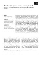
To ensure the inactivation of BLNK, RT-PCR was performed can be seen in Fig. 2 (lower panel), BLNK/SLP-65-deficient
mice lack a population of B220
ϩ
CD43
–
pre-B cells that wason bone marrow and spleen samples with primers that
correspond to the 5 Ј and 3Ј portions of the BLNK cDNA. As reduced by 5-fold compared to wild-type animals. In addition,
there was a 2-fold accumulation of B220
ϩ
CD43
ϩ
pro-B cellsshown in Fig. 1(C), no BLNK message was detected in the
samples obtained from mutant mice compared to those from in the bone marrow of these mutant mice. The increase in the
proportion of pro-B cells in BLNK
–/–
mice reflects an increasewild-type control. Thus, the BLNK loci have been disrupted.
Initial flow cytometric analyses using cell surface markers in the number of these cells as compared to wild-type mice
(Table 1). Thus, the inactivation of BLNK/SLP-65 results in asuggest that there were no detectable defects in the develop-
ment of macrophages, T, NK or dendritic cells (data not severe inhibition of pro-B to pre-B cell transition. However,
the block in B cell development is incomplete as a small poolshown), consistent with the fact that BLNK is not shown to be
expressed in these cell types (7–9). The major defect of of IgM
ϩ
B cells is generated.
BLNK
–/–
mice lies in the development and function of the B
BLNK is not required for the maintenance of peripheral B cells
lineage cells, and that is the focus of our subsequent analyses.
Flow cytometric analyses of spleen and lymph nodes of
Severe but incomplete block in B cell development in the
BLNK
–/–
mice indicate that B220
ϩ
IgM
ϩ
B cells can be found
bone marrow of BLNK/SLP-65-deficient mice
in the peripheral lymphoid tissues although they are reduced
in numbers considerably (Figs 3 and 4). This suggestsTo determine the effect of BLNK/SLP-65 inactivation in early
B cell development, we analyzed bone marrow cells of mutant that developing BLNK
–/–
B cells can exit the bone marrow
environment and seed the peripheral lymphoid organs.and wild-type mice by flow cytometry. As shown in Fig. 2
(upper panel) and Table 1, immature B220
ϩ
IgM
ϩ
B cells can Expression of a BCR is required for the persistence of B
400 Immunodeficiency in BLNK/SLP-65 knockout mice
Fig. 2. Flow cytometry analyses of bone marrow cells from wild-type
and BLNK
–/–
mice. Cells were obtained from the femur and tibia of
8-week-old mice, and stained with FITC–anti-IgM, PE–anti-B220 and
biotin–anti-CD43 (S7) mAb. The latter was revealed by streptavidin–
CyChrome. The upper panel depicts the B220 versus IgM staining
of bone marrow cells, whereas the lower panel depicts the B220
versus CD43 profiles of IgM
–
bone marrow cells. B cells at different
stages of development are indicated. Numbers indicate percentage
of cells in the lymphocyte gate. Representative of Ͼ10 analyses.
Table 1. Bone marrow B cell populations ( ϫ10
6
) in wild-type
and BLNK
–/–
mice
Genotypen Pro-B Pre-B Immature B Re-circulating
B
ϩ/ϩ 6 0.91 Ϯ 0.24 2.17 Ϯ 0.16 1.43 Ϯ 0.20 0.81 Ϯ 0.27
–/– 4 2.20 Ϯ 0.50 0.56 Ϯ 0.29 0.57 Ϯ 0.20 0.21 Ϯ 0.09
Cells were obtained from one femur and tibia of mice that were 6–
8 weeks old.
The number of B cells at each developmental stage was estimated
on the basis of total cell count and flow cytometric analyses as shown
in Fig. 2.
cells in the peripheral lymphoid tissue (5). It is postulated that
the BCR provides a low-level survival signal to the peripheral
B lymphocytes that is distinct from the signal that is required
Fig. 3. Flow cytometry analysis of spleen and lymph node cells from
to activate them (18). To determine whether signal transduced
wild-type and BLNK
–/–
mice. (A) Spleen and lymph node cells of 3-
by BLNK is required for the maintenance of B cells in the
and 12-week-old wild-type and BLNK
–/–
mice were stained with FITC–
periphery, we examine the number of B cells in the spleens
anti-IgM and PE–anti-B220 mAb to resolve for the presence of B
of BLNK
–/–
mice of varying age. As shown in Fig. 3(A and B),
lineage cells in the periphery. Numbers indicate the percentage of
the number of B cells that are found in 3-week-old BLNK
–/–
cells in the lymphocyte gate. Representative of more than three
analyses of mice for each age group. (B) Number of B cells found
mice is drastically reduced by 30-fold compared to control
in the spleens of 3-, 7- and 12-week-old wild-type and BLNK
–/–
mice
mice of similar age. However, as the mice grow older, the
as estimated by total splenic cell count and flow cytometry analyses
reduction in B cell numbers compared to control mice of
as shown in (A). The fold difference in the number of B cells between
equivalent age decreases, such that by 6 and 12 weeks of
wild-type and mutant mice of similar age is indicated for each age
group. Analyses include more than four mice for each age group.
age, BLNK mutant mice have only 7- and 2-fold fewer B cells
than wild-type animals of comparable age respectively. This
Immunodeficiency in BLNK/SLP-65 knockout mice 401
Fig. 5. Absence of CD5
ϩ
IgM
ϩ
cells in the peritoneal cavity of BLNK
–/–
Fig. 4. Severe reduction of IgM
low
IgD
high
Fraction I cells in the spleen
mice. Peritoneal cavity cells were obtained from 3-month-old wild-
and lymph node of BLNK
–/–
mice. Spleen and lymph node cells from
type and BLNK
–/–
mice and stained with FITC–anti-IgM and PE–anti-
8-week-old wild-type and BLNK
–/–
mice were stained with FITC–
B220 (upper panel) or FITC–anti-IgM and PE–anti-CD5 (lower panel)
anti-IgD and PE–anti-IgM mAb to depict cells form Fractions I
mAb. Numbers indicate percentage of cells in the lymphocyte gate.
(IgM
low
IgD
high
), II (IgM
high
IgD
high
) and III (IgM
high
IgD
low
). Numbers
Representative of more than five analyses.
indicate percentage of cells in the lymphocyte gate. Representative
of more than five analyses.
Absence of CD5
ϩ
B cells in the peritoneal cavity of BLNK/
suggests that the number of peripheral B cells in BLNK mutant
SLP-65-deficient mice
mice can accumulate with age. Thus BLNK is apparently not
required for the maintenance of B cells in the periphery. The Other than the conventional or B-2 cells found in the spleen
and lymph nodes, another major subset of B cells, designatedaccumulation of B cells in BLNK
–/–
mice with age contrasted
sharply with the situation in mb-1∆c/∆c mice (4) that have a B-1 cells, exists, and these cells are found mainly in the pleural
and peritoneal cavities. These cells can be distinguished fromtruncation of the cytoplasmic tail of Igα leading to the expres-
sion of a compromised BCR. In mb-1∆c/∆c mice the peripheral conventional B cells by their cell surface phenotype. In
contrast to B-2 cells that express high levels of B220 andB cell pool remains diminished regardless of the age of
the animals. IgD, and moderate levels of IgM, B-1 cells express low levels
of B220 and IgD, and high levels of surface IgM. In addition,
they express CD5, a marker found on T cells, and do not
express CD23 (21).
Severe reduction of IgM
low
IgD
high
(Fraction I) B cells in the
Flow cytometric analyses of 6-week-old BLNK
–/–
mice indi-
spleen and lymph nodes of BLNK/SLP-65-deficient mice
cate a scarcity of B cells in the peritoneal cavity of these
mice compared to wild-type control (data not shown). SincePeripheral B cells can be subdivided into Fractions I, II and
III on the basis of differential IgM and IgD expression, and B-1 cells accumulate with age, we examine the peritoneal
cavity cells of 3-month-old BLNK
–/–
mice. Detail analysesrepresent different stages of B cell maturation (19). Cells in
Fractions III (IgM
high
IgD
low
) and II (IgM
high
IgD
high
) are the revealed that most of the cells present in BLNK
–/–
mice are
conventional or B-2 cells, with the noticeable absence of thenewly emigrating or transitional B cells, whereas cells in
Fraction I (IgM
low
IgD
high
) are the mature B lymphocytes (20). B220
low
IgM
high
and CD5
ϩ
IgM
ϩ
B cells (Fig. 5). This suggests
that BLNK is required for the generation of B-1 cells.Interestingly, as seen in Fig. 4, most of the peripheral B cells
present in the spleen of an 8-week-old BLNK
–/–
mouse have
BLNK/SLP-65-deficient B Cells cannot be activated and do
an immature phenotype, and are found mainly in Fractions III
not proliferate in response to anti-IgM stimulation in vitro
and II. This is in contrast to wild-type mice in which the
majority of the peripheral splenic B cells are found in Fraction Cross-linking of BCR activates B lymphocytes, resulting in
their up-regulation of co-stimulatory and activation molecules.I. This block in peripheral B cell maturation is even more
evident in the lymph nodes of BLNK
–/–
mice compared to To determine whether BLNK
–/–
B cells are functional and
responsive to external stimuli, we treated purified splenic Bcontrol animals (Fig. 4) where in the latter all cells are found
in Fraction I. Thus, although BLNK
–/–
B cells can seed the cells from wild-type and BLNK
–/–
mice with anti-IgM mAb
in vitro. As shown in Fig. 6(A), anti-IgM activated wild-type Bperipheral lymphoid tissues and accumulate in numbers, the
majority of them do not differentiate to the IgM
low
IgD
high
cells up-regulate their expression of CD69, an early activation
marker, as well as the co-stimulatory molecule, CD86 (B7-2).mature B cell stage.
In contrast, BLNK
–/–
B cells did not up-regulate either the
402 Immunodeficiency in BLNK/SLP-65 knockout mice
mice with NP conjugated to CG (NP-CG). As shown in Fig.
7(B) and in contrast to the situation for the T cell-independent
antigen, BLNK
–/–
mice can mount an effective immune
response to NP-CG that is comparable to wild-type animals,
both in terms of IgM and IgG1 secretion. Thus taken together,
the data indicate that BLNK
–/–
mice have an impaired immune
response to T-independent but not T-dependent antigens.
Discussion
Mice deficient for the adaptor protein BLNK/SLP-65 exhibit a
severe block in early B cell development at the pro-B to pre-
B cell transition stage where the pre-BCR is expressed. This
is consistent with the notion that a signal from the pre-BCR
is required for the progression of early B lymphopoiesis (1)
and presumably BLNK is needed for the transduction of such
a developmental signal. However, this early developmental
block is incomplete as sIgM
ϩ
B cells do develop that could
eventually seed the peripheral lymphoid tissues. It is not
known currently why and how these sIgM
ϩ
B cells could
bypass the developmental block at the pro-B to pre-B cell
transition stage. It is intriguing to speculate that perhaps
these sIgM
ϩ
B cells encode for polyreactive antibodies that
recognize certain environmental or self ligands with higher
affinities and this heightened interaction provides the signal
that could compensate for BLNK deficiency during the devel-
Fig. 6. BLNK
–/–
B cells cannot be activated in vitro. (A) BLNK
–/–
B
opmental process. This possibility can be readily tested by
cells do not up-regulate CD86 and CD69 in response to anti-IgM
breeding Ig heavy and light chain transgenic mice bearing
stimulation. Purified splenic B cells from wild-type and BLNK
–/–
mice
polyreactive or autoreactive antibodies with BLNK
–/–
mice.
were incubated in medium alone or with 10 µg/ml goat anti-mouse
Such experiments are in progress in the laboratory.
IgM F(ab)Ј
2
fragment overnight, and stained with anti-B220 and anti-
CD86 (B7-2) or anti-CD69 mAb. Representative of three separate
Interestingly, BLNK
–/–
mice failed to generate a population
experiments. (B) BLNK
–/–
B cells do not proliferate in response to
of IgM
low
IgD
high
B cells in the periphery and lack B-1 cells in
BCR cross-linking. Purified B cells from wild-type and BLNK
–/–
mice
the peritoneal cavity. In addition, BLNK
–/–
mice could not
were stimulated with increasing concentrations of goat anti-mouse
mount an effective humoral immune response to T cell-
IgM F(ab)Ј
2
fragment for 48 h and cell proliferation was quantified in
a MTT colorimetric assay.
independent antigens while maintaining a normal T cell-
dependent immune response. While the current work is in
progress, two other groups have also generated mice deficient
in BLNK or SLP-65 (22,23). The phenotypes of the three
independently generated BLNK mutant mice (22,23 andexpression of CD69 or CD86, indicating that they are non-
responsive to activation via BCR cross-linking. current study) are comparable and together confirmed the
physiological role of BLNK in B cell development. However,Activated wild-type B cells also undergo cell proliferation
upon BCR cross-linking in a manner proportional to the we differ with respect to the inability of our BLNK
–/–
B cells
to up-regulate the expression of the activation markers CD69concentration of stimulating anti-IgM mAb present (Fig. 6B).
However, BLNK
–/–
B cells do not proliferate even in the and CD86 upon anti-IgM stimulation in vitro. This difference
could be due to the use of an enriched population of B cellpresence of increasing amount of stimulant given. Thus, these
data indicate that BLNK
–/–
B cells are non-responsive to BCR in our assay as compared to the use of total splenocytes by
the other groups (22,23). It is conceivable that in the latter,cross-linking
in vitro.
other factors such as the availability of T cell help in the form
Impaired T cell-independent but not T cell-dependent immune
of secreted cytokines might overcome the inability of BLNK
–/–
responses in BLNK
–/–
mice
B cells to respond to anti-IgM stimulation in vitro. Another
possible explanation for the difference in our data could beAntigens that elicit an antibody response from B cells can be
classified either as T independent or T dependent according the difference in the timing of assessment of the activation of
BLNK
–/–
B cells. In our study, we examine the up-regulationto their dependency on CD4
ϩ
T cell help. To examine whether
BLNK
–/–
mice can mount an efficient immune response to of the activation markers after an overnight stimulation of
Ͻ18 h. Since BLNK is an adaptor molecule that facilitates theexogenous antigens, we first immunized mice with NP coupled
to Ficoll (NP-Ficoll), a T cell-independent antigen. The primary interaction of other proteins, the absence of BLNK may simply
lead to a slower kinetic of activation of mutant B lymphocytesantibody response to NP-Ficoll is mainly of the IgM and IgG3
class. As can be seen in Fig. 7(A), BLNK
–/–
mice showed compared to wild-type cells. Indeed, further experiments will
have to be conducted to determine the kinetics (if any) andundetectable IgM or IgG3 antibody response to NP-Ficoll 8
days after immunization compared to the wild-type control. parameters of activation of BLNK
–/–
B cells in vitro. Finally,
our data indicating the inability of BLNK
–/–
B cells to proliferateFor the T-cell-dependent immune response, we immunized
Immunodeficiency in BLNK/SLP-65 knockout mice 403
Fig. 7. BLNK
–/–
mice have impaired immune response to T cell-independent but not T cell-dependent antigens. Groups of three wild-type (j)
and BLNK
–/–
mice (d) were immunized i.p. with (A) 10 µg NP-Ficoll, a T cell-independent antigen, and (B) 100 µg alum-precipitated NP-CG,
a T cell-dependent antigen. Sera were collected 8 days after immunization and quantified for the presence of NP-specific antibodies of various
Ig classes in an ELISA using NP-BSA as the coating antigen. The immune sera were diluted several fold and the absorbance values for the
indicated dilution (e.g. 1:100 for IgG3 in the T cell-independent immune response) were plotted. Pre-immune sera were negative for the
presence of NP-specific antibodies and are not shown.
and be activated by anti-IgM stimulation in vitro would correl- major substrate phosphorylated by Syk that leads to calcium
mobilization by PLC-γ2 (11). Recently, it has been shown thatate much better with the inability of these mutant B cells to
mount a T cell-independent immune response
in vivo. BLNK can also bind to the SH2 domain of Btk (10). Perhaps,
it is this disruption in Btk–BLNK interaction that is responsibleIt has been shown that BCR expression is required for the
maintenance of peripheral B cells (5). Our additional data on for the lack of B-1 cells in BLNK
–/–
mice. It remains to be
established whether B-1 cells failed to be generated or, ifthe accumulation of peripheral B cells in BLNK
–/–
mice with
age suggest that signals transduced by BLNK are not involved generated, fail to be maintained in BLNK
–/–
mice.
Btk mutation is responsible for X-linked agammaglobuline-in the persistence or maintenance of lymphocytes. This is in
contrast to the situation in mb-1∆c/∆c mice that have a mia (XLA), a human immunodeficiency syndrome (34). A
recent report indicates that mutation in BLNK can also causecompromised BCR and in which the peripheral B cell pool
does not expand with increasing age of the animals (4). human immunodeficiency (35). Since BLNK
–/–
mice resemble
xid mice, it is now of great interest to establish whetherThese data together would imply that the cell survival signal
mediated by Igα in the BCR complex is not propagated by mutation in BLNK may be responsible for a large proportions
of the human immunodeficiency that is not associated with aBLNK. Although BLNK
–/–
B cells can accumulate in the
periphery, the majority of these cells failed to mature to a mutation in Btk. This is of particular significance as BLNK is
expressed only in B cells and, unlike Syk (27,28) or PI-3KIgM
low
IgD
high
stage, suggesting that BLNK-transduced signal
is needed for the final maturation of B lymphocytes in the (29,30), its deficiency does not result in embryonic lethality.
Finally, disruption of SLP-76 in T cells leads to a completesecondary lymphoid tissues.
The developmental and functional defects in BLNK
–/–
mice block in T cell development (14,15), whereas IgM
ϩ
B cells
can develop in the absence of BLNK/SLP-65 (22,23 andbear striking resemblance to
xid mice that lack Btk (24–26).
Both mutant mice have a block in primary B lymphopoeisis, current study). This would suggest that although the ordered
pathways for the development of T and B are quite similar,lack B-1 and mature B cells but accumulate immature
IgM
high
IgD
low
and IgM
high
IgD
high
cells in the periphery; and their mechanisms for the control and regulation of maturation
might be quite different in some aspect (36). The availabilityare unable to mount immune responses to T-independent
antigens. To a lesser extent, the B cell developmental defect of BLNK
–/–
mice will no doubt aid in the further study of the
B cell differentiation process.in BLNK
–/–
mice is also similar to that of mutant mice lacking
the tyrosine kinase Syk (27,28) and to mice with disruption of
Acknowledgements
the p85α subunit of phosphoinostitide 3-kinase (PI-3K)
(29,30). All these mutant mice had a block in the pro-B to
We thank Dr Leonore Herzenberg for advice, Edwin Oh and Siew-
Cheng Wong for insight discussion, and the In Vivo Model Unit of
pre-B cell transition and lack the IgM
low
IgD
high
peripheral
IMCB for the care and maintenance of mice. This work is supported
B cell fraction. BLNK
–/–
mice also bear a similarity in phenotype
by grants from The National Science and Technology Board (NSTB)
to mice that lack the proto-oncogene Vav (31–33) in that they
of Singapore.
both have increased number of IgM
high
IgD
low
cells and lack
Abbreviations
B-1 cells. The similarity in the phenotypes of these various
mutant mice is not surprising considering that Syk, Btk, Vav,
BCR B cell receptor
BLNK and perhaps also PI-3K could interact with each other
BLNK B cell linker protein
Btk Bruton’s tyrosine kinase
biochemically (7,8,10,11). BLNK has been identified as the
404 Immunodeficiency in BLNK/SLP-65 knockout mice
CG chicken globulin pre-pro-B cell stages in normal mouse bone marrow. J. Exp.
Med.
173:1213.ES embryonic stem cell
NP 4-hydroxy-3-nitrophenyl acetyl 18 Neuberger, M. S. 1997. Antigen receptor signaling gives
lymphocytes a long life.
Cell 90:971.PI-3K phosphoinostitide 3-kinase
PLC phospholipase C 19 Hardy, R. R., Hayakawa, K., Haaijman, J. and Herzenberg, L. A.
1982. B-cell subpopulations identified by two color fluorescenceSLP SH2 domain-containing leukocyte protein
xid X-linked immunodeficiency analysis.
Nature 297:589.
20 Cariappa, A., Kim, T. J. and Pillai, S. 1999. Accelerated emigrationXLA X-linked agammaglobulinemia
of B lymphocytes in the Xid mouse.
J. Immunol. 162:4417.
21 Kantor, A. B. and Herzenberg, L. A. 1993. Origin of murine B cell
lineages.
Annu. Rev. Immunol. 11:501.
References
22 Jumaa, H., Wollscheid, B., Mitterer, M., Wienands, J., Reth, M.
1 Benschop, R. J. and Cambier, J. C. 1999. B cell development:
and Nielsen, P. J. 1999. Abnormal development and function of
signal transduction by antigen receptors and their surrogates.
B lymphocytes in mice deficient for the signaling adaptor protein
Curr. Opin. Immunol. 11:143.
Slp-65.
Immunity 11:547.
2 Kitamura, D., Roes, J., Kuhn, R. and Rajewsky, K. 1991. A B cell-
23 Pappu, R., Cheng, A. M., Li, B., Gong, Q., Chiu, C., Griffin, N.,
deficient mouse by targeted disruption of the membrane exon of
White, M., Sleckman, B. P. and Chan, A. C. 1999. Requirement
the immunoglobulin µ chain gene.
Nature 350:423.
for B cell linker protein (BLNK) in B cell development.
Science
3 Kitamura, D., Kudo, A., Schaal, S., Muller, W., Melchers, F. and
286:1949.
Rajewsky, K. 1992. A critical role of λ5 protein in B cell
24 Hardy, R. R., Hayakawa, K., Parks, D. R. and Herzenberg, L. A.
development.
Cell 69:823.
1983. Demonstration of B cell maturation in X-linked
4 Torres, R., Flaswinkel, H., Reth, M. and Rajewsky, K. 1996.
immunodeficient mice by simultaneous three color
Aberrant B cell development and immune response in mice with
immunofluorescence.
Nature 306:270.
a compromised B cell antigen receptor.
Science 272:1804.
25 Khan, W. N., Alt, F. W., Gerstein, R. M., Malynn, B. A., Larsson,
5 Lam, K P., Kuhn, R. and Rajewsky, K. 1997.
In vivo ablation of
I., Rathbun, G., Davidson, L., Muller, S., Kantor, A. B. and
surface immunoglobulin on mature B cells by inducible gene
Herzenberg, L. A. 1995. Defective B cell development and
targeting results in rapid cell death.
Cell 90:1073.
function in Btk-deficient mice.
Immunity 3:283.
6 Campbell, K. S. 1999. Signal transduction from the B cell antigen-
26 Kerner, J. D., Appleby, M. W., Mohr, R. N., Chien, S., Rawlings,
receptor
. Curr. Opin. Immunol. 11:256.
D. J., Maliszewski, C. R., Witte, O. N. and Perlmutter, R. M. 1995.
7 Fu, C., Turck, C. W., Kurosaki, T. and Chan, A. C. 1998. BLNK: a
Impaired expansion of mouse B cell progenitors lacking Btk.
central linker protein in B cell activation.
Immunity 9:93.
Immunity 3:301.
8 Wienands, J., Schweikert, J., Wollscheid, B., Jumma, H., Nielsen,
27 Turner, M., Mee, P. J., Costello, P. S., Williams, O., Price, A. A.,
P. J. and Reth, M. 1998. SLP-65: a new signaling component in
Duddy, L. P., Furlong, M. T., Geahlen, R. L. and Tybulewicz, V. L.
B lymphocytes which requires expression of the antigen receptor
1995. Perinatal lethality and blocked B-cell development in mice
for phosphorylation.
J. Exp. Med. 18:791.
lacking the tyrosine kinase Syk.
Nature 378:298.
9 Goitsuka, R., Fujimura, Y., Mamada, H., Umeda, A., Morimura, T.,
28 Cheng, A. M., Rowley, B., Pao, W., Hayday, A., Bolen, J. B. and
Uetsuka, K., Doi, K., Tsuji, S. and Kitamura, D. 1998. BASH, a
Pawson, T. 1995. Syk tyrosine kinase required for mouse viability
novel signaling molecule preferentially expressed in B cells of
and B cell development.
Nature 378:303.
the bursa of Fabricius.
J. Immunol. 161:5804.
29 Suzuki, H., Terauchi, Y., Fujiwara, M., Aizawa, S., Yazaki, Y.,
10 Hashimoto, S., Iwamatsu, A., Ishiai, M., Okawa, K., Yamadori, T.,
Kadowaki, T. and Koyasu, S. 1999. Xid-like immunodeficiency in
Matsushita, M., Baba, Y., Kishimoto, T., Kurosaki, T. and Tsukada,
mice with disruption of the p85α subunit of phosphoinositide 3-
S. 1999. Identification of the SH2 domain binding protein of
kinase.
Science 283:390.
Bruton’s tyrosine kinase as BLNK—functional significance of Btk-
30 Fruman, D. A., Snapper, S. B., Yballe, C. M., Davidson, L., Yu, J.
SH2 domain in B-cell antigen receptor-coupled calcium signaling.
Y., Alt, F. W. and Cantley, L. C. 1999. Impaired B cell development
Blood 94:2357.
and proliferation in absence of phosphoinositide 3-kinase p85α.
11 Ishiai, M., Kurosaki, M., Pappu, R., Okawa, K., Ronko, I., Fu, C.,
Science 283:393.
Shibata, M., Iwamatsu, A., Chan, A. C. and Kurosaki, T. 1999.
31 Tarackhovsky, A., Turner, M., Schaal, S., Mee, P. J., Duddy, L. P.,
BLNK required for coupling Syk to PLCγ2 and Rac1-JNK in B
Rajewsky, K. and Tybulewicz, V. L. 1995. Defective antigen
cells.
Immunity 10:117.
receptor-mediated proliferation of B and T cells in the absence
12 Jackman, J. K., Motto, D. G., Sun, Q., Tanemoto, M., Turck, C.
of
vav. Nature 374:467.
W., Peltz, G. A., Koretzky, G. A. and Findell, P. R. 1995. Molecular
32 Zhung, R., Alt, F. W., Davidson, L., Orkin, S. H. and Swat, W.
cloning of SLP-76, a 76-kDa tyrosine phosphoprotein associated
1995. Defective signaling through the T- and B-cell antigen
with Grb2 in T cells.
J. Biol. Chem. 270:7029.
receptors in lymphoid cells lacking the
vav proto-oncogene.
13 Yablonski, D., Kuhne, M. R., Kadlecek, T. and Weiss, A. 1998.
Nature 374:470.
Uncoupling of nonreceptor tyrosine kinases from PLC-γ1inan
33 Fischer, K. D., Zmuldzinas, A., Gerdner, S., Barbacid, M.,
SLP-76-deficient T cell.
Science 281:413.
Bernstein, A. and Guidos, C. 1995. Defective T-cell receptor
14 Pivniouk, V., Tsitsikov, E., Swinton, P., Rathbun, G., Alt, F. W. and
signaling and positive selection of
vav-deficient CD4
ϩ
CD8
ϩ
Geha, S. 1998. Impaired viability and profound block in thymocyte
thymocytes.
Nature 374:474.
development in mice lacking the adaptor protein SLP-76.
Cell
34 Vihinen, M., Kwan, S. P., Lester, T., Ochs, H. D., Resnick, I.,
94:229.
Valiaho, J., Conley, M. E. and Smith, C. I. 1999. Mutations of the
15 Clements, J. L., Yang, B., Ross-Barta, S. E., Eliason, S. L., Hrstka,
human BTK gene coding for Bruton tyrosine kinase in X-linked
R. F., Williamson, R. A. and Koretzky, G. A. 1998. Requirement
agammaglobulinemia.
Hum. Mutat. 13:280.
for the leukocyte-specific adaptor protein SLP-76 for normal T
35 Minegishi, Y., Rohrer, J., Coustan-Smith, E., Lederman, H. M.,
cell development.
Science 281:416.
Pappu, R., Campana, D., Chan, A. C. and Conley, M. E. 1999.
16 Lam, K P. and Stall, A. M. 1994. Major histocompatibility complex
An essential role for BLNK in human B cell development.
class II expression distinguishes two distinct B cell developmental
Science 286:1954.
pathways during ontogeny.
J. Exp. Med. 180:507.
36 Townsend, S. E., Weintraub, B. C. and Goodnow, C. C. 1999.
17 Hardy, R. R., Carmack, C. E., Shinton, S. A., Kemp, J. D. and
Growing up on the streets: why B-cell development differs from
T-cell development.
Immunol. Today 20:217.Hayakawa, K. 1991. Resolution and characterization of pro-B and