DOSR a response regulator essential for hypoxic dormancy in mycobacterium bovis BCG 2
Bạn đang xem bản rút gọn của tài liệu. Xem và tải ngay bản đầy đủ của tài liệu tại đây (3.76 MB, 145 trang )
Chapter 1
Introduction
Tuberculosis (TB), a disease caused by the bacterium Mycobacterium tuberculosis
(MTB) had been an agent of death and suffering for mankind since the days of the
pharaohs, with a mortality rate of 40 to 60% if left untreated (Iseman and Madsen
1989). A new hope to eradicate the disease emerged when a live attenuated strain of
Mycobacterium bovis was first used to vaccinate a child whose mother had died of TB
(Calmette 1927). This vaccine called BCG or bacille Calmette-Guérin, named after its
creators, is now the world’s most widely administered vaccine (WHO 1991). A further
reprieve from the killer disease was granted by the introduction of antibiotics in the
1950’s, which significantly reduced the mortality rate. Armed with a new vaccine and
an arsenal of seemly invincible drugs, an atmosphere of complacency prevailed as
shown by the U.S surgeon general statement “to close the book on infectious disease”
cited in (Bloom and Murray 1992). This was a grave mistake. Now TB is the leading
cause of death among infectious diseases with global fatality rate of 23% and an
estimated 2 billion people (one-third of the world’s population) infected with MTB
(Dye, Scheele et al. 1999). This problem is further compounded by the inconsistent
protective efficacy of the BCG vaccine in adults (McKinney, Jacobs et al. 1998).
Obviously, a more effective vaccine is required. Furthermore, the pressing problems
of multiple drug resistant TB strains (MDR TB) that have identical mortality rates to
untreated TB has to be addressed (Iseman and Madsen 1989). Currently, a patient has
to be treated for six months with a cocktail of drugs (isoniazid, rifampicin, ethambutol
1
and pyrazinamide) before greater than 90% cure rates can be achieved (Bloom and
Murray 1992). This long treatment time inevitably resulted in poor patient
compliance. One consequence is the emergence of MDR TB strains. As such,
shortening treatment time is of prime importance in the global control of TB [Global
TB Alliance, 2001 #200]. The key question is: ‘Why does it take so long?’
1.1 Bacilli Persist in vivo
MTB is predominantly transmitted through the inhalation of bacilli-containing
droplets. Once inhaled, the bacilli are internalized by macrophages and an infection of
the lungs is established. In an immunocompetent host, the infection is usually
contained by the immune system and the bacilli are walled-off in granulomas. The
patient does not show any clinical symptoms and the disease enters a latent phase
(Parrish, Dick et al. 1998). Due to the denseness of the granulomas, the bacilli may be
exposed to a hypoxic environment. MTB is an obligate aerobe and as such requires
oxygen for growth. This suggests that the bacilli may enter a quiescent state within the
granulomas. Work by Wayne et al. demonstrated that decade old granulomas
contained acid-fast bacilli and up to 40% of them contains viable bacilli as shown by
plating and viable count determination (Hurford and Valentine 1957),(Wayne and
Salkin 1956). Since mRNA is rapidly degraded in dead cells due to its short half-life,
it is a good indicator for viability and the presence of metabolic activity. Indeed,
expression of several MTB genes was detected in lung granulomas via RNA-RNA in
situ hybridization (Fenhalls, Stevens et al. 2002). Taken together, these findings
suggest that bacilli are able to persist in the host for long periods of time in a viable
2
but non-growing state. There is a 2 to 23% chance of reactivation of the disease in a
lifetime but the risk increases to 5 to 10% per year if the host becomes
immunocompromised, for example upon co-infection with HIV (Parrish, Dick et al.
1998). If bacilli residing in the granulomas are indeed non-growing, it is conceivable
that conventional TB drugs, which target growth related functions such as cell wall
synthesis (Schroeder, de Souza et al. 2002), are not effective against them. In other
words, they might show phenotypic drug resistance as opposed to a genetic mutation
or acquisition of a drug resistant gene (Wayne 1994). Thus, the development of
phenotypic drug resistance could be a major contributing factor to prolonged
chemotherapy. Therefore it is conceivable that by targeting these bacilli, or preventing
the occurrence of persistence, the lengthy chemotherapy regimes can be shorten (Dick
2001; Wayne and Sohaskey 2001). This requires detailed understanding of the
molecular mechanisms of persistence for the identification of drug targets. Hence, the
next question is: “What is the trigger, and does hypoxia play a role in phenotypic drug
resistance?’
1.2 Hypoxia could be a Factor in Persistence
As stated above, one working model suggests that non-growing bacilli in granulomas
reside in hypoxic environments. Here, they wait for the opportunity to reactivate when
oxygen becomes available, perhaps during the liquification of the granulomas.
Interestingly, oxygen levels can be low in the lungs, even outside of granulomas, as
superinfection with obligate anaerobes can occur in the tubercular cavities (Garay
1996). This indicates that anoxic environments exists in the latter. In a review (Segal
3
1984), Segal had shown that MTB directly harvested from host tissues, shifted from
oxygen dependent pathways to anaerobic metabolic pathways. Indeed, the complete
genome sequence of MTB H37Rv strain revealed several genes encoding components
of anaerobic respiration. They include narGHIJ and nirBD, for nitrate reduction and
nitrite reduction respectively, frdABCD for furamate reduction and narX, encoding a
‘fused’ nitrate reductase (Cole, Brosch et al. 1998). NarX was shown recently to be
expressed by bacilli residing in human granulomas via RNA-RNA in situ
hybridization (Fenhalls, Stevens et al. 2002) and under hypoxic conditions in vitro
(Hutter and Dick 1999; Sherman, Voskuil et al. 2001). A study of the narGHJI cluster
demonstrated that nitrate respiration is an important factor contributing to the
virulence of BCG in immunodeficient SCID mice (Weber, Fritz et al. 2000). Here, a
mutant lacking the nitrate reductase subunit encoded by narG was generated.
Histological analysis showed that mice infected with the narG mutant had small
granulomas with reduced numbers of bacilli when compared to those infected with the
wild type strain. In addition, infection of SCID mice with the latter resulted in death
within 85 days while those infected with the narG mutant still survived after more that
200 days. As such, nitrate respiration may be of importance to the virulence of MTB
in vivo. Further evidence that the bacilli in vivo encounter hypoxic condition came
from work on the drug metronidazole, which is known to be selectively active against
anaerobes (Edwards 1979) and MTB grown under hypoxic conditions (Wayne and
Sramek 1994; Wayne and Hayes 1996; Lim, Eleuterio et al. 1999). Brooks et al.
infected mice with MTB via aerosol administration and fed them metronidazole at
15mg/kg/day for 30 days (Brooks, Furney et al. 1999). When treatment was started
during the established disease state (100 days post infection), a moderate but
significant decrease in bacterial counts was observed. This is expected as
4
metronidazole is only active against the bacilli under strictly anoxic conditions
(Wayne 1994; Wayne and Hayes 1996). It is unlikely that anoxic environments are
predominant in vivo, but rather the bacilli encounter varying degree of hypoxia. In
addition, mice are not good models of human tuberculosis due to the difference in the
pathology of the disease, for instance in the nature of the granulomatous response
(McMurray, Collins et al. 1996; Barry 2001). Therefore, this small bactericidal effect
of metronidazole observed by Brooks and co-workers is intriguing and argues that
MTB encounters severe hypoxia in the mouse model of the disease. Thus, these data
do indicate that a small proportion of the bacilli resides in anoxic pockets in vivo and
further supports the working model that hypoxia is encountered in vivo.
In conclusion, these in vivo data point to a probable relationship between hypoxic
environments and persistence. However, it is difficult to identify and thus experiment
with these persistent bacilli in vivo due to a lack of molecular markers. Even if this
could be achieved, the small amount of materials available makes this exercise rather
difficult. Hence, a culture system that can generate a large amount of these persistent
bacilli in vitro has to be developed before the molecular dissection of hypoxia-induced
persistence can begin.
1.3 Discovery of the Dormancy Response: Wayne ’76
Culture Model
Early observations by Fisher and Kirchheimer (Fisher and Kirchheimer 1952) found
that standing cultures of tubercle bacilli grown in Dubos Tween Albumin and only
5
agitated on occasion, displayed a net rate of arithmetic multiplication. Volk and
Myrvik (Volk and Myrvik 1953) later showed that this was a direct consequence of
oxygen limitation as cultures grown with constant aeration exhibit logarithmic growth.
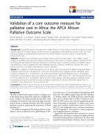
It is apparent that bacilli in standing cultures would slowly settle to the bottom of the
culture vessel, forming a sediment (see figure 1.3.1). As the latter thickens, it is
unavoidable that the bacilli in the lower levels of the sediment encounter self-
generated hypoxic conditions. Through labelling of cells with
14
C- uridine, it was
revealed that bacilli in the upper liquid phase displayed a logarithmic decrease in
radioactivity counts, signifying that the bacilli there are growing logarithmically
(Wayne 1976). However, replication proceeded at a much slower generation time of
33 hours. This slowing down in replication is attributed to the bacilli settling through a
spatial oxygen gradient where oxygen levels decrease to very low levels as they
approach the sediment. In contrast, the sediment showed a short rise in radioactive
counts (probably due to settling of labelled cells) followed by a stable plateau of
counts. As such these cells are not replicating or do so at a very slow rate (Wayne
1976). Thus, the net arithmetic growth observed is a product of the combinatorial
effect of slow logarithmic growth of settling bacilli in the upper phase and non-
replication in the sediment (Wayne 1976). In addition, when the sediment bacilli are
reconstituted in fresh oxygen rich media, re-growth was observed, thereby
demonstrating that the bacilli in sediment are viable (Wayne 1977). Since sudden
exposure to hypoxia is lethal (Wayne 1976), it appears that the bacilli adapt to hypoxia
during their journey across the spatial oxygen gradient. In addition, when the sediment
bacilli are exposed to isonazid and rifampin, a very limited effect was observed as
compared to 90% death in logarithmic cultures (Wayne and Sramek 1994). However,
they developed sensitivity to metronidazole (Wayne 1976). Thus, using the standing
6
culture system one could generate hypoxia induced non-replicating, drug resistant
bacilli that can grow when oxygen is provided. The physiological state of these in
vitro generated bacilli could be similar to those found in hypoxic environments in
vivo.
Further examination of the bacilli in the sediment yielded several interesting findings.
A unique antigen (URB) was discovered when protein extracts from hypoxic non-
replicating bacilli was subtracted with anti-sera from aerobic logarithmically cells
(Wayne and Sramek 1979). It was later identified as the 16kDa α-crystallin
homologue (Yuan, Crane et al. 1998; Desjardin, Hayes et al. 2001). In addition,
Wayne not only detected the resumption of growth when the sediment bacilli were
diluted in fresh media, he observed that the growth was synchronous (Wayne 1977).
Detailed analysis of the phenomenon by monitoring incorporation of tritium labeled
uridine revealed that the first eight hours post reconstitution showed a marked increase
in RNA synthesis before the first cell division. However, DNA synthesis was seen
only after the latter was completed. This suggests that a set of molecules is required to
be synthesized before cell division can proceed and that the bacilli probably arrested
growth as diploids (Wayne 1977).
It is likely that specific genes are expressed during the adaptation phase such that an
orderly shut down of the cell machinery could be completed before growth arrest and
that the bacilli have everything in place, for instance, stress protection and enzymes to
satisfy metabolic needs during prolong survival under hypoxic conditions. Indeed, a
five-fold increase in the specific activity of isocitrate lysase, the first enzyme in the
glyoxylate by-pass that converts isocitrate to glyoxylate and succinate, was detected in
7
non-replicating synchronous bacilli (Wayne and Lin 1982). This enzyme was later
shown to be important for persistence and virulence in vivo as mice infected by bacilli
lacking this gene displayed reduced lung pathology and survived the infection
(McKinney, Honer zu Bentrup et al. 2000). The detection of a novel enzyme, glycine
dehyhdrogenase in MTB, which catalyses the reductive amination of glyoxylate to
glycine using NADH, prompted Wayne and colleagues to investigate its occurrence in
sediment cells. This is because the pathway may be employed by the bacilli to
maintain the NAD pool such that sufficient ATP is generated for the completion of a
final round of DNA replication before total shutdown of the cellular machinery
(Wayne and Lin 1982). The fact that reconstituted bacilli only synthesizes DNA after
the first cell division supports this concept. Indeed, a 10-fold induction in specific
activity of glycine dehydrogenase was observed (Wayne and Lin 1982). Further
analysis revealed that a 60:1 ratio of NAD to NADH is sufficient to decrease the rate
of NADH reduction by 50% as compared to 400:1 ratio of glycine to glyoxylate. This
suggests that the aim of the reaction is to produce NAD rather that glycine. Taken
together, the induction of these two enzymes gave a first glimpse of alternative
metabolic pathways that may be utilized by MTB to maintain a sufficient level of
metabolic activity required for long term survival.
In conclusion, the transition from an actively replicating cell to non-replication can be
viewed as an entry to a new developmental state. It involves the induction of
alternative metabolic pathways, antigenic differences and the population of non-
growing bacilli is arrested at an identical stage of the cell cycle (i.e. the culture is
synchronous). In this defined physiological state, the bacilli maintain viability for
extended periods of time and can be reactivated. As such, these bacilli can be
8
described as dormant where dormancy is defined as: ‘a reversible state of low
metabolic activity in which cells can persists for extended periods without division’
(Kaprelyants, Gottschal et al. 1993).
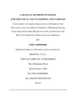
Figure 1.3.1 The Wayne’76 standing culture model (Wayne 1976). The bacilli settle
through a spatial oxygen gradient allowing adaptation to hypoxic conditions. The
bacilli in the sediment are non-replicating, drug resistant and synchronized.
1.4 Temporal Analysis of Dormancy: Wayne’96 Dormancy
Culture Model
The work of Wayne and colleagues on standing cultures described above provided
valuable insight to dormancy. However, the Achilles' heel of the system is its
heterogeneous nature due to the presence of two (or more) distinct populations of cells
that exists in different physiological states within one culture tube. Only the sediment
9
consists of a homogenous population of cells (Wayne and Hayes 1996). Therefore, the
standing culture system does not allow for the temporal dissection of the dormancy
response. Hence, in 1996 Wayne et al. developed the Wayne in vitro dormancy culture
system (Wayne and Hayes 1996), which brought the study of dormancy to a new
dimension – Time.
The strength of the Wayne’96 dormancy culture system is that the culture at any time
point is homogenous (Wayne and Hayes 1996). The bacilli are grown in sealed tubes
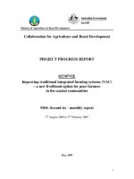
with stirring which cause the creation of a temporal self-generated oxygen gradient
that allows for adaptation to hypoxia. This results in the shift down to a non-
replicating dormant state (see figure 1.4.1). As such, parameters that will affect the
oxygen diffusion kinetics and its availability have to be defined and kept constant for
the culture system to be reproducible. The bacilli are grown with Dubos Tween-
albumin broth in sealed tubes measuring 20 by 125mm that have a total capacity of
25.5ml. The logarithmically growing bacilli are diluted to A
600
0.005 and dispensed
into the tubes as 17 ml aliquots. Thus, 8.5 ml of headspace remains, which
corresponds to a 0.5 ratio of air to liquid volume. In addition, the bacilli are uniformly
distributed by constant stirring with 7mm magnetic stirring bars at a speed of 120rpm.
At this rate, the oxygen diffusion rate is slow and comparable to those of standing
cultures, as the liquid surface is undisturbed. With these conditions, three distinct
growth phases were observed (Wayne and Hayes 1996). Initially, aerobic logarithmic
growth (generation time of 18hrs) was sustained for 58 hours, after which the culture
enters a transition phase when a microaerobic threshold is reached (10% oxygen
saturation). This growth phase is characterized by a slow increase in turbidity which
occurs without replication. This phenomenon can be attributed to a 33 % increased in
10
cell size. This transition phase termed non-replicating persistence stage 1 (NRP1) is
further distinguished by an increased rate of nitrate reduction that is independent of
substrate concentration (Wayne and Hayes 1998).
The bacilli enter a stationary phase (non-replicating persistence stage 2, NRP2) when
oxygen saturation levels dropped to 0.06% and turbidity reaches a plateau. After 100
hours in the hypoxic stationary phase, the culture becomes anoxic (detection limit
0.02%) and is characterized by the complete decolorization of the oxygen indicator
methylene blue. The entry into the two NRP stages is induced by hypoxia and not
caused by the depletion of nutrient. This is shown by the extension of the transition
phase when the tubes were loosely capped to allow for free exchange of air to take
place. As such, NRP2 is referred to as the hypoxic stationary phase. It was shown that
termination of DNA synthesis took place during the transition phase, which is
consistent with the observed cessation of replication. In addition, viability is
maintained during the hypoxic stationary phase. As such, the latter is the result of
termination of growth and not due to the combined effect of death and cryptic growth.
Although, the bacilli had stopped growing, they were still metabolically active. Pulse
–labelling experiments demonstrated that bacilli in the transition phase and hypoxic
stationary phase continue to synthesize RNA. Further, a 10 fold increased in glycine
dehydrogenase activity was detected during the transition phase. Reconstitution of
bacilli in the late hypoxic stationary phase with fresh media resulted in the resumption
of growth in a synchronous fashion. In addition, bacilli in the late hypoxic stationary
phase are resistant to isonazid and rifampicin but developed sensitivity to
metronidazole.
11
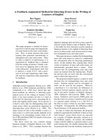
Figure 1.4.1 The Wayne’96 in vitro dormancy culture model. The bacilli are adapted
to hypoxia through a self-generated temporal oxygen gradient. After 20 days the
bacilli are non-replicating, drug resistant and synchronized (Wayne and Hayes 1996).
In conclusion, the bacilli in the late hypoxic stationary phase are identical to those in
the sediment of standing cultures. They possess the hallmarks that are attributed to
dormant cells where its physiological state is defined by synchronicity, drug resistance
and sustained metabolic activity in a non-replicating persistent state. Taken together,
Wayne’s in vitro works showed that MTB is able to adapt and survive hypoxia by
shifting to a non-replicating (‘dormant’) state. The existence of hypoxia in vivo is
supported by several lines of evidence and is discussed extensively above (see section
1.2). Hence, it appears that the bacilli generated in vitro are similar to the persistent
bacilli in hypoxic environments in vivo. However, an important question remains: Do
12
hypoxic dormant cells contribute to persistence in vivo? Correlations can be drawn
from observations made with the two culture systems described above and infection
studies performed in animal models. The phenotype of attenuated virulence
(McKinney, Honer zu Bentrup et al. 2000; Weber, Fritz et al. 2000) and persistence of
the isocitrate lyase and narG mutants is consistent with the induction of isocitrate
lyase activity and nitrate reduction in hypoxic dormancy (Wayne and Lin 1982;
Wayne and Hayes 1998). Further, the transcript of narX that was shown to be induced
in hypoxic dormant cells in vitro via northern blot analysis (Hutter and Dick 1999)
was also observed to be induced in granulomas (Fenhalls, Stevens et al. 2002). In
addition, the 16KD α-crystallin homologue (URB antigen) had been shown to be
involved in long-term viability and is required for growth in macrophages (Yuan,
Crane et al. 1996; Yuan, Crane et al. 1998). These correlations (although
circumstantial) provide evidence which suggest that hypoxic dormant bacilli exist in
vivo and that they play a role in persistence. Thus, the Wayne dormancy culture
system could be viewed as an in vitro model of persistence. Further, its capability to
allow for temporal studies makes it a powerful tool for unrevealing the sequence of
events leading to dormancy and hence allow for the definition of persistence at the
molecular level.
1.5 Other ‘Hypoxia’ Culture Systems
Work by groups employing other culture systems provided valuable data on the
process of in vitro adaptation to hypoxia and long-term survival of the bacilli. Yuan et
al. detected a rapid increase in synthesis of the 16 kD α-crystallin homologue (HspX)
13
in agitated cultures of MTB grown to late stationary phase (Yuan, Crane et al. 1996).
In addition, an induction in transcription of the gene (35 fold) was detected via
promoter lacZ fusions when oxygen levels are lowered to 0.7% (less than 5% oxygen
saturation) (Yuan, Crane et al. 1998). This appears to suggest that the induction of
HspX is specific to hypoxia as subjecting the bacilli to other stresses such as cold, heat
and ethanol shock, did not result in induction (Yuan, Crane et al. 1996). These
observations were consistent with later work where the hspX mRNA and protein was
shown to be drastically up regulated in the microaerophilic transition phase of the
Wayne dormancy culture system and in hypoxic dormant bacilli (Boon, Li et al. 2001;
Desjardin, Hayes et al. 2001). Overexpression of HspX resulted in increased survival
of the bacilli in stationary phase. Intriguingly, the promoter of hspX was reported to be
active in THP-1 cells, a human monocytic cell line via fusion of the promoter to a
GFP reporter (Yuan, Crane et al. 1998). This may suggest that the environment in the
macrophages may be hypoxic. Consistent with this, the hspX mutant displayed
impaired replication in macrophages (Yuan, Crane et al. 1998). This suggests a role
for HspX for growth in macrophages.
A role for the stringent response in hypoxia was shown by Primm and co-workers
(Primm, Andersen et al. 2000). The authors showed that bacilli lacking the MTB RelA
homologue had an increased loss of viability compared to the wild type strain, when
subjected to hypoxic conditions. In addition, the relA mutant was unable to reduce the
amount of stable RNA and ribosomal proteins, which is required to control growth
rate. As such, this defect may contribute to the mutant’s failure to slow growth for the
adaptation to hypoxia and hence affect its ability for long-term survival (Primm,
Andersen et al. 2000).
14
Hu et al. observed in their (poorly characterized) version of the standing culture model
that hspX mRNA decreases and HspX protein increases during transition from
growing to stationary phase (Hu and Coates 1999). As such, they speculated that the
expression of hspX is regulated post-transcriptionally. The contradictory data provided
by the authors can be attributed to the uncharacterized culture system that they
employed. It is important to note that, a slight disruption in equilibrium between
settling and replication that may occur by simply changing the type of culture vessels
or amount of media used would have affected the dynamics of the culture system and
hence generate unreliable results. In addition, for standing cultures the samples should
be obtained from sediments only where the bacilli are homogenous and exist in a
define physiological state. Hence, it is possible that their observations are due to
‘contamination’ from growing cells because samples were obtained from entire
cultures. As such, data generated by undefined culture conditions (Hu, Butcher et al.
1998; Hu and Coates 1999; Florczyk, McCue et al. 2001; Hu and Coates 2001) has to
be interpreted with caution.
1.6 The Hypoxia Induced in vitro Dormancy Response is
Poorly Understood
HspX belongs to the Hsp20 family of small molecular weight heat shock proteins
(smHSP) (Caspers, Leunissen et al. 1995). They are known to be usually
developmentally regulated and possess ATP-independent chaperone activity (Jakob,
Gaestel et al. 1993; Nicholl and Quinlan 1994; Buchner 1996). These properties,
15
together with its induction during hypoxia, raise the possibility that HspX may play a
role in hypoxia-induced stationary phase survival. Indeed, overexpression of HspX
provided limited protection from autolysis during the stationary phase of agitated
cultures (Yuan, Crane et al. 1996). In addition, HspX is able to prevent the
precipitation of alcohol dehydrogenase at 48
o
C, which is comparable to the protection
provided by the HspX homologue from bovine eye tissue (Horwitz 1992). Thus, HspX
may play a role in maintaining long-term survival of MTB by enhancing the stability
of proteins (Yuan, Crane et al. 1996). It had been shown recently that the function of
HspX is not solely limited to its chaperone activity. Through analysis using colloidal
gold immunoelectron microscopy, Cunningham and Spreadbury found that HspX is
frequently located at the cell membrane and cell wall skeleton of BCG, suggesting a
possible role in stabilization of a thicken cell wall observed in hypoxic stationary
phase (Cunningham and Spreadbury 1998). HspX role in vivo was further characterize
when a MTB deletion mutant in the gene displayed impaired growth in both THP-1
cells and bone marrow derived macrophages (Yuan, Crane et al. 1998). However,
viability of the bacilli was not affected. As such, HspX may be required for growth
during initial infection by MTB (Yuan, Crane et al. 1998). Work by Manabe et al.
revealed that the expression of hspX in MTB is modulated by the alternative sigma
factor SigF (Manabe, Chen et al. 1999). They demonstrated that deletion of sigF from
the genome results in the complete loss of HspX expression (Chen, Ruiz et al. 2000).
In addition, mice infected with bacilli lacking the sigF gene had a mean survival time
that is 85 days longer than those infected with the wild type strain, signifying a role
for sigF in virulence. Further, exposing the bacilli to hypoxia causes a 150-fold
induction in the expression of sigF and a further increase was observed when
metronidazole was added to the hypoxic cultures (Michele, Ko et al. 1999). As such,
16
SigF may be involved in the regulation of genes required for survival and adaptation
to hypoxic conditions encountered in vivo.
Taken together, it is apparent that our understanding of the molecular biology
involved in the hypoxia-induced dormancy response (at the beginning of this thesis
work) is limited to HspX, SigF and NarX. A good deal of work is required before a
complete picture of the dormancy response can be established. However, there are
several experimental disadvantages associated with the pathogenic nature of MTB.
The latter is a biosafety level III organism. Hence, it is difficult to conduct
experiments involving large amount of cultures, thereby severely limiting the amount
of raw material (e.g. protein, RNA) available for experimentation. This prompted Lim
et al. to investigate the possibility of using BCG as a non-pathogenic model for
dormancy. They showed that the molecular and physiological behaviour of BCG
grown in the Wayne dormancy culture system is similar to MTB (Lim, Eleuterio et al.
1999). Thus, their work established BCG as a non-pathogenic model for the study of
dormancy in MTB.
It is now evident that the molecular mechanism of the hypoxic dormancy response is
poorly understood, as only a few molecules have been discovered (Figure 1.6.1 for
summary). No genes have yet been identified to be essential and specific for
dormancy survival. Identification of more dormancy molecular markers is important
so as to provide further conclusive evidence that bacilli similar to those generated in
the Wayne’s in vitro dormancy culture system do exist in vivo. In addition, generation
of in vitro dormancy mutants is important to demonstrate the relevance of the in vitro
dormancy response in animal models.
17
Figure 1.6.1 Hallmarks of bacilli grown in the Wayne’76 and’96 in vitro dormancy
culture models (Wayne and Sohaskey 2001).
18
In this work, the in vitro dormancy response was analysed by employing proteomics.
The Wayne’96 dormancy culture system was chosen as it permits the temporal
analysis of this response. Further, BCG was selected as the working organism as it
allows large-scale experiments to be conducted easily without the restrictions of
biosafety level III work. The aim of the work is to reveal new dormancy specific
proteins that may serve as molecular markers for hypoxic dormant bacilli and to ask
the question: Are these proteins essential for dormancy survival in vitro?
19
Chapter 2
Materials and Methods
2.1 Materials
2.1.1 Vectors
pJQ200 SK
This vector was designed by (Quandt and Hynes 1993). It is a
suicide plasmid that can be used for targeted gene disruptions
in Mycobacterium. It confers gentamycin resistance and has a
sacB gene as a counter selectable marker.
pNBV1
A shuttle vector that replicates in both E.coli and
Mycobacterium (Howard, Gomez et al. 1995). It contains the
hyr gene from Streptomyces hygroscopicus that confers
hygromycin resistance.
pNEB193
This is an E.coli high copy plasmid (New England Biolabs). It
contains a lacZ
α
gene that allows for blue/white selection and
uses ampilcilin as an antibiotic selection agent.
pUCK4
This is a plasmid containing the kanamycin resistant cassette
used here in replacing gene coding sequences (Amersham
Biosciences)
20
2.1.2 Bacterial Strains
DH5α
E.coli strain used for general cloning work (Gibco).
XL1 Blue MRF’
E.coli strain used as host cells for λ phage supplied by
Stratagene in the λ ZAP II cloning kit. It was used for screening
of the BCG genomic λ phage library constructed by Murugasu-
Oei, B. and Dick, T. (unpublished)
SOLR Cells
E.coli strain used as host cells for the excision of the
pBluescript phagemid from λ ZAP vectors during genomic
library screens.
M.bovis BCG Pasteur
ATCC 35734
All Mycobacterium experiments were performed with this
strain.
2.1.3 Chemicals and Reagents
Company Chemicals And Reagents
Amersham Biosciences
Bromophenol Blue, CHAPS, Dry Strip Cover
Fluid.
BDH Laboratories Supplies
β-mecaptaethanol, Butanol, Chloroform,
Ethanol, Formaldehyde, Formamide, Glacial
Acetic Acid, HCl, Isopropanol, Methanol,
NaCl, Phenol: Chloroform:Isoamyl Alcohol,
SDS, Sodium Carbonate Anhydrous,
Sucrose, TEMED, Triton X-100.
Bio-RAD
Acrylamide, AG® 501-X8 (D) Resin,
Agarose, Boric Acid, Glycine, Ethidium
Bromide, PDA, Urea, Xylene cyanol.
21
New England Biolabs
100mM dNTP, Apa I, Bam HI, Calf
Intestinal Phosphatase, DNA Ligase, Dra III,
Eco R I, Eco RV, Klenow Fragment, Kpn I,
Not I, Pst I, T4 DNA Polymerase, Xba I, X-
gal, Xho I.
PIERCE
Neutralized TCEP Bond Breaker.
Gibco
1kb plus ladder, High Grade Glycerol
ROCHE Molecular Biochemicals
Leupeptin, Pefabloc, Pepstatin A, DNase
RNase Free, RNase A, RNase DNase Free,
RNase Inhibitor.
Sigma
Ammonium Persulphate, Ampilcillin
Sulphate, BSA, Dextran Sulphate, DEPC,
DTT, EDTA, Ficoll, Gentamycin Sulphate,
Hygromycin B Sulphate, Idoacetamide,
Kanamycin Sulphate, Methylene Blue,
MOPS, NaH
2
PO
4
(monobasic), Na
2
HPO
4
(dibasic), Ovalbumin, Polyvinylpyrrolidone,
Salmon Sperm DNA, Silver Nitrate, Sodium
Thiosulphate, Sodium Citrate, Trima®
BASE, Tween 20.
2.1.4 Growth Media
Dubos Tween-albumin broth
6.5g Dubos broth base
(Becton Dickinson) in 180 ml of
distilled water. Autoclaved. 20 ml of Dubos medium albumin
(Becton Dickinson) was added before use.
Dubos Oleic-albumin agar
4g Dubos oleic agar base
(Becton Dickinson) in 180 ml of
distilled water. Autoclaved. 20 ml Dubos oleic albumin
complex
(Becton Dickinson) was added before use.
LB broth 1.0% bacto tryptone, 2% yeast extract, 1.0% NaCl. Autoclaved.
22
LB Agar 1.5% bacto agar dissolved in LB. Autoclaved.
2.2 Mycobacterial Culture
2.2.1 The Wayne Dormancy Culture Model
BCG was grown as described in Lim et al and Wayne et al. (Wayne and Hayes 1996;
Lim, Eleuterio et al. 1999). Aerobic exponentially growing precultures of A
600nm
0.2 to
0.3 were diluted to A
600nm
0.005 with Dubos Tween-albumin broth. They were
dispensed in 17 ml aliquots into 20 mm by 125mm screw-cap test tubes (Wheaton)
that have a total volume of 25.5ml and contain a 8mm Teflon coated magnetic stirrer
(Wheaton). The tubes were sealed tightly by screwing down caps with latex liners.
The cultures were then stirred gently at 170 rpm on magnetic platforms (Variomag).
Oxygen levels were monitored via decolorization of the redox indicator methylene
blue where 17 µl of 500 µg/ml stock solution was added to give a final concentration
of 1.5 µg/ml (Wayne and Hayes 1996; Lim, Eleuterio et al. 1999). Daily turbidity
measurements were determined in a Bacharach Coleman model 35 photometer
(Thomas Scientific).
2.2.2 Aerated Stationary Phase Culture Model
An aerobic exponentially growing preculture of A
600
0.2 to 0.3 was diluted to A
600
0.05 with Dubos Tween-albumin broth. The diluted culture was dispensed in 100 ml
23
aliquots into 100 mm by 140 mm roller bottles (Corning) and rolled at 1 rpm in a
roller bottle incubator (Bellco Biotechnology) at 37
o
C. The bottles were opened daily
to allow for exchange of air (Boon, Li et al. 2001). Daily turbidity measurements were
determined in an Ultrospec 3000 spectrophotometer (Amersham Biosciences).
2.2.3 Determination of C.F.U
Dilutions of the culture were made in 10 fold increments with Dubos Tween-albumin
broth. Twelve well tissue culture plates (Falcon) containing 3 ml of Dubos Oleic-
albumin agar were prepared. The wells were then inoculated in triplicate with 20 µl of
diluted culture and incubated at 37
o
C. Colonies were then counted after 2 to 3 weeks
using a dissection microscope (Nikon).
Bacilli grown in the aerated stationary phase culture system were sonicated for 15
seconds 5 times with 1 minute ice breaks at level 2 of an Ultrasonic processor XC
sonicator (Misonix) before the dilutions were made. This served to disperse the
observed bacilli clumps.
2.3 DNA and RNA Methods
All DNA and RNA methods described in the following sections were performed using
standard protocols (Sambrook, Fritsch et al. 1989) and according to manufacturer
recommendations.
24
2.3.1 Restriction Digest of DNA
The reaction was performed in a final volume of 40 µl. It contains 4 µl of 10x reaction
buffer (manufacturer supplied), 1 to 2 µg of DNA and 1 µl (10-20 units) of
appropriate restriction enzyme. When necessary 4 µl of 10X BSA (manufacturer
supplied) was added to the reaction. The remaining volume of the reaction was made
up with distilled water. The digest was performed at 37
o
C for two hours for plasmid
DNA or overnight for genomic DNA and PCR fragments. After which, the enzyme
was deactivated by heat according to manufacturer recommendations. If heat
denaturation was not possible, the enzyme was removed by agarose gel
electrophoresis (see 2.3.5) and the digested fragment was recovered via elution from
the agarose gel (see 2.3.6)
2.3.2 Blunt Ending of DNA Fragments
Blunt ending of 5’ and 3’ overhangs were performed with T4 DNA polymerase in 50
µl reaction volumes. This consists of digested DNA, 1µl (10 units) of the enzyme, 5 µl
of supplied 10X reaction buffer, and 25 mM of each DNTP. The reaction was
incubated at 12
o
C for 15 minutes. Subsequently, the enzymes were inactivated by
heating to 75
o
C for 20 minutes. DNA was concentrated via ethanol precipitation (see
2.3.7).
25