Tissue engineering approaches to tendon repair studies on the use of bone marrow stromal cells and knitted poly (d, l lactide co glycolide scaffold
Bạn đang xem bản rút gọn của tài liệu. Xem và tải ngay bản đầy đủ của tài liệu tại đây (8.41 MB, 174 trang )
TISSUE ENGINEERING APPROACHES TO TENDON REPAIR:
STUDIES ON THE USE OF BONE MARROW STROMAL CELLS AND
KNITTED POLY (D, L-LACTIDE-CO-GLYCOLIDE) SCAFFOLD
OUYANG HONGWEI
(Bachelor of Medicine/Bachelor of Surgery)
A THESIS SUBMITED
FOR THE DEGREE OF
DOCTOR OF PHILOSOPHY
DEPARTMENT OF ORTHOPAEDIC SURGERY
NATIONAL UNIVERSITY OF SINGAPORE
2003
Acknowledgements i
Acknowledgements
I wish to express my deepest gratitude and heartfelt thanks to my supervisors:
Professor Lee Eng Hin, Dean, Faculty of medicine, National University of
Singapore, and Associate Professor James Goh Director of research, Department of
Orthopedic Surgery, National University of Singapore, for their constant
encouragement, invaluable guidance and infinite patience throughout the course of
this study.
I would like to express my sincere thanks to Professor K Satkunanantham Head,
Department of Orthopedic Surgery, National University of Singapore, for his support.
Without the excellent facilities, this work would not have been possible.
I owe my thanks to Professor Teoh Swee Hin, Assistant Professor Dietmar
Hutmacher, Dr Mo Xui-Mei, Division of Bioengineering, NUS for their assistance
in the provision of biomaterials and reagents, as well as technique assistance in
manufacturing of scaffolds.
I would also like to express my appreciation to the following staff members Ms
Chong Sue-Wee, Ms Julee Chan, Mr Ashvin Thambyah, Ms Grace Lee, Mr
Barry P Pereira, Ms Jessie Tan, Mr Yong Soon Chiong, Mr Dominic Tey, Dr Li
Li, Dr Ge Zi-Gang, and Dr Shao Xin-Xin for their kind help.
I will always remember my friends in Singapore for their constant encouragement and
kind help. This work was support by a grant from NMRC, Singapore.
Acknowledgements ii
Last but not least, I am grateful to my families, my parents, my wife Zou Xiao-Hui
and my baby OuYang Xin-yi for their understanding and love during the years of my
Ph.D. pursuit.
. Table of Content iii
Table of Content
I. Acknowledgement…………………………………………………………………i
II. Table of content………………………………………………………………… iii
III. Summary………………………………………………………………………….ix
IV. Publications………………………………………………………………………xii
Chapter I: Introduction and Literature Review……………………………………………1
Chapter II Literature Review…………….……………………………………………… 5
2.0. Introduction to Literature Review……………………………………….… … 6
2.1 Tendon Anatomy, Physiology And Injury…………………………………… …6
2.1.1 Tendon Anatomy……………………………………………………………… 6
2.1.2 Tendon Composite…………… …………………………………….… ………8
2.1.3 Tendon to Bone Insertion…… …………………………………… ….….……13
2.1.4 Tendon Biomechanics…… …………………………………………… ……14
2.1.4.1 Tendon Mechanical Properties………… …………… .…… 14
2.1.4.2 Effect of Biomechanical Load on Tendon…………………………………….16
2.2 Tendon Injury Healing and Current Therapy………………………………… 18
2.2.1 Tendon Injury Healing ……………………………………………………… 18
2.2.2 Several Concerns about Tendon Healing…………………………………… 20
2.2.2.1 The Amount of Reparative Cells………………………………………………20
2.2.2.2 The Mobilization of Reparative Tendon…………………………………….…22
. Table of Content iv
2.2.2.3 The Evaluation of Tendon Healing……………………………………………22
2.2.3. Current Therapy……………………………………………………………… 23
2.3. Tissue Engineering Approaches To Improve Tendon Healing……………… 24
2.3.1. Tissue Engineering Principles………………………………………………….24
2.3.2. Cell Source…………………………………………………………………… 25
2.3.3. Biomaterials and Scaffold……………………………………………… 29
2.3.4. Biomolecules………………………………………………………………… 33
2.3.5. Animal Model………………………………………………………………….36
2.3.6. Tissue Engineering Techniques for Tendon Insertion Healing……………… 39
2.4 Hypotheses and Objective of This study……………………… …….…… 40
Chapter III: Materials and Methods…………………………………………….… ……42
3.0 Introduction to Material and Methodology…………………………….………43
3.1 Stage1. bMSCs Differentiation Study……………………………….…………43
3.1.1 Isolation And Culture Of Bone Marrow Stromal Cells……………………… 43
3.1.2 Osteogenesis Induction and Detection……………………………….……… 44
3.1.2.1 In vitro Osteogenesis Induction ……………………… …………….……… 44
3.1.2.2 Vonkossa Staining…………………………………………………………… 45
3.1.3 Chondrogensis Induction And Detection………………………………………46
3.1.3.1 In Vitro Chondrogenesis Induction ……………… ………………….…….…46
3.1.3.2 Collagen Type II Immunoassaying………………………………………….…47
3.1.4 Adipogenesis Induction And Detection……………………………………… 48
3.1.4.1 In Vitro Adipogenesis Induction …………………………………… ……….49
. Table of Content v
3.1.4.2 Oil Red Staining……………………………………………………………….50
3.2 Stage II. Trace Study On The Fate of bMSCs After Implantation…………….51
3.2.1. Animal Model ……………………………………………………………… 51
3.2.2. Cells Labeling And Detection………………………………………………….52
3.2.2.1 CFDA Labeling And Detection……………………………………………… 52
3.2.2.2 GFP Gene Transfection And Detection……………………………………….53
3.2.3. Tissue Preparation For Cryostat Section……………………………………….54
3.3 Stage 3. Study On The behavior of bMSCs On Various Polymer Films …… 55
3.3.1. Materials……………………………………………………………………….55
3.3.2. Polymer Film Manufacture…………………………………………………….56
3.3.3. Polymer film Sterilization and Prewetting…………………………….……….56
3.3.4. Water Contact Angle Test…………………………………………………… 57
3.3.5. bMSCs Adhesion Assay ………………………………………………………57
3.3.6. bMSCs Proliferation Assay…………………………………………………….57
3.3.7. MTS Assay For Cell Proliferation…………………………………………… 58
3.3.8. Statistic Analysis Method…………………………………………………… 58
3.4 Stage 4. Study On The Effect Of bMSCs Seeded knitted PLGA For Achilles Tendon
Repair……………………………………………………………………………….……59
3.4.1 Animal Surgery……………………………………………………………… 59
3.4.2 Knitting PLGA Fiber Scaffold…………………………………………………61
3.4.3 Tissue Preparation For Paraffin Section……………………………………….61
3.4.4 H&E Staining…………………………………………………………… …….62
3.4.5 Immunohistochemical Staining……………………………………………… 63
. Table of Content vi
3.4.6 Transmission Electronic Microscopy……………………………………… ….64
3.4.7 Biomechanical Test………………………………………………………….…64
3.5. Stage 5. Study on the Effect Of bMSCs On the Tendon Insertion Healing… … 66
3.5.1. Animal Surgery………………………… …………………………….……… 66
3.5.2. Hard Tissue preparation For Paraffin Section……… ………………….………67
3.5.3. H&E & Immunohistochemical Staining ……………………………………… 67
Chapter IV: Results………………………………………………………………………68
4.1 Stage 1.The Differentiation Of bMSCs………… ………………………….….69
4.1.1. bMSCs Isolation……………………………………………… …69
4.1.2. Osteo-lineage Differentiation………………………………………………… 70
4.1.3. Chondro-lineage Differentiation ………………………………………… … 71
4.1.4. Adipo-lineage Differentiation………………………………………… … ……72
4.2 Stage 2. The Fate of bMSCs After Implantation…… …………………… ……73
4.3 Stage3. The Adhesion and Proliferation of bMSCs on Various Polymer
Films…………………………………………………………………………………… 76
4.3.1. Polymer Films……………………………………………………………………76
4.3.2. Cell Adhesion…………………………………………………………………….77
4.3.3. Cell Proliferation ………………………………… …………………….………79
4.3.4. Cell Morphology………………………………………… …………….……….81
4.4 Stage4. The Efficacy Of Allogeneic bMSCs Seeded Knitted PLGA Scaffold For
Achilles Tendon Repair…………………………………………………………… ….82
4.4.1. The Histology of Tendon Repair………………………………………… ……82
. Table of Content vii
4.4.2. The Immunohistology of Tendon Repair……………………………… ………90
4.4.3. The Biomechanics of Tendon Repair…………………………… …… … … 91
4.5 Stage5. The Effect of bMSCs on Tendon Insertion Healing………… …….….94
5.5.1. The Natural Healing of Tendon Insertion……………………………… ….… 94
5.5.2. The Effect of bMSCs on Tendon Insertion Healing …………………… … …96
Chapter V: Discussion…………………………………………………………………99
5.1. The Isolation and Differentiation of bMSCs…………………….…………… 100
5.1.1. The isolation of bMSCs………………………………………………….…… 100
5.1.2. The Multipotential of bMSCs…………………………………………….…….101
5.1.3. Bone Marrow Stromal Cells as Tendon progenitor Cells………………….… 102
5.2. The Fate of bMSCs After Implantation………………………………… … 103
5.2.1. The Methods of Cell Trace Study………………………………………… … 103
5.2.2. The Fate of bMSCs after Implantation into Tendon Wound Site….……… .…103
5.2.3. The Possibility of Allogeneic bMSCs for Tissue Engineering…………….… 105
5.2.4. The Potential of bMSCs For Gene Delivery……………………… …….…….107
5.3. The Behavior of bMSCs on Various Polymer Films……………… ……… …108
5.3.1. Method for Characterizing Cell-Polymer Interaction……………………… ….108
5.3.2. The Effect of Cell Source on The Cell-Polymer Interaction………………… 109
5.3.3. The Effect of Substrate on The bMSCs Adhesion And Proliferation……… 110
5.4. The Efficacy of bMSCs and Knitted PLGA scaffold for Achilles Tendon
Repair……………………………………………………………………………… … 113
5.4.1. The Effect of Knitted PLGA on Tendon Repair…………………………… ….113
. Table of Content viii
5.4.2. The Effect of bMSCs on Tendon Repair…………………………………… 116
5.4.3. Tendon Repair versus Tendon Regeneration……………………………… …118
5.5. The Effect of bMSCs on Tendon Insertion Healing ……………………… …120
5.5.1. The Natural Healing of Tendon Insertion………………………………… ….120
5.5.2. The Effect of bMSCs on Tendon Insertion Healing……………………… ….121
Chapter VI Conclusion and Recommended Future Study……………………… ….…124
ChapterVII References………………………………………………………… …… 128
Summary ix
Summary
Background: Unlike bone that is able to heal by regenerating normal bone in most
cases, tendons often heal by forming scar tissue. The limited capacity for injured
tendon to regenerate poses a great challenge and creates an opportunity for
engineering new tendons. To date, less work has been done on tendon tissue
engineering compared to the extensive work on the bone and cartilage tissue
engineering. Several studies have investigated the use of gel and braided scaffold
with or without cells for tendon repair; however, the inferior mechanical strength of
gel carrier and the poor tissue ingrowths associated with the braided fiber scaffold has
limited the success. Many other problems have yet to be addressed: the fate of
implanted bone marrow stromal cells (bMSCs) at the tendon site has not yet been
studied; no general principles have been established to select material for bMSCs
delivery; the role of bMSCs in tendon repair has been arguable due to the lack of
appropriate controls in previous studies, and limited attention has been placed on the
tendon-to-bone healing when engineering tendon graft for tendon repair.
Hypotheses: The main hypothesis is that tissue engineered graft composed of bMSCs
and knitted PLGA can improve tendon repair. To support this hypothesis: (a) bMSCs
should have multipotential and be good candidates of cell source for tendon repair.
(b) The knitted PLGA should be promising for bMSCs delivery and tendon repair.
Summary x
Methods: With the aim to verify the above hypotheses, five sequential stages of
experiments were designed. They are as follows:
• Stage 1: The bMSCs differentiation test to determine the potential of the
bMSCs used in this study to differentiate into different tissues.
• Stage 2: Cell trace test to determine whether the allogeneic MSCs can
survive and differentiate into tenocytes after implantation.
• Stage 3: Cell- matrix interaction test to determine the appropriate material
for MSCs’ delivery.
• Stage 4: Tendon repair with bMSCs/knitted PLGA composites to evaluate
the efficacy of this treatment modality.
• Stage 5: The effect of bMSCs on the healing of tendon to bone.
Results: The stage 1 experiment showed that the bMSCs isolated by short-term
plastic adhesion were able to differentiate into multi-mesenchymal lineages such as
osteo-, chondro- and adipo-lineages. The stage 2 experiment illustrated that the
implanted allogeneic bMSCs could survive as long as 8 weeks and was able to
differentiate into spindle-shape cells 5 weeks after implantation at rabbit patella
tendon window wound site. The stage 3 experiment selected optimal material for
bMSCs delivery by verifying that PLGA was more likely to allow bMSCs to adhere
and grow as compare to other five synthetic biodegradable polymers. The stage 4
experiment exhibited that the composite of bMSCs and knitted PLGA scaffold
could improve the structure and biomechanics of tendon repair in rabbit Achilles
model. Besides showing the ability to accelerate tendon tissue formation in the
Summary xi
stage 5, the bMSCs exhibited the potential to restore the native structure at the
tendon to bone interface healing in the stage 5 experiment.
Conclusion: In all, these sequential experiments proved that the bMSCs were able
to be the seed cells for tendon repair; the knitted PLGA scaffolds possess optimal
material and structural properties for bMSCs delivery and tendon tissue formation;
and the composite of bMSCs and knitted PLGA could be an ideal substitute for
tendon repair. This work could be logically extended to ligament repair.
Publications xii
Publications
The following papers/abstracts have resulted from the present paper.
International Journal:
1. Ouyang HW
, Goh JCH, Mo XM, Teoh SH, Lee EH. Characterization of anterior
cruciate ligament cells and bone marrow stromal cells on various biodegradable
polymeric films. Mat Sci Eng C-BIO S 20 (1-2): 63-69 Sp. Iss. SI MAY 31 2002.
2. Ouyang HW, Goh JCH, Mo XM, Teoh SH, Lee EH. The efficacy of bone marrow
stromal cell-seeded knitted PLGA fiber scaffold for Achilles tendon repair. Ann N
Y Acad Sci. Jun; 961:126-9. 2002.
3. Ouyang HW, Goh JCH, Thambyah A, Teoh SH, Lee EH. The use of knitted
PLGA and MSCs for Achilles tendon repair in rabbit model. Tissue Engineering.
Vol 9, No.3, 431-439, 2003
4. Goh JCH, Ouyang HW, Chan CK, Teoh SH, Lee EH. Tissue engineering
approaches to tendon and ligament repair and regeneration. Tissue Engineering
Vol.9 Sup1, 31-44, 2003
5. Ouyang HW, Goh JCH, Lee EH. The effect of MSCs on the tendon to bone
healing. Am J Sport Med (In Press) 2003
6. Ouyang HW,
Goh JCH, Lee EH. Viability of allogeneic bone marrow stromal
cells following local delivery into patella tendon in rabbit model. J Orthop Res
(submitted). 2003.
International Conference
:
1 Goh JCH, Ouyang HW
, Lee EH. Survivability and functionality of bone
marrow stromal cells following implantation in tendon regeneration in rabbit
model. 49
th
annual meeting of orthopedic research society. New Orleans, LA,
USA. Feb 2-5, 2003.
2 Ouyang HW
, Goh JCH, Lee EH. Application of mesenchymal stem cells in the
repair of tendon and ligament. 11
th
international conference of biological and
medical engineering. 4
th
to 7th December 2002, Singapore.( Finalist in Young
Investigator’s Award)
Publications xiii
3 Ouyang HW, Bini TB, Mo XM, Yang F, Wang S, Ramakrishna S, Goh JCH,
Teoh SH, Lee EH. Fibrous scaffolds in regeneration of tendon and nerve
tissues. BECON 2001 Repair Medicine: Growth Tissue and Organs, Jun 25-26,
2001, NIH, Maryland, USA.
4 Goh JCH, Ouyang HW
, Mo XM, Teoh SH, Lee EH. The efficacy of bone
marrow stromal cell-seeded knitted PLGA fiber scaffold for Achilles tendon
repair. BECON 2001 Repair Medicine: Growth Tissue and Organs, Jun 25-26,
2001, NIH, Maryland, USA.
5 Ouyang HW
, Goh JCH, Mo XM, Teoh SH, Chong SW, Wang Z, Lee EH. Cell-
material systems for ACL regeneration: The behavior of biodegradable
polymer films. ICMAT 2001, Singapore
6 Ouyang HW, Goh JCH, Chong SW, Wang Z, Lee EH. Effect of marrow
stromal cells on the interface healing of tendon to bone. ICMAT 2001,
Singapore
7 Ouyang HW, Goh JCH, Mo XM, Teoh SH, Lee EH. The efficacy of bone
marrow stromal cell-seeded knitted PLGA fiber scaffold for Achilles tendon
repair. ICMAT 2001, Singapore
8 Goh JCH, Ouyang HW, Lee EH. Use of tissue engineering techniques in the
repair of tendon and ligament. Conference on biomedical engineering
technology. Taiwan 2001.
Chapter I Introduction 1
Chapter I
Introduction
Chapter I Introduction 2
Musculoskeletal conditions are increasingly becoming one of the major health
concerns because of an aging population and increased occurrence of sport-related
injuries. To date, orthopedic surgeons still rely on conventional methods of repair, such
as the use of autograft, allograft and prostheses. Although these procedures have been
fairly successful, the various shortcomings of these procedures have prompted surgeons
and scientists to look for viable alternatives. Recently developments in tissue
engineering have shown great potential in solving the problems associated with tissue or
organ transplantation. As compared to the extensive work on bone and cartilage tissue
engineering, tendon tissue engineering has not received much attention.
Tendon is arguably the least complex of the connective tissues with respect to its
composition and architecture. This might reasonably lead to the expectation that it would
be more amenable to tissue engineering approaches than other tissues. However, decades
of experience have showed how difficult it is for tendon to regenerate after treatment.
This limited capability of tendon injuries to regenerate poses a challenge to tendon tissue
engineering, and emphasizes the importance of developing a procedure to do so.
So far, few works have been done on tendon tissue engineering compared to the
extensive work on the bone and cartilage tissue engineering. Several studies have
investigated the use of gel and braided scaffold with or without cells for tendon repair;
however, the inferior mechanical strength of the gel carrier and the poor tissue ingrowths
associated with the braided fiber scaffold has limited its success. Many other issues have
not yet to be addressed, for example: 1) the fate of implanted bone marrow stromal cells
(bMSCs) at the tendon site has not yet been studied; 2) no general principles have been
Chapter I Introduction 3
established to select material for bMSCs delivery; 3) the role of bMSCs in tendon repair
has been arguable due to the lack of appropriate controls in previous studies, and 4)
limited attention has been placed on the tendon-to-bone healing when engineering tendon
graft for tendon repair.
It is clear that the continued development of tendon tissue engineering will depend
upon identification and characterization of appropriate sources of cells as well as the
development of new scaffolds. The identification of an optimal cell source for a particular
tissue engineering application will depend on rigorous characterization with regards to
plasticity, propagation, and control of differentiation. To guide the organization, growth,
and differentiation of cells in tissue engineered constructs, appropriate scaffolds are
needed to provide mechanical support as well as physical, chemical, and mechanical cues
in forming functional tissues.
The main hypothesis of this study is that tissue engineered graft composed of bMSCs
and knitted PLGA can improve tendon repair. Specific mechanistic hypotheses on why
bMSCs can be the seed cells and knitted PLGA scaffolds can be the vehicle and template
for tendon repair are as follows:
1). bMSCs can differentiate into multimesenchymal tissues including teno-lineage.
2). bMSCs can survive and differentiate into tenocytes after local delivery into tendon
site.
3). bMSCs attach and grow faster on PLGA substrate than other five polymers.
4). Knitted PLGA scaffold can allow a large number of cells infiltration and connective
fibrous tissue ingrowths; and bMSCs/knitted PLGA graft can accelerate tissue
formation and regenerate stronger neotendon as compare to scaffold alone.
Chapter I Introduction 4
5). bMSCs can restore physical structure of tendon-to-bone interface by inducing
fibrocartilage zone formation at the insertion.
This thesis includes seven chapters. In chapter I, I briefly introduced the background of
tendon repair, tissue engineering, the hypotheses of this study, and the scope/ structure of
this thesis.
The literature review (Chapter II) of this thesis covers the tendon anatomy, physiology,
biomechanics, injury healing, clinical therapies and current tendon tissue engineering
researches. Chapters III, IV and V of this thesis respectively describe the material &
methodology, results and discussion of the five sequential experiments which were
designed to verify the hypotheses of this study. In Chapter VI, a conclusion is drawn and
suggestions for future studies are proposed. The references are in Chapter VII.
Chapter II. literature review 5
Chapter II
Literature Review and hypotheses
Chapter II. literature review 6
2.0. Introduction to Literature Review
Understanding of the cellular and extracellular components of tendon is essential for
determining the methodology required to successfully engineer tissue, and provide useful
information in the development of analogs of the extracellular matrix as implants to
facilitate tendon regeneration. Insights into tendon biomechanical properties and the effect
of load on tendon structure can help design the tissue engineering graft with appropriate
biomechanical properties. Moreover, knowledge of the process of spontaneous healing of
tendon injuries and the results of selected treatment modalities can provide a guide to
strategies for tissue engineering. Finally, a review of the principles of tissue engineering
and current tissue engineering studies in developing grafts to facilitate tendon regeneration
can serve as the foundation for this tendon tissue engineering work.
2.1 Tendon Anatomy, Physiology And Injury
2.1.1 Tendon Anatomy
Tendon is a specialized dense connective tissue that links muscle to bone and allows for
transmission of muscle contraction forces to the bone for skeletal locomotion; for example,
the Achilles tendon, one of the largest in the body, links the triceps surae muscle, the
grouping of gastrocnemius, the soleus, and the plantaris muscle to the calcaneus bone. As
organs, tendons consist of three parts: the muscle attachment region, the substance of the
tendon itself, and tendon to bone insertion region. These three parts vary in their cellular
Chapter II. literature review 7
composite, histology, and function. This study will focus on the midsubstance of the tendon
and give some insight into the healing of tendon insertion.
Healthy tendons are brilliant white in color and fibroblastic in texture, showing great
resistance to mechanical loads. Besides a few tendons in the hands and feet that have the
two-layer tendon sheaths, most of the tendons are surrounded by loose areolar connective
tissue called paratenon. The main components of the paratenon are collagen type I and III
fibrils( Kvist, 1985), elastic fibrils and synovial cells that form the inner lining ( Williams,
1986). Paratenon functions like an elastic sleeve and permits free movement of the tendon
against the surrounding tissues (Hess et al 1989). Under the paratenon, the entire tendon is
surrounded by a fine connective tissue sheath called the epitenon or peritenon. On its outer
surface, the epitenon is continuous with the paratenon and on its inner surface with the
endotenon. The epitenon is a relatively dense fibrillar network of collagen with strands of 8
nm to 10 nm in thickness (Jozsa, 1991). The endotenon is a thin reticular network of
connective tissue inside the tendon that has a well-developed crisscross pattern of collagen
fibrils (Jozsa, 1982, Kastelic, 1978, Rowe, 1985). The endotenon fibrils connect tendon
fibers and bind fibers together. A bunch of collagen fibrils forms a collagen fiber, which is
the basic unit of a tendon and is aligned from end to end in a tendon, and a bunch of tendon
fibers forms a primary fiber bundle (subfascicle), and groups of these bundles form
secondary bundles (fascicles). A group of secondary bundles forms tertiary bundles, which
make up the tendon.
Chapter II. literature review 8
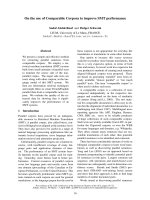
Fig2. The hierarchical organization of the tendon structure from collagen fibrils (20nm-
150 nm) to collagen fiber (1 µm -50 µm), fascicle and the entire tendon.( Adapted from
Josza 1997)
2.1.2 Tendon Composite
Tendons have relatively few cells but a large amount of extracellular matrixes that give
them extremely high tensile strength. The extracellular tendon matrix is composed of
collagen and elastin fibers, the ground substance, and the inorganic components. Collagen
constitutes approximately 90% of the total protein of the tendons or 65-75% of the dry
mass of tendons. Elastin accounts for only about 2 % of the dry mass of tendons (Hess,
1989, Jozsa 1989b). The ground substance, which surrounds the collagen, consists of
proteoglycan, glycosaminoglycans(GAGs), structural glycoproteins and a wide variety of
other small molecules. These elements are produced by tenoblasts and tenocytes that are
the elongated fibroblasts and fibrocytes lying between the collagen fibers.
Chapter II. literature review 9
All collagen molecules are long stiff rods consisting of a triple helix of three polypeptide
chain. To date, 13 different types of collagen molecules have been found in the mammalian
body ( Karpakka 1991) The 13 types are named by roman numbers. The relative proportion
of each type of collagen in each tissue is characteristic and specific. In tendon and
ligament, type I collagen predominates, but small amounts of other types of collagen are
also found (see table 1).
Table 1. The chemical composition, amount, and location of the different collagen types in
tendon.
Collagen
type
Molecular
formula
Molecular
Weight(Dalto
n)
Amount in
tendon ( %)
Location in tendon
I [1( I )]2 2 95.000 97-98 Collagen fibers,
endotenon, epitenon
II [1( II)]3 95.000 0.2-0.8 Cartilage Zone of
tendon to bone
insertion
III [1( III )]3 95.000 1.0-1.5 Endotenon, epitenon
,vascular walls,
Muscle tendon
junction
IV [pro-1( IV )]3 180.000 < 0.2 Vascular basement
membrane, Muscle
tendon junction
V [A1( B )2] 300.000 < 0.2 Vascular walls,
Muscle tendon
junction
Type I collagen has predilection to form parallel fibers. One collagen type I molecule
consists of three helical polypeptide alpha-chains that wind around each other to form a
right-hand triple helix that extends collinear through out the length of the tropocollagen
molecule or microfibril (Ghadially 1988). A number of tropocollagen molecules or
microfibrils form the collagen fibril. The diameter of a native collagen fibril is usually
Chapter II. literature review 10
between 20 nm and 150 nm (Demel et al 1982). Within a fibril, the microfibrils are
surrounded by proteoglycans and GAGs. A bunch of collagen fibrils then form a collagen
fiber whose diameter may range between 1 and 50 um. Within a fiber, proteoglycan and
GAGs surround the fibrils. So each unit in the collagen fiber structure is surrounded by
proteoglycan and GAGs. Type I collagen has high tensile strength with limited elasticity
and is therefore suitable for force transmission. Other collagen types constitute only a small
part of tendinous collagen. Their roles are not entirely clear. With the advancement in basic
science of collagen biochemistry, the present concepts of collagen types and their functions
may well be changed.
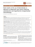
Fig 3. Schematic figure of a mature type I collagen fibril (Adapted from Ghadially, 1998)
Elastic fibers are barely present in human tendons (Carlstedt, 1987). The function of
elastic fibers is not entirely clear, but they may contribute to the recovery of the wavy
configuration of the collagen fibers after tendinous stretch (Buter, 1978). Proteoglycans are
composites of a protein core in which one or more glycosaminoglycan (GAG) are
Chapter II. literature review 11
covalently attached. They are large (WM 10
6
daltons), negatively charged hydrophilic
molecules that can retain water 50 times their weight. By virtue of their high fixed charge
density and charge-to- charge repulsion force, proteoglycans are stiffly extended to provide
the collagen fibrils with high capacity to resist high compressive and tensile forces
(Karpakka, 1991). In addition, the proteoglycans enable rapid diffusion of water soluble
molecules and migration of cells. In tendon, a wide variety of inorganic components have
been detected(Lappalainen 1982). Calcium is found in the highest concentrations, with
concentration of 0.001% to 0.01% of tendon dry weight in the tensional area of a normal
tendon( Josza 1989b). Other detected components have been magnesium, manganese,
cadmium, cobalt, copper, zinc, nickel, lithium, lead, fluoride, phosphor, and silicon(
Spadaro 1970, Strehlow 1969).
The organization of the extracellular matrix molecules of the tendon is the principal
determinant of the physical function of this tissue. The degree to which tissue engineering
approaches are successful will depend on the degree to which the normal composition and
architecture of the extracellular matrix is restored.
Apart from the extensive work on the biology of the extracellullar components of
tendons, there are also many studies that focused on understanding the biology of tendon
cells. Tendons have a variety of cell types, including fibroblasts, fibrocartilage cells and
occasional fat cells. Fibroblasts are the most common and can be found in all regions of
tendons and ligaments. They are typically arranged in elongated rows within the parallel
bundles of collagen fibers. In longitudinal section, the cells are elongated and have spindle
shape nuclei. In addition to the responsibility of producing extracellular matrix, fibroblasts
also mediate the tendons response’ to biomechanical load.