Development of mediatorless glucose sensing strategies for blood glucose monitoring in diabetes
Bạn đang xem bản rút gọn của tài liệu. Xem và tải ngay bản đầy đủ của tài liệu tại đây (5.39 MB, 145 trang )
DEVELOPMENT OF MEDIATORLESS GLUCOSE
SENSING STRATEGIES FOR BLOOD GLUCOSE
MONITORING IN DIABETES
ZHENG DAN
(M.Eng., South China University of Technology, China)
A THESIS SUBMITTED
FOR THE DEGREE OF DOCTOR of PHILOSOPHY
DEPARTMENT OF CHEMISTRY
NATIONAL UNIVERSITY OF SINGAPORE
2014
Declaration
I herebydeclarethat this thesisis my original work and it hasbeenwritten by
me in its entirety, under the supervision of SHEU Fwu-Shan, (in the
laboratoryBiolab at T-Lab), NUSNNI-Nanocore,National University of
Singapore,
between2010Januaryatd20l3 December.
I haveduly acknowledged
all the sourcesof informationwhich havebeenused
in thethesis.
This thesis has also not been submittedfor any degreein any university
previously.
The contentof the thesishasbeenpartlypublishedin:
1) D. Zheng,S.K. Vashist,M.M. Dykas, S. Saha,K. Al-Rubeaan,E. Lam,
J.H.T. Luong, F.-S. Sheu, Graphene versus Multi-Walled Carbon
Nanotubesfor ElectrochemicalGlucoseBiosensing,Materials,2013, 6,
r0tl-1027.
2) D. Zheng, S.K. Vashist, K. Al-Rubeaan,E. Lam, S, Hrapovic, J.H.T.
Luong, F.-S. Sheu, Effect of 3-Aminopropyltriethoxysilaneon the
Electrocatalysis
of CarbonNanotubesfor Reagentless
GlucoseBiosensing,
Journalof Nanopharmaceutics
andDrugDelivery,2013,l,1,64-73.
i) O. Zheng, S.K. Vashist, K. Al-Rubeaan,J.H.T. Luong, F.-S. Sheu,
Mediatorless amperometric glucose biosensing using
3aminopropyltriethoxysilane-functionalized
graphene,Talanta 2012, 99,
22-28.
4) D. Zheng,S.K. Vashist,K. Al-Rubeaan,J.H.T. Luong, F.-S. Sheu,Rapid
and simplepreparationof a reagentless
glucoseelectrochemical
biosensor,
Analyst,2012,137,3800-3805.
5) S.K. Vashist, D. Zheng, K. Al-Rubeaan,J.H.T. Luong, F.-S. Sheu,
Technologybehind commercialdevicesfor blood glucosemonitoring in
. diabetesmanagement:
A review,AnalyticaChimica Acta,20ll, 703, 124136.
ZhengDan
Name
An-"] - zot+
Date
Acknowledgements
First and foremost, I would like to thank my supervisor Prof. Sheu Fwu
Shan for his continuous support and guidance in my Ph.D. study and research.
His patience, motivation, and immense knowledge in science have been
inspiring me during my study. I could not have my thesis completed without
his help and advice.
I am grateful to Prof. Loh Kian Ping for he was willing to be my cosupervisor so that I could pursue my Ph.D. in Department of Chemistry. I am
also deeply influenced by his energy and enthusiasm in science and research.
My most sincere gratitude also goes to Dr. Vashist Sandeep, the former
post-doctoral in Nanocore Laboratory. He has helped me in many ways and
has molded me to be a better researcher. I am truly blessed to have such a
collaborator during the first two years of my Ph.D.
I also would like to thank Prof. Luong John from National Research
Council Canada, who has provided enthusiastic assistance in guiding me
electrochemical experiments and revising manuscripts during my Ph.D.
I am grateful to Prof. Venkatesan Venky for his support and help in my
doctoral study. My gratitude also extends to Dr. Noort Danny Van, Dr. Saha
Surajit and Dykas Michal for their assistance and advice on the scanning
electron microscopy, Raman spectroscopy and helium ion microscopy.
I would like to thank all my colleagues working in NUSNNI-Nanocore for
their help during my time in Nanocore Laboratory.
Also, I owe sincere and earnest thankfulness to my friends in NUSCCF and
HCMC who have been generously providing me help whenever I needed them.
Last but not least, I am truly indebted to my husband Sha Zhou, my parents
Mr. Zheng Jianping and Mrs. Chen Suhua who have been ever supportive
throughout the course of my Ph.D. To them I dedicate this thesis.
i
Table of Contents
Acknowledgements………………………………………………………..….i
Table of Contents…………………………………….…………………....…ii
Summary………………………………………………………………..…....vi
List of Tables…………………………………………………………..…...ix
List of Figures………………………………………………………..............x
List of Symbols……………………………………………………….........xiv
List of Abbreviations…………………………….………………………xv
List of Publications…………………………………………………….......xvii
Chapter 1 Introduction……………………………………………………....1
1.1 Traditional blood glucose monitoring in diabetes: overview……………...3
1.1.1 Methods used for glucose detection in BGM: electrochemistry versus
other methods………………………………………………………………3
1.1.2 Enzymatic versus non-enzymatic glucose detection……………......8
1.1.2.1 Enzymes used in BGMDs………………………………….......10
1.1.3 Mediator-based glucose detection………………………………......12
1.2 Mediatorless glucose sensing strategies: literature review……………..14
1.2.1 Nanomaterial-based glucose biosensors…………………………...14
1.2.1.1 CNT-based glucose biosensors…………………………….......14
1.2.1.2 Graphene-based glucose biosensors……………………….......17
1.2.1.3 Glucose biosensors based on other types of nanomaterials…....19
1.2.2 Glucose biosensors developed without using nanomaterials…….....22
1.3 The mechanisms of glucose detection by mediatorless glucose biosensors
………………………………………………………………………………24
1.4 Objectives and significance of the study………………………………...26
ii
1.4.1 Research gaps of the study……………………………………….....26
1.4.2 Aim and objectives of the study…………………………………..27
1.4.3 Significance and scope of the study……………………………....28
1.4.4 Overview of the thesis…………………………………………….29
Chapter 2 Experimental…………………………………………..………..30
2.1 Electrochemical analysis……………………………………..………….30
2.1.1 Cyclic voltammetry……………………………………..………….30
2.1.2 Amperometry…………………………………………..…………...31
2.1.2.1 Detection of glucose and blood glucose……………………...31
2.1.2.2 Effect of interfering substances on glucose detection………..32
2.1.2.3 Production reproducibility of glucose sensing strategies…….32
2.1.2.4 Stability of glucose biosensors stored under various conditions
………………………………………………………………………33
2.1.2.5 Continuous glucose monitoring…………………………..…...33
2.1.2.6 Effect of biofouling on glucose detection…………………......33
2.2 Bicinchoninic acid protein assay………………………………….....33
2.3 Helium ion microscopy……………………………………….……...35
2.4 Scanning electron microscopy………………………………..…........35
2.5 Energy-dispersive X-ray spectroscopy………………………..……...36
2.6 Raman spectroscopy…………………………………………..……...36
2.7 Infrared spectroscopy…………………………………………..…….37
2.8 Chemicals and materials………………………………………..…….37
Chapter 3 Effect of 3-aminopropyltriethoxysilane on the electrocatalysis
of carbon nanotubes for mediatorless glucose biosensing…………..........39
3.1 Introduction……………………………………………………………..39
iii
3.2 Preparation of various glucose biosensing formats……………………...41
3.3 Results and discussion…………………………………………………...42
3.3.1 Development and characterization of various glucose biosensing
formats ……………………….…………………………………….….…42
3.3.2 Effect of APTES on electrochemical glucose biosensing………......47
3.3.3 Analytical performance of the MWCNT (dispersed in DMF)
format …………………………………………………..............................50
3.4 Conclusions……………………………………………………………..53
Chapter 4 Mediatorless amperometric glucose biosensing using 3aminopropyltriethoxysilane-functionalized graphene……………….…..54
4.1 Introduction………………………………………………………….…..54
4.2 Preparation of graphene-based glucose biosensor……………………….56
4.3 Results and discussion……………………………………………….…..56
4.3.1 Development of graphene-based glucose biosensor……………......56
4.3.2 Detection of glucose and blood glucose……………………….…...59
4.3.3 Effect of interfering substances………………………………….….68
4.3.4 Analytical performance of the graphene-based glucose biosensor....69
4.4 Conclusions……………………………………………………………...71
Chapter 5 Rapid and simple preparation of a mediatorless glucose
electrochemical biosensor…………………………..................................72
5.1 Introduction……………………………………………………………..72
5.2 Preparation of the simple and rapid glucose biosensor………………….74
5.3 Results and discussion…………………………………………………..74
5.3.1 Development of the simple and rapid glucose biosensor…...….....74
5.3.2 Detection of glucose and blood glucose ………………………..….76
iv
5.3.3 Effect of interfering substances on glucose sensing …….…..…...77
5.3.4 Biosensor performance of the simple and rapid glucose sensing
strategy…………………………………………………………………….79
5.4 Conclusions……………………………………………………………..81
Chapter 6 Graphene versus multi-walled carbon nanotubes for
electrochemical glucose biosensing……………………………….….….....83
6.1 Introduction………………………………………………………….…...83
6.2 Preparation of graphene- and MWCNT-based glucose biosensors…….85
6.3 Results and discussion……………………………………………….…...85
6.3.1 Dev elopm ent of graphene- and M WCNT-bas ed glucos e
biosensors……………………………………………………………….85
6.3.2 Evaluation of direct electron transfer…………………………...….90
6.3.3 Evaluation of glucose oxidation ….…………………………….....94
6.3.4 Amperometric detection of commercial and blood glucose……....96
6.3.5 Effect of interfering substances…………………………………...100
6.4 Conclusions…………………………………………………………….101
Chapter 7 Conclusions and Recommendations………………….……...103
Reference…………………………………………………………………...108
v
Summary
Tremendous efforts have been made in developing mediatorless glucose
biosensors because of the potential hazards of mediator-based glucose sensing
methods. However, most of these studies which employed tedious and lengthy
preparation procedures, failed to detect the entire pathophysiological glucose
range, or lacked systematic analysis of sensor performance. Therefore, the aim
of this study was to develop simple, cost-effective and advanced strategies for
constructing mediatorless electrochemical glucose biosensors based on the
usage of 3-aminopropyltriethoxysilane (APTES), glucose oxidase (GOx) and
carbon-based nanomaterials (carbon nanotubes or graphene). In addition, the
developed glucose biosensors could precisely detect glucose in the diabetic
pathophysiological range of 1-30 mM and would be free from interference.
In the first experiment, the concentration effect of APTES on the
electrocatalysis of three mediatorless glucose sensing formats (with and
without using multi-walled carbon nanotubes (MWCNTs)) was studied. It was
indicated that the concentration of APTES considerably affected the glucose
sensing results of the three formats in different patterns. This study provided a
guided insight into the optimization of APTES-based chemistry applied in
electrochemical glucose biosensor.
In the second experiment, a graphene-based mediatorless glucose biosensor
was constructed by covalent binding GOx to an APTES-graphene
functionalized glassy carbon electrode (GCE). This biosensor was able to
detect 1-30 mM glucose at -0.45 V (vs. Ag/AgCl) and its anti-interference
capability was also demonstrated. This strategy was the first to apply APTES
in dispersing and functionalizing graphene for the preparation of mediatorless
vi
glucose biosensor. Furthermore, the excellent production reproducibility of
this strategy may be beneficial for the mass production of glucose biosensor.
In the third experiment, a rapid and highly simplified strategy for the
immobilization of GOx on GCE surface in a leach-proof pattern was proposed.
Besides its superior performance on glucose sensing, the constructed biosensor
was able to preserve its initial activity for at least 4 weeks when stored at room
temperature in dry state. Additionally, this strategy was the most rapid method
to prepare a robust and stable glucose biosensor compared to the reported
methods so far.
In the last experiment, MWCNT- and graphene-based glucose biosensors
were prepared and the glucose sensing performance of MWCNTs and
graphene was compared for the first time. The cyclic voltammogram showed
that the direct electron transfer between GOx and GCE surface was only
observed on the MWCNT-based biosensor, which may be attributed to
shortened tunneling distance facilitated by the unique structure of MWCNTs.
The results of this experiment suggested that graphene might not be more
advanced than CNTs in developing biosensors.
In conclusion, this study proposes several highly convenient and stable
mediatorless electrochemical glucose biosensing strategies for blood glucose
monitoring. Some of the strategies are proved to be suitable for continuous
glucose monitoring owing to their high stability and excellent anti-biofouling
capability. This study may be practically beneficial to the fabrication of
various mediatorless biosensors for determining analytes of interest. Moreover,
the systematic investigation of sensor performance in this study should
vii
provide valuable guidelines for the development of non-invasive glucose
sensor.
viii
List of Tables
Table 1-1. Advantages and limitations/challenges of diverse techniques used
in glucose sensing…………………................................................................5
Table 3-1. Elemental analyses (weight %) of different electrodes………...42
Table 4-1. Comparison of our graphene based electrochemical glucose
biosensing method with other published graphene based formats………….64
Table 5-1. Stability of the developed Nafion/APTES-GOx/GCEs stored under
various conditions………………………………………..……….….…...80
Table 6-1. Detailed comparison between this work and recently reported
graphene- and CNT-based glucose biosensors.…………………………....98
Table 6-2. The effect of therapeutic levels of interfering substances on the
graphene- and MWCNT-base biosensors…………………………………101
ix
List of Figures
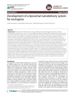
Fig. 1-1. Number of articles published in the past few decades pertaining to
blood glucose monitoring. Data was taken on Nov 21, 2012
fromwww.scopus.com using “blood glucosemonitoring” in the advanced
search option………………..………………………………………………..2
Fig. 2-1. Schematic diagram of the electrochemical system used for the
electroanalytical experiments in this research. WE: working electrode; RE:
reference electrode; CE: counter electrode. The electrodes and the gas tube
were inserted through holes in the cell cover……………..………………….31
Fig. 2-2. The formation of purple complex with BCA and cuprous ion…..…35
Fig. 3-1. Schematic of various APTES based electrochemical glucose
biosensing formats. The notations 1, 2, and 3 refer to direct GOx, MWCNT
(dispersed in DMF), and MWCNT (dispersed in APTES) based formats…40
Fig. 3-2. ATR-FTIR spectra of different reaction intermediates deposited on
the GCE surface…………………….……………………………..………....43
Fig. 3-3. Pictures of MWCNTs dispersed in (A) water and (B) APTES
(0.125%), and the SEM images of (C) MWCNT (dispersed in DMF)-, and (D)
MWCNT (dispersed in APTES)-functionalized glassy carbon………...……45
Fig. 3-4. (A) CVs of Nafion/APTES(2%)/GCE (dash-dot-dot), direct GOx
(dash), MWCNT (dispersed in DMF) (solid), and MWCNT (dispersed in
APTES) (dot) formats in 50 mM PBS at 100 mV s-1 in the presence of
nitrogen. (B) CVs of 0 mM (dash) and 4 mM (solid) glucose detected on
MWCNT (dispersed in DMF) format. (C) Amperometric curves of varying
concentrations of glucose on the MWCNT (dispersed in DMF) format in 50
mM PBS at -0.45 V vs. Ag/AgCl………………….........................................46
Fig. 3-5. Effect of varying APTES concentrations on the performance of three
different glucose biosensing formats: (A) Direct GOx; (B) MWCNT
(dispersed in DMF); and (C) MWCNT (dispersed in APTES) formats. (D)
Overlay plot of various formats based on the optimized APTES concentration
for a particular format. The error bars shown in (A, B, C, D) represent
standard deviation (SD)…………………………………………………...….47
Fig. 3-6. Use of MWCNT (dispersed in DMF) based format for
electrochemical glucose sensing. (A) Detection of Streck blood glucose
linearity standards. The steady current Isteady/µA (ordinate) is presented as a
function of the log scale of glucose concentration Log[Gluc]/mM (abscissa).
(B) The effect of physiological interferences and medications on the specific
detection of glucose. The error bars shown in (A, B) represent SD.................50
Fig. 3-7. The bioanalytical performance of the MWCNT (dispersed in DMF)
glucose sensing format. (A) Production reproducibility on 25 GCEs. (B) BCA
x
protein assay for the determination of GOx binding on electrode for 9 weeks.
(C) Stability of the electrode that was stored at RT in dry state for 5 weeks. (D)
Continuous monitoring of 4 mM glucose for 150 times using the same
electrode. The error bars shown in (A, B, C) represents SD............................52
Fig. 4-1. Schematic diagram of the graphene based glucose biosensor…....55
Fig. 4-2. FTIR spectra of (a) pH 9 APTES-graphene, (b) APTES-graphene and
(c) pristine graphene in the region of (A) 4000-400, (B) 3100-2800 and (C)
1800-400 cm-1 ……………………………………………….....................57
Fig. 4-3. Graphene dispersed in (A) APTES, and (B) water. (C) SEM image of
graphene-functionalized glassy carbon substrate (the inlet is the SEM image of
blank glassy carbon)………………………….……………………...............59
Fig. 4-4. (A) (a) C Vs of Nafion/graphene-APTES/GCE and (b)
Nafion/GOx/graphene-APTES/GCE in N2-saturated 50 mM PBS. (B) CVs of
Nafion/GOx/graphene-APTES/GCE in 0, 1 and 4 mM glucose solutions in the
presence of oxygen. Scan rate: 100 mV s -1…………………………….60
Fig. 4-5. (A) Optimization of applied potential for the electrochemical
detection of 4 mM glucose using Nafion/GOx/graphene-APTES/GCE. (B)
Amperometric detection of 1-32 mM glucose at -0.45 V in the presence of
oxygen. The error bars shown in (A) represent SD ………………….………61
Fig. 4-6. (A) Effect of APTES concentration on the electrochemical glucose
sensing by the graphene-based glucose biosensor. (B) Comparison of
electrochemical glucose sensing by Nafion/GOx/graphene-APTES/GCE and
Nafion/GOx/GCE. (C) Detection of Sugar-Chex whole blood glucose linearity
standards by the Nafion/GOx/graphene-APTES/GCE. The error bars shown in
(A, B, C) represent SD …………………………………………………….62
Fig. 4-7. Effect of interfering substances on the specific detection of 6.8 mM
blood glucose by the developed glucose biosensor. The error bars represent
SD……..………………………………………………………...…………....68
Fig. 4-8. (A) Production reproducibility of the graphene-based glucose
biosensing procedure. (B) Determination for stability of
Nafion/GOx/graphene-APTES/GCEs that were stored at RT in dry. (C) BCA
protein assay for the determination of GOx binding on electrodes for 9 weeks.
(D) Determination of the effect of biofouling. The error bars shown in (A, B,
C, D) represent SD …………………………………………………………..69
Fig. 5-1. Schematic diagram for the preparation of a simple and rapid GOxbound GCE……………………………………………...…………………....74
Fig. 5-2. The (A) cyclic voltammograms and (B) amperometric response of
the developed glucose biosensor for varying glucose concentrations (0.5-32
mM) in 50 mM PBS in the presence of oxygen. The scan rate in (A) was 100
mV s-1, while the applied potential in (B) was -0.45 V……………….……...76
xi
Fig. 5-3. Detection of (A) commercial glucose, and (B) Sugar-Chex blood
glucose linearity standards by the simple and rapid glucose biosensor. The
error bars shown in (A, B) represent SD …………….………………………77
Fig. 5-4. The effect of interfering substances on the detection of blood glucose
by the glucose biosensor. The error bars represent SD…………………........78
Fig. 5-5. (A) Production reproducibility of the developed simplified procedure
for the preparation of Nafion/APTES-GOx/GCEs. (B) Effect of biofouling on
the electrochemical sensing of glucose by the developed glucose biosensor. (C)
BCA protein assay for the determination of GOx bound to the Nafion/APTESGOx/GCEs for 12 weeks. The error bars shown in (A, B, C) represent
SD..………………………………………………………………………..….80
Fig. 6-1. The preparation of graphene- and MWCNT-based glucose
biosensors…………………………………………………………………….84
Fig. 6-2. High resolution images of (A) graphene-GOx, (B) Nafion/grapheneGOx, (C) MWCNT-GOx and (D) Nafion/MWCNT-GOx modified glassy
carbon substrates using HIM. The scale bars for (A)/(B) and (C)/(D) are 10
μm and 200 nm, respectively.…………...………….…………………..........86
Fig. 6-3. Raman spectra of (a) APTES-functionalized and (b) pristine
graphene. The Raman shift range is 1000~3500 cm-1, 1300~1650 cm-1, and
2300~3500 cm-1 for (A), (B), and (C)………………………………….…….87
Fig. 6-4. Raman spectra of (a) APTES-functionalized and (b) pristine
MWCNTs. The Raman shift range is 1000~3500 cm-1, 1300~1650 cm-1, and
2300~3500 cm-1 for (A), (B), and (C)….…………………………………….87
Fig. 6-5. The FTIR spectrum of (a) the KOH-treated GCE modified with
APTES and MWCNTs dispersed in DMF; (b) the KOH-treated GCE modified
with MWCNTs dispersed in APTES; (c) same as (b) followed by conjugation
with GOx……………………………………………………………………89
Fig. 6-6. The FTIR spectrum of (a) the KOH-treated GCE modified with
“layered by layered” APTES and graphene dispersed in DMF; (b) the KOHtreated GCE modified with graphene dispersed in APTES; and (c) same as (b)
followed by conjugation with GOx……………….………………………….90
Fig. 6-7. CVs of (A) graphene- and (C) MWCNT-functionalized GCE at
different scan rate in 5 mM Fe(CN)63-. Scan rate: 20-200 mV s-1. (B) and (D):
Linear relation between I p and v 1/2 (B: graphene-GCE; D: MWCNTGCE)…………………………………………………………………….……91
Fig. 6-8. (A) CVs of various modified electrodes in N2-saturated PBS at 100
mV s−1; (B) The effect of scan rate (20, 50, 100, 150 and 200 mV s−1) on the
DET of GOx on Nafion/MWCNT-GOx/GCE in N2-saturated PBS. Inlet: the
linear relation between Ipc (or Ipa) and v; (C) The relation between the E0
xii
(observed on Nafion/MWCNT-GOx/GCE) and different pH values: 5.65, 6.36,
7.2, 7.72, 8.29. v = 100 mV s−1; (D) Plot of Ep (of the Nafion/MWCNTGOx/GCE) vs. log v, v = 0.1, 0.2, 0.3, 0.4, 0.5, 0.7, 0.9, 1.1, 1.3, 1.5, 1.7, 1.9
V s−1. Inlet: the relation between Epa (or Epc) and log v……………………93
Fig. 6-9. CVs of (A, C) Nafion/graphene-GOx/GCE and (B, D)
Nafion/MWCNTs-GOx/GCE in PBS containing 0, 1 and8mM glucose in the
presence of (A, B) nitrogen and (C, D) oxygen. Scan rate: 100 mV s-1……95
Fig. 6-10. (A) The amperometric response of Nafion/MWCNT-GOx/GCE for
the detection of 0.5 to 32 mM glucose at −0.45 V in the presence of O2; (B)
Assay curves for the detection of commercial glucose by the graphene- and
MWCNT-based electrodes; (C) Assay curves for the detection of Sugar-Chex
whole blood glucose linearity standards by both electrodes; (D) The effect of
interfering substances on the electrochemical detection of 6.8 mM blood
glucose standard by both electrodes. The error bars shown in (B, C, D)
represent SD…..……………………………………………………………...97
xiii
List of Symbols
A
effective area
α
charge transfer coefficient
C
concentration
CO
bulk concentration of oxidizing agent
DO
diffusion coefficient of oxidizing agent
E
potential
Ep
peak potential
ΔEp
potential difference
Epa
anodic peak potential
Epc
cathodic peak potential
E0
formal potential
I
current
Ip
current response, peak current
Ipa
anodic peak current
Ipc
cathodic peak current
Ipmax
maximum current response
Isteady
steady current
Kd
dissociation constant
ks
heterogeneous transfer rate constant
n
number of electrons transferred
v
scan rate
xiv
List of Abbreviations
APTES
3-aminopropyltriethoxysilane
ATR-FTIR
attenuated total reflectance fourier transform infrared
BCA
bicinchoninic acid
BGM
blood glucose monitoring
BGMD
blood glucose monitoring device
CGMS
continuous glucose monitoring systems
CNTs
carbon nanotubes
CV
cyclic voltammetry
DET
direct electron transfer
DMF
dimethylformamide
DPV
differential pulse voltammetry
EDC
1-ethyl-3-[3-dimethylaminopropyl]carbodiimide
hydrochloride
EDX
energy-dispersive X-ray spectroscopy
FAD
flavin adenine dinucleotide
GCE
glassy carbon electrode
GDH
glucose dehydrogenase
GOx
glucose oxidase
HIM
helium ion microscope
HPLC
high performance liquid chromatography
IL
ionic liquid
IR
infrared
LbL
layer-by-layer
MES
2-(N-morpholino)ethanesulfonic acid
xv
MWCNT
multi-walled carbon nanotube
NAD
nicotinamide adenine dinucleotide
NMR
nuclear magnetic resonance
NPs
metal nanoparticles
PBS
phosphate buffer solution
PQQ
pyrroloquinoline quinone
RT
room temperature
SEM
scanning electron microscopy
SD
standard deviation
SHE
standard hydrogen electrode
SWCNTs
single-walled carbon nanotubes
vs.
versus
xvi
List of Publications
1. D. Zheng, S.K. Vashist, M.M. Dykas, S. Saha, K. Al-Rubeaan, E.
Lam, J.H.T. Luong, F.-S. Sheu, Graphene versus Multi-Walled Carbon
Nanotubes for Electrochemical Glucose Biosensing, Materials, 2013,
6, 1011-1027.
2. D. Zheng, S.K. Vashist, K. Al-Rubeaan, E. Lam, S. Hrapovic, J.H.T.
Luong, F.-S. Sheu, Effect of 3-Aminopropyltriethoxysilane on the
Electrocatalysis of Carbon Nanotubes for Reagentless Glucose
Biosensing, Journal of Nanopharmaceutics and Drug Delivery, 2013,
1, 1, 64-73.
3. D. Zheng, S.K. Vashist, K. Al-Rubeaan, J.H.T. Luong, F.-S. Sheu,
Rapid and simple preparation of a reagentless glucose electrochemical
biosensor, Analyst, 2012, 137, 3800-3805.
4. D. Zheng, S.K. Vashist, K. Al-Rubeaan, J.H.T. Luong, F.-S. Sheu,
Mediatorlessamperometricglucosebiosensingusing3aminopropyltriethoxysilane-functionalizedgraphene, Talanta, 2012,
99, 22-28.
5. S.K. Vashist*, D. Zheng*, K. Al-Rubeaan, J.H.T. Luong, F.-S. Sheu,
Technology behind commercial devices for blood glucose monitoring
in diabetesmanagement: A review, Analytica Chimica Acta, 2011, 703,
124-136.
*These authors contributed equally
PCT patent application
1. Sandeep Kumar VASHIST, Dan ZHENG, Fwu-Shan SHEU, Khalid
Ali AL-RUBEAAN; Title: A Mediator-less Electrochemical Glucose
Sensing Procedure Employing the Leach-proof Covalent Binding of
Enzyme to the Electrodes and Products Thereof; International
Publication Number: WO 2013/165318 A1.
xvii
Chapter 1 Introduction
The human body naturally tightly regulates blood glucose levels within the
normal range of 4-8 mM. Nevertheless, a persistently high level above the
normal range is referred to as diabetes mellitus, which is a disease related to
failure of blood sugar regulation. Additionally, diabetes is incurable but
manageable. Therefore, maintaining the blood glucose level within the normal
range is essential for the healthcare of diabetics by avoiding diabetesassociated complications such as kidney damage, blindness, neuropathy,
adverse effect on circulatory system, amputations, etc. Indeed, diabetes has
been declared as a global epidemic by World Health Organization owing to its
unprecedented increase worldwide. Presently, there are about 285 million
diabetics, while the number is anticipated to multiply by 1.54 folds by the end
of 2030 [1]. Also, there are about 3.96 million deaths per year caused by
diabetes, which is about 6.8% of the mortality in the age group of 20-79 years
[2]. Moreover, diabetes monitoring and management is a heavy financial
burden for the society as approximately 11.6% of the total global healthcare
expenditure in 2010 (estimated to be US$ 376 billion) was spent on diabetes.
It is expected that the spending on treating and preventing diabetes and
associated complications will increase to US$ 490 billion by 2030 [3].
As mentioned, regular blood glucose monitoring (BGM) is a key
requirement for diabetics. Actually, self-glucose monitoring should be used in
patients on intensive insulin therapy at least three times daily. About 10 billion
glucose assays are performed worldwide yearly. Therefore, there have been
continuously increasing research efforts in the area of BGM as shown by the
tremendous increase in the number of articles published in the previous
1
decades (Fig. 1-1). Notably, the market of blood glucose monitoring device
(BGMD) accounts for about 85% of the total biosensors market [4]. Such a
huge market is monopolized by a few diagnostic companies such as Abbott,
Roche Diagnostics, Bayer, Minimed and LifeScan [5]. Despite their various
features, most commercial BGMDs measure blood glucose concentrations
based on the electrochemical redox reaction occurring between the enzyme
and the substrate (i.e. glucose). More specifically, artificial electron mediator
is employed in all of the commercial products.
Fig. 1-1. Number of articles published in the past few decades pertaining to
blood glucose monitoring. Data was taken on Nov 21, 2012
fromwww.scopus.com using “blood glucose monitoring” in the advanced
search option.
However, there are several problems in the mediator-based devices such as
the toxicity [4] and leakage of the mediator [6]. To overcome these challenges
is especially important to the development of implantable devices. In addition,
the active mediator can easily interact with some of the interfering substances
coexisting in the blood even at a low potential applied to detect glucose [4].
As a result, the safety, accuracy and reliability of blood glucose measurement
2
are badly compromised. Therefore, a mediatorless glucose sensing strategy
that can selectively detect glucose without being interfered by the electroactive
substances will be an ideal solution to overcome the shortcomings of the
traditional mediator-based devices. Moreover, the development of accurate,
cost-effective, and safe mediatorless glucose sensing strategies is of great
economic importance. During the past decade, mediatorless glucose
biosensors have been extensively developed based on the utilization of carbonbased nanomaterial (e.g., carbon nanotubes (CNTs) and graphene), metal
nanoparticles (NPs) and other materials. The subsequent sections provide a
brief overview of the mediator-based devices and more attention will be given
to the latest development of mediatorless-based glucose sensing strategies.
1.1 Traditional blood glucose monitoring in diabetes: overview
In this section, some of the essential techniques behind traditional BGMDs
are introduced. These techniques include the glucose determination methods,
enzymes used in BGMDs and the glucose sensing principle of mediator-based
devices. The limitations of mediator-based devices are also discussed.
However, the other aspects of the techniques used in BGMDs (such as glucose
limiting membrane, test strip design, etc.) are not covered in this section since
they are not the research emphasis in this thesis. The comprehensive
discussion of the techniques applied in commercial BGMDs can be found in
many review articles [5, 7-9].
1.1.1
Methods used for glucose detection in BGM: electrochemistry
versus other methods
A wide range of diversified analytical methods such as high performance
liquid chromatography (HPLC) [10], fluorescence [11-14], infrared (IR) [15],
3
UV-visible (photometry) [16-19] and nuclear magnetic resonance (NMR)
spectroscopy [20] have been reported for glucose monitoring. Among these
methods, HPLC, fluorescence and UV-visible spectroscopy can provide
glucose quantification information with different sensitivities, while IR and
NMR spectroscopy are normally used for offering structural information of
glucose (especially when glucose is mixed with other types of sugars) rather
than detecting its concentration. The advantages and limitations (or challenges)
of these techniques in glucose sensing are listed in Table 1-1.
Regarding the glucose sensing in commercial BGMDs, two methods are
mainly employed, which are photometry and electrochemistry. Indeed
photometric glucose sensors preceded the electrochemical sensors. The
principle of a photometric glucose sensor is based on the enzymaticallycatalyzed electron transfer from glucose to light-absorbing dye molecules and
the subsequent reflectance measurement [8]. Nevertheless, since reflectance
measurement request the exclusion of red blood cells and large assayed area of
at least a few square millimeters [8], electrochemical glucose sensors are
prevailingly used due to their ability to overcome the shortcomings of
photometric devices. Furthermore, compared to the devices based on the
above methodologies, electrochemical glucose sensors are preferable as they
are rapid, easy to be simplified and highly cost-effective.
4
Table 1-1. Advantages and limitations/challenges of diverse techniques used in glucose sensing
Technique used in
glucose sensing
HPLC
Advantages
Limitations/Challenges
1. It is easy to separate sugar mixtures and relatively free
from interference. 2. It is accurate for quantification.
1. It can be quite costly to use HPLC in glucose
sensing (e.g. filtration of samples). 2. It is very time
consuming in analyzing samples with HPLC. 3. The
sensitivity of glucose sensing considerably depends
on the types of column and HPLC used.
Fluorescence
spectroscopy
1. It is a powerful technique for fast, mediatorless and
noninvasive glucose sensing. [14] 2. It is highly sensitive for
detection of low glucose concentrations in body fluid
samples such as tears [11].
1. Active study is particularly needed in the area of
exploring potential interferents, and designing sensor
with wide detection range, high signal response,
reproducibility and stability, extended lifetime,
satisfactory accuracy and excellent biocompatibility.
2. Besides, research efforts are also required to
overcome the interference problems due to the
“optical window” of the skin, to develop fluorescent
transdermal glucose monitoring. [12, 14]
IR spectroscopy
1. It offers fast and reliable result. 2. The continuing
improvements in hardware and software design make it a
promising routine method during industrial process control
[15].
This technique is mainly used for quality control of
glucose but not quantification [15].
UV-visible
It is simple, selective and sensitive for glucose quantification 1. The detection range of glucose is normally below 1
5
spectroscopy
(Photometry)
[16-19].
mM, which requires dilution of sample if its glucose
concentration is above the detection range. 2.
Biological samples may need filtration before
detection. 3. Large assayed area is needed.
NMR spectroscopy
It is a great characterization tool for identification of
metabolites of glucose metabolism [20].
Electrochemistry
1. Accuracy of blood glucose reading can be delayed
at alternate sites during times of rapidly changing
blood glucose, making it more difficult to identify
hyperglycemia or hypoglycemia. 2. Most
electrochemical BGMDs based on invasive test. 3.
1. It is rapid, convenient and highly cost-effective compared
Although minimally invasive and non-invasive
to other techniques. 2. BGMDs are available commercially
glucose meters such as MiniMed Continuous Glucose
and new technique such as Alternative Site Testing instead of
Monitoring System and Cygnus GlucoWatch
Fingerstick allows patients experience painless BGM.
Biographer are commercially available, the results of
these devices are not meant to be used as
replacements for fingerstick-based tests, so patients
must confirm these results with a standard glucose
meter before corrective action is taken.
6
This technique is not used for glucose quantification.