ANTI CANCER EFFECTS OF THYMOQUINONE IN BREAST CANCER CELLS INVOLVEMENT OF NON HOMOLOGOUS END JOINING AND TELOMERE TELOMERASE HOMEOSTASIS
Bạn đang xem bản rút gọn của tài liệu. Xem và tải ngay bản đầy đủ của tài liệu tại đây (24.36 MB, 114 trang )
ANTI-CANCER EFFECTS OF THYMOQUINONE IN
BREAST CANCER CELLS: INVOLVEMENT OF NONHOMOLOGOUS END-JOINING AND TELOMERETELOMERASE HOMEOSTASIS
LIM SHI NI
(B.Sc.(Hons.), NUS)
A THESIS SUBMITTED
FOR THE DEGREE OF MASTER OF SCIENCE
DEPARTMENT OF PHYSIOLOGY
YONG LOO LIN SCHOOL OF MEDICINE
NATIONAL UNIVERSITY OF SINGAPORE
2012
DECLARATION
I hereby declare that the thesis is my original work and it has been written by me in its
entirety. I have duly acknowledged all the sources of information which have been
used in the thesis.
This thesis has also not been submitted for any degree in any university previously.
Lim Shi Ni
10th July 2012
ACKNOWLEDGEMENTS
Many people had provided assistance, knowledge and motivation over the last
two years and they deserve the recognition and thanks.
First and foremost, I would like to extend my sincere appreciation to my
supervisor, Associate Professor M. Prakash Hande, for the opportunity to join his
laboratory team as a graduate student and completion of this research and dissertation.
The two years spent in the graduate student program were one of the most formative
and fulfilling experiences. Not only was I involved in my own research project, I had
an opportunity to undertake a research collaboration with KK Women’s and
Children’s Hospital (KKH) and attend an overseas conference.
I would also like to express gratitude to the past and present Genome Stability
Laboratory colleagues, whose knowledge, wisdom, memories and experiences have
supported, enlightened and entertained me over the many years of friendship
cultivated within and outside of NUS. Special thanks to Dr Resham Lal Gurung for
his generous time, expertise and insights to better my research and writing efforts over
the years. I sincerely thank them for their contributions and good-natured support.
I am very grateful for the unflagging encouragement and wise advice from
family and friends throughout the two years as a graduate student. Lastly, many
thanks to the Department of Physiology for their timely coordination of administrative
matters that made it possible for me to graduate.
TABLE OF CONTENTS
DECLARATION ............................................................................................................ i
ACKNOWLEDGEMENTS ........................................................................................... ii
TABLE OF CONTENTS .............................................................................................. iii
SUMMARY .................................................................................................................. vi
LIST OF FIGURES .................................................................................................... viii
ABBREVIATIONS ....................................................................................................... x
LIST OF PUBLICATIONS ........................................................................................ xiv
LIST OF CONFERENCES......................................................................................... xiv
CHAPTER 1 .................................................................................................................. 1
1. Introduction ................................................................................................................ 1
1.1 DNA damage and repair ....................................................................................... 1
1.2 DNA repair pathway – Non-homologous end-joining (NHEJ)............................ 3
1.2.1 Major players in the NHEJ pathway ......................................................................... 3
1.3 Telomeres and its structure................................................................................... 7
1.3.1 Telomeric end-replication problem ........................................................................... 8
1.4 Telomerase – a regulator of telomere length ...................................................... 10
1.4.1 Regulation of telomerase ........................................................................................ 12
1.5 Regulation of telomere function ......................................................................... 14
1.5.1 Telomere binding proteins – regulators of telomere function ................................. 14
1.5.2 DNA repair proteins involvement in telomere maintenance ................................... 15
1.5.2.1 ATM and telomere maintenance .......................................................................... 15
1.5.2.2 DNA-PKcs and telomere maintenance ................................................................ 16
1.5.2.3 PARP-1 and telomere maintenance ..................................................................... 16
1.6 Dysfunctional telomere-induced genomic instability in cancer ......................... 17
1.7 Trends in breast cancer ....................................................................................... 19
1.7.1 Current treatment for breast cancer ......................................................................... 20
1.8 Possible development of telomerase inhibition in cancer therapeutics .............. 21
1.9 Natural plant products in cancer therapy............................................................ 21
1.9.1 Thymoquinone ........................................................................................................ 22
1.9.1.1 Reported biological effects of TQ ........................................................................ 23
1.10 Motivation and significance ............................................................................. 24
1.11 Breast cancer cells as the model of study ......................................................... 25
1.12 Objectives ......................................................................................................... 27
CHAPTER 2 ................................................................................................................ 28
2. Materials and Methods ............................................................................................. 28
2.1 Cell lines and drug treatment.............................................................................. 28
2.2 Cell viability ....................................................................................................... 29
2.3 Wound healing assay .......................................................................................... 29
2.3 Cell cycle analysis .............................................................................................. 29
2.5 Alkaline single cell gel electrophoresis (comet) assay ....................................... 30
2.6 Telomeric Repeat Amplification Protocol (TRAP) assay .................................. 31
2.7 Population doubling (PD) study ......................................................................... 32
2.8 Telomere Restriction Fragment (TRF) length analysis ...................................... 32
2.9 Immunofluorescence staining for H2AX ......................................................... 33
2.10 Immunofluorescence staining for telomere dysfunction .................................. 34
2.11 Western blot analysis........................................................................................ 34
2.12 Gene expression analysis ................................................................................. 35
2.13 Statistical analysis ............................................................................................ 36
CHAPTER 3 ................................................................................................................ 37
3. Results ...................................................................................................................... 37
3.1 Effects of TQ on proliferative ability of normal and breast cancer cells ........... 37
3.1.1 Breast cancer cells are sensitive to the anti-proliferative effects of TQ .................. 37
3.1.2 TQ causes deficiencies in cell cycle checkpoint function in breast cancer cells .... 41
3.1.3 Changes in cell cycle protein expressions in TQ-treated breast cancer cells .......... 44
3.2 DNA damaging effects of TQ in normal and breast cancer cells....................... 47
3.2.1 TQ induces significantly greater DNA damage in breast cancer cells .................... 47
3.2.2 TQ induces DNA double strand breaks with subsequent inefficient/delayed repair
in breast cancer cells ........................................................................................................ 50
3.2.3 Increased expression levels of p-DNA-PKcs and PARP-1 in TQ-treated breast
cancer cells ....................................................................................................................... 52
3.3 Immediate effects of TQ on telomerase expression and activity ....................... 55
3.3.1 TQ reduces telomerase activity only in MDA-MB-231 cells ................................. 55
3.3.2 TQ alters c-myc regulatory pathway of hTERT expression in breast cancer cells
and affects TRF2 expression levels ................................................................................. 57
3.4 Long-term effects of TQ on cell proliferation and telomere-telomerase
homeostasis .............................................................................................................. 60
3.4.1 Prolonged TQ exposure reduces proliferative capacity of breast cancer cells ........ 60
3.4.2 Telomere shortening in breast cancer cells at 2 weeks of TQ treatment ................. 62
3.4.3 Prolonged exposure to TQ alters hTERT and TRF2 expression levels in breast
cancer cells ....................................................................................................................... 64
3.5 Possible relationship between DNA damage and telomeres .............................. 66
3.5.1 TQ induces DNA double strand breaks at telomeric regions in breast cancer cells 66
3.6 Gene expression profiles of normal and breast cancer cells .............................. 68
3.6.1 Differential gene expression profiles in breast cancer cells .................................... 68
CHAPTER 4 ................................................................................................................ 72
4. Discussion ................................................................................................................ 72
CHAPTER 5 ................................................................................................................ 85
5. Limitations and Future Directions ........................................................................... 85
CHAPTER 6 ................................................................................................................ 87
6. Conclusion ............................................................................................................... 87
REFERENCE LIST ..................................................................................................... 88
SUMMARY
Recent trends in cancer management have sparked a growing interest in
discovering novel natural compounds that aim to effectively and specifically target
cancer cells with minimal toxicity in normal cells. The anti-neoplastic effects of
thymoquinone (TQ), a main active constituent of Nigella Sativa seeds, had been
demonstrated in various in vitro and in vivo cancer models with minimal toxicity in
normal cells. However, studies till date have only examined the proliferative ability of
breast cancer cells upon TQ treatment and the possible underlying mechanisms of
action of TQ are not well understood.
Recently, our laboratory had shown that TQ induced telomere shortening,
DNA damage and apoptosis in glioblastoma cells. Based on the foregoing accounts,
this study investigated the anti-cancer potential of TQ in breast cancer cells, MDAMB-231 and MCF-7. Reduced proliferative capacity was observed only in breast
cancer cells, which showed inefficient or delayed repair of TQ-induced
deoxyribonucleic acid (DNA) damage in comparison to normal epithelial cells.
Specifically, TQ-induced DNA double strand breaks (DSBs) in the breast cancer cells
could possibly involve the non-homologous end-joining (NHEJ) pathway as the main
DNA DSB repair mechanism in this study.
However, the regulation of telomere-telomerase homeostasis by TQ in MDAMB-231 and MCF-7 cells appeared to be dissimilar. In MDA-MB-231 cells, the
observations were likely associated with telomerase inhibition via c-myc regulatory
pathway of telomerase reverse transcriptase (hTERT) expression with concomitant
telomeric repeat-binding factor-2 (TRF2) down-regulation and subsequent telomere
shortening. The acute effects of such de-regulation have been shown to induce DSBs
at telomeric sites and also ataxia telangiectasia mutated (ATM)-independent activation
of DNA-protein kinase catalytic subunit (DNA-PKcs) via mediation of NHEJ repair
pathway. On the other hand, in MCF-7 cells, telomerase inhibitory effects were
evident only at high TQ doses and upon chronic low dose exposure for up to 8 weeks.
The inhibitory effects could possibly involve indirect modulation of the c-myc
regulatory pathway of hTERT expression with subsequent progressive telomere
shortening. Likewise in MDA-MB-231 cells, there was subsequent activation of
DNA-PKcs via mediation of NHEJ repair pathway.
Taken together, our findings suggest that the common activation of DNAPKcs in TQ-treated breast cancer cells could serve as an important observation for
future possible combinatory treatment with TQ and the potential of translating this
nature endowed compound for cancer treatment in humans.
LIST OF FIGURES
Title
Page
Figure 1
Possible sources of DNA damage, DNA repair
mechanisms and subsequent consequences of immediate
and sustained DNA damage.
2
Figure 2
Double strand break recognition and repair pathways
6
Figure 3
The proposed structure of telomeres and their associated
proteins
8
Figure 4
The telomere-telomerase hypothesis of cell aging and
immortalization
10
Figure 5
Simplified structure of
maintenance mechanism
telomere
12
Figure 6
Multiple mechanisms for the transcriptional regulation of
hTERT gene
13
Figure 7
The ten hallmarks of cancer and specific therapeutic
targeting each of the cancer hallmarks
18
Figure 8
Ten most frequent cancers in Singapore females (20052009)
20
Figure 9
Black cumin seeds (left) and chemical structure of
thymoquinone (right)
23
Figure 10
Growth inhibitory effects of TQ on breast cancer cells
39, 40
Figure 11
Growth inhibition of breast cancer cells following 48 h
TQ exposure is largely attributed to changes in cell cycle
profiles
42, 43
Figure 12
Changes in expression levels of cell cycle proteins in TQtreated breast cancer cells
45, 46
Figure 13
TQ induced greater amount of DNA damage in breast
cancer cells
49
telomerase
and
Figure 14
TQ induced significant DNA double strand breaks with
subsequent inefficient/delayed repair in breast cancer cells
51
Figure 15
Activation of DNA-PKcs and PARP-1 in TQ-treated
breast cancer cells
53, 54
Figure 16
Effects of TQ on telomerase activity
Figure 17
Alteration of c-myc. hTERT and TRF2 expression levels
upon TQ treatment
58, 59
Figure 18
Prolonged exposure to TQ reduced proliferative capacity
of breast cancer cells
61
Figure 19
TQ induced telomere attrition in breast cancer cells upon
prolonged exposure for up to 8 weeks
62, 63
Figure 20
Continued exposure to TQ for 8 weeks altered hTERT and
TRF2 expression levels in breast cancer cells
65
Figure 21
TQ induced co-localisation of -H2AX with telomeres in
breast cancer cells
67, 68
Figure 22
Differential gene expression profiles in MDA-MB-231
and MCF-7 cells
70, 71
Figure 23
Systematic summary for the functional interaction of the
different signalling pathways studied
84
56
ABBREVIATIONS
(6-4)PP
6-4 photoproduct
ADP
adenosine diphosphate
ALT
alternative lengthening of telomeres
A-T
ataxia telangiectasia
ATM
ataxia telangiectasia mutated
ATP
adenosine triphosphate
BARD1
BRCA1-associated RING domain-1
BER
base excision repair
BFB
breakage fusion bridge
BRCA1
breast cancer type 1 susceptibility protein
BSA
bovine serum albumin
CDK
cyclin-dependent kinase
Cis-Pt
cisplatin
CPD
cyclobutane pyrimidine dimer
DIG
digoxgenin
DMSO
dimethylsulfoxide
DNA
deoxyribonucleic acid
DNA-PK
DNA-protein kinase subunit
DNA-PKcs
DNA-protein kinase catalytic subunit
EDTA
ethylenediaminetetraacetic acid
ER
estrogen receptor
ER(+)
estrogen receptor-positive
ER
estrogen receptor alpha
FACS
fluorescence-activated cell sorter
FITC
fluorescein isothiocyanate
FL
fluorescence emission
HCl
hydrochloric acid
HEK
human embryonic kidney
HER2 or Neu
human epidermal growth factor receptor 2
HR
homologous recombination
IRAK4
interlukin-1 receptor-associated kinase-4
MDC1
mediator of DNA damage checkpoint protein-1
MEFs
mouse embryonic fibroblasts
MMC
mitomycin
MPF
Mitotic promoting factor
MRN
Mre11/ Rad50/ Nbs1
NER
nucleotide excision repair
NF- B
nuclear factor-kappa B
NHEJ
non-homologous end-joining
PAGE
polyacrylamide gel electrophoresis
PARP-1
poly (ADP-ribose) polymerase-1
p-ATM
phosphorylated ataxia telangiectasia mutated
PBS
phosphate buffered saline
PCA
principle component analysis
PCR
Polymerase chain reaction
PD
population doubling
PDN
population doubling number
p-DNA-PKcs
phosphorylated DNA-protein kinase catalytic subunit
PI-3K
phophatidylinositol 3-kinase
PNA
peptide nucleic acid
POT1
protection of telomeres-1
PPAR-
peroxisome proliferator activated receptor-gamma
PR(+)
progesterone receptor-positive
pRB
retinoblastoma
Q-FISH
quantitative-fluorescence in situ hybridisation
R
sulforhodamine emission
RING
really interesting new gene
RNA
ribonucleic acid
RNF8
ring finger protein-8
SDS
sodium dodecyl sulphate
SEM
standard error mean
TEP1
telomerase-associated protein-1
TEP3
telomerase-associated protein-3
TERC
telomerase RNA component
TERT
telomerase reverse transcriptase
TGF-
transforming growth factor-beta
TIF
telomere dysfunction induced foci
TIN2
TRF1-interacting protein-2
TQ
thymoquinone
TRAP
telomeric repeat amplification protocol
TRF
terminal restriction fragment
TRF1
telomeric repeat-binding factor-1
TRF2
telomeric repeat-binding factor-2
WINMDI
windows multiple document interface
LIST OF PUBLICATIONS
1. Gurung RL, Lim SN, Khaw AK, Soon JF, Shenoy K, Mohamed Ali S, Jayapal
M, Sethu S, Baskar R, Hande MP. Thymoquinone induces telomere
shortening, DNA damage and apoptosis in human glioblastoma cells (2010).
PLoS One. Aug 12; 5(8)
LIST OF CONFERENCES
1. Lim SN, Gurung RL, Hande MP. Effects and mechanisms of action of
Thymoquinone in breast cancer cells. 70th Annual meeting of the Japanese
Cancer Association. October 3-5, 2011, Nagoya, Japan.
CHAPTER 1
1. Introduction
1.1 DNA damage and repair
Deoxyribonucleic acid (DNA) is a stable macromolecule, which carries the
genetic material essential for all processes of life and maintenance of cellular
functions. Nevertheless, DNA can be damaged when exposed to environmental
agents, oxidative stress and spontaneous degradation (Friedberg E. C., 2005a;
Hoeijmakers, 2001). This may manifest as single or double strand breaks (DSBs),
base deletions, insertions or point mutations, instability of hydrogen bonds between
complementary strands and even formation of base adducts (Fig. 1A) (Hoeijmakers,
2001). However, the most common DNA lesions are the formation of strand breaks,
especially DSBs which is considered the most deleterious form of DNA damage
(Jackson, 2002). When a DNA lesion is detected in cells, cell regulatory mechanisms
are activated to allow correction of any possible DNA or chromosomal defects
(Hartwell and Kastan, 1994; Hartwell and Weinert, 1989). Inadequate or unsuccessful
repair may lead to the accumulation of DNA lesions culminating to apoptosis or in
rare instances progressing to cancer (Fig. 1B) (Hoeijmakers, 2001). Due to the
essential roles of DNA as aforementioned, it is the only biological macromolecule that
undergoes repair when damaged so as to preserve genomic integrity and hence
stability.
Cells with resistance to DNA damaging agents are likely associated with
increased cellular repair activities. On the other hand, defective DNA repair pathways
contribute to hypersensitivity to these agents. Interestingly, somatic or inherited
mutations in DNA repair proteins in tumour cells have been reported to rely much
more than normal cells on the remaining functional DNA repair mechanisms for
damage repair (Damia and D'
Incalci, 2007).
A.
B
.
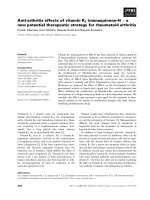
Figure 1. Possible sources of DNA damage, DNA repair mechanisms and
subsequent consequences of immediate and sustained DNA damage. (A) Common
DNA damaging agents (top), which are capable of inducing the various types of DNA
lesions (middle) and can be repaired by specific repair pathways (bottom). (B)
Immediate effects of unrepaired DNA damage causes cell cycle arrest (top) or
apoptosis (middle), while continued accumulation of DNA damage is likely to lead to
permanent changes in DNA sequences and hence, cancer. Abbreviations: cis-Pt and
MMC, cisplatin and mitomycin C, respectively; (6-4) PP and CPD, 6-4 photoproduct
and cyclobutane pyrimidine dimer, respectively; BER and NER, base- and nucleotideexcision repair, respectively; HR, homologous recombination; EJ, end joining.
Reproduced from Nature: Genome maintenance mechanisms for preventing cancer.
(Hoeijmakers, 2001)
1.2 DNA repair pathway – Non-homologous end-joining (NHEJ)
Non-homologous end-joining (NHEJ) pathway is one of the major DNA DSB
repair pathways in mammalian cells (Jackson, 2002; Lieber et al., 2004). NHEJ
mediates the repair of DSBs by directly re-joining the broken ends, which will
ultimately cause deletions of small DNA sequences at the sites of DNA breakages
(Jackson, 2002). Although NHEJ is active in all cell cycle phases, it has been shown
to be particularly important for recombination repair in G0 and G1 cell cycle phases as
such cells do not possess a homologous chromosome, which is required for repair of
DNA damage (Hendrickson, 1997; Rothkamm et al., 2003). However, the human
genome is complex and only a small percentage encodes for proteins. Therefore the
risks associated with such an error-prone repair pathway are not as detrimental as cells
entering S phase with unrepaired DSBs.
1.2.1 Major players in the NHEJ pathway
The NHEJ pathway is governed by a highly regulated protein complex
comprising of a large DNA-dependent protein kinase catalytic subunit (DNA-PKcs)
and its regulatory Ku 70 and 80 subunits (Burma and Chen, 2004; Smith et al., 1999;
Smith and Jackson, 1999). The importance of the protein complex in NHEJ has been
shown by several studies, which reported greater occurrences of chromosomal
aberrations and genomic instability in mouse embryonic fibroblasts (MEFs) and
mammalian cells lacking in either DNA-PKcs, Ku70 or Ku80 proteins (Barnes et al.,
1998; Ferguson et al., 2000; Gao et al., 1998a; Gao et al., 1998b; Gu et al., 2000;
Taccioli et al., 1998).
In a cellular response to DSBs (Fig. 2), DNA end-binding protein Ku70/80
complex recognizes and binds to each of the DSB sites (Mahaney et al., 2009; Yuan et
al., 2010). This consequentially signals the recruitment of DNA-PKcs, which
stimulates its catalytic activity via phosphorylation of ser-2056 or Thr-2609 clusters
(Chan et al., 2002; Chen et al., 2005; Ding et al., 2003). Compromised DSB repair
function of DNA-PKcs has been shown to occur when these two identified cluster
sites were mutated (Chan et al., 2002; Ding et al., 2003). The interaction of DNAPKcs and Ku70/80 forms the DNA-PK complex, which aids in synapsis of the DSB
sites (Mahaney et al., 2009). The XRCC4-DNA ligase IV is recruited for ligation of
the double strand ends for completion of the repair process. The kinase activity of
DNA-PKcs can be regulated by auto-phosphorylation of Ser-2056 cluster, which leads
to inactivation of its kinase activity and subsequent dissociation of the DNA-PK
complex after damage repair (Chan et al., 2002; Ding et al., 2003). Serine/threonine
phosphorylation sites are commonly present in DNA repair proteins and are cognate
substrates of phophatidylinositol 3-kinase (PI-3K) members (Mahaney et al., 2009;
Poltoratsky et al., 1995).
Ataxia telangiectasia-mutated (ATM) protein kinase, which also belongs to the
PI-3K super family, is a general DNA damage sensor (Shiloh, 2006). In the event of
DNA damage, ATM will be activated to phosphorylate downstream targets involved
in cell cycle arrest, DNA repair and stress response (Riballo et al., 2004; Shiloh,
2006). Although the involvement of ATM in homologous recombination (HR) of
DSBs repair has been well-established, recent evidence has revealed a possible
complementary involvement of ATM in mediating DNA-PKcs phosphorylation at the
Thr-2609 cluster upon detection of DSBs contributing to the NHEJ pathway (Chen et
al., 2007).
ATM and DNA-PKcs have been shown to be separately involved in regulating
the phosphorylation of H2AX (An et al., 2010; Burma et al., 2001; Park et al., 2003;
Stiff et al., 2004). H2AX is a component of chromatin and comprises of a central
globular domain, an N-terminal tail and a unique C-terminal tail with a conserved
motif connected by a linker of variable sequence and length (Bonner et al., 2008). The
conserved motif contains the omega-4 serine 139 that becomes phosphorylated to
generate gamma-H2AX ( -H2AX) (Rogakou et al., 1998). -H2AX is a specific and
efficient coordinator in the early response for DNA DSB repair (Kinner et al., 2008).
It is a well-established biomarker employed in immunofluorescence experiments for
detection of DSBs (Kinner et al., 2008).
Once the initial DNA damage sensor proteins as described previously becomes
activated, a nucleation reaction is initiated with the recruitment of MDC1 and
continuing with that of the MRN (Mre11/Rad50/NBS1) complex to further activate
DNA-PK and ATM (Yuan and Chen, 2010). This generates a feedback loop that leads
to further phosphorylation of H2AX and chromatin modifications required for the
recruitment of 53BP1 (Lee et al., 2010; Yuan and Chen, 2010). The activation cascade
culminates with the recruitment of RNF8 to phosphorylated MDC1 and the
polyubiquitinylation of H2AX to recruit BRCA1/BARD1 (Wei et al., 2008).
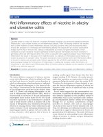
Figure 2. Double strand break recognition and repair pathways. Ku70/80
heterodimer recognizes and binds directly to broken DNA double strand ends.
Recruitment of DNA-PKcs initiates a series of phosphorylation events, including
generation of -H2AX. General DNA damage sensor, ATM, can also activate DNAPKcs. Subsequent nucleation reaction completes the repair process. Reproduced from
FEBS Letters: Focus on histone variant H2AX: To be or not to be. (Yuan et al., 2010)
1.3 Telomeres and its structure
Telomeres are chromosomal end-capping structures first discovered by
Hermann Muller in 1938 (Rodier et al., 2005). These specialized nucleoprotein
complexes function to protect from end-to-end chromosomal fusions, prevent the
recognition of chromosomal ends as DSBs and also from nuclease degradation
(Greider and Blackburn, 1985; Shay and Wright, 2006)
Mammalian telomeres consist of repetitive non-coding sequences of TTAGGG
with a single-stranded 3’ G-rich overhang, which invades into the duplex telomeric
region forming a secondary structure (Rodier et al., 2005). The secondary structure
consists of a telomere-loop (T-loop) and a displacement-loop (D-loop), which aid to
stabilize and cap telomeric DNA (Fig. 3). Specific protein complexes such as
protection of telomeres-1 (POT1), telomeric repeat-binding factor-1 (TRF1), TRF2,
TRF1-interacting protein-1 (TIN1), TIN2, TINF2-interacting protein (TPP1) and
transcriptional repressor/activator protein (RAP1) form the shelterin complex, which
play additional roles in protecting telomeres and hence maintaining its function (de
Lange, 2004). Telomere-specific proteins are able to interact and bind directly or
indirectly to either the single- or double-stranded telomeric DNA (Shay and Wright,
2006).
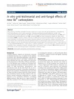
Figure 3. The proposed structure of telomeres and their associated proteins.
TRF1 and TRF2 interact with the double-stranded duplex telomeric DNA. Indirectlybinding proteins bind to telomeric DNA through interaction with directly-binding
proteins, especially through TRF1 and TRF2. Abbreviations: POT1, protection of
telomeres-1; RAP1, transcriptional repressor/activator protein; TRF1 and TRF2,
telomeric repeat-binding factor-1 and telomeric repeat-binding factor-2, respectively;
TIN2, TRF1-interacting protein-2, respectively; NBS1, nijmegen breakage syndrome
1; TANK1 and TANK2, tankyrase 1 and tankyrase 2, respectively; hnRNPs,
heterogeneous nuclear ribonucleoproteins. Reproduced from Nature Reviews:
Molecular Cell Biology 5. (de Lange, 2004)
1.3.1 Telomeric end-replication problem
Considering the important roles that telomeres are involved in as
aforementioned, it is thus necessary to maintain functional telomeres for continued
cell proliferation.
Each round of DNA replication in normal somatic cells leads to progressive
loss of terminal telomeric sequences of approximately 50 to 200 base pairs with each
cell division (Lansdorp, 2000). The loss of telomeric repeats is primarily attributed to
the end replication problem of the lagging strand or in some cases due to the presence
of exonucleases (Xin and Broccoli, 2004). The end replication problem arises due to
the intrinsic inability of DNA repair mechanisms to fill in the 5’ gap contributed by
the short RNA primers, which are required for initiating replication by DNA
polymerase (Rodier et al., 2005). As a result, progressive telomere attrition occurs
with each cell division and thus, telomeres are known to serve as mitotic clocks
recording proliferative history.
However, continued telomere shortening will eventually lead to the triggering
of multiple safeguard mechanisms in cells under normal circumstances. Cells stop
dividing and undergo permanent G0 ,a process also known as replicative senescence or
mortality stage 1 (M1) (Fig. 4) (Hayflick, 1965). This prevents deregulation of
proliferation pathway that may otherwise predispose cells to the development of
cancer.
However, when a mutation imparts a selective survival advantage, further
mutations such as those involving the dominant gain-of-function of proto-oncogenes
or recessive loss-of-function of tumour suppressor genes will tend to accumulate
(Lengauer et al., 1998; Loeb et al., 2003a). Therefore, cells may bypass M1 when
somatic mutations that inactivate retinoblastoma (pRB) or p53 tumour suppressor
genes occur. Consequently, cells continue to proliferate until telomeres are critically
shortened and are unable to form the secondary telomere structure. This telomere
dysfunction will then lead to a continuous break-fusion-bridge (BFB) cycle resulting
in massive gene dosage changes and genetic instability. Eventually, cells will enter
cellular crisis or mortality stage 2 (M2) (DePinho, 2000), which serves as a potential
barrier for the road to immortalization.
As rare event prior to M2, telomerase reactivation or up-regulation allows cells
to escape crisis and proliferate indefinitely with subsequent progression to invasion
and metastasis of cancerous cells (Fig. 4) (Greider and Blackburn, 1985). Majority of
advanced human malignant tumour cells undergo telomerase reactivation enabling
cancer cells the capacity for unlimited proliferation.
proliferation.
Hence, up-regulation
regulation of
telomerase can be considered as an almost universal marker for most (approximately
85 to 90 %) tumour cells in diagnostics. However, an alternative pathway exists for
cancerr cells to attain immortality, which is through recombination via alternate
lengthening of telomeres (ALT) (Shay
Shay and Bacchetti,
Bacchetti, 1997).
1997
Figure 4.. The telomere-telomerase
telomere telomerase hypothesis of cell aging and immortalization.
immortalization
Telomerase activity persists in germ line cells but not in somatic cells; hence
telomeres shorten with age and time in normal somatic cells but not in germ line cells.
In normal somatic cells, when a critical telomere length is reached, a rare event causes
the cell to reactivate or up-regulate
up regulate telomerase. This allows the cell to maintain the
length of telomeres and attain immortalization. Modified and reprodu
reproduced
ced from Cold
Spring Harbour Symposium: Quantitative Biology. (Harley
Harley et al., 1994
1994)
1.4 Telomerase – a regulator of telomere length
The holoenzyme, telomerase, was first purified by Greider and Blackburn
(1985). It is a cellular ribonucleic acid (RNA)-dependent
(RNA) dependent DNA polymerase consisting
of three major components: (1) the telomerase RNA (TERC) subunit, (2) the catalytic
telomerase reverse transcriptase (TERT) subunit and the (3) protein dyske
dyskerin,
rin, with