Cell based screening assay for inhibitors of porcine circovirus type2 (PCV2) replication
Bạn đang xem bản rút gọn của tài liệu. Xem và tải ngay bản đầy đủ của tài liệu tại đây (9.12 MB, 59 trang )
CELL-BASED SCREENING ASSAY FOR INHIBITORS OF
PORCINE CIRCOVIRUS TYPE 2 (PCV2) REPLICATION
CARLA BIANCA LUENA VICTORIO
YONG LOO LIN SCHOOL OF MEDICINE
NATIONAL UNIVERSITY OF SINGAPORE
&
SWISS TROPICAL AND PUBLIC HEALTH INSTITUTE
UNIVERSITY OF BASEL
2010
CELL-BASED SCREENING ASSAY FOR INHIBITORS OF
PORCINE CIRCOVIRUS TYPE 2 (PCV2) REPLICATION
CARLA BIANCA LUENA VICTORIO
(BSc. Molecular Biology and Biotechnology)
University of the Philippines Diliman
A THESIS SUBMITTED FOR THE DEGREE OF
JOINT MASTER OF SCIENCE IN INFECTIOUS DISEASES,
VACCINOLOGY AND DRUG DISCOVERY
YONG LOO LIN SCHOOL OF MEDICINE
NATIONAL UNIVERSITY OF SINGAPORE
&
SWISS TROPICAL AND PUBLIC HEALTH INSTITUTE
UNIVERSITY OF BASEL
2010
Acknowledgements
This research project wouldn’t have been possible without the support and funding from
Temasek Life Science Laboratories (TLL) and the expertise of the researchers at the Animal Health
Biotechnology Group headed by Prof. Jimmy Kwang. My special thanks to my supervisor, Prof. Kwang, co‐
supervisor, Prof. Justin Chu, and mentor Mr. Anbu Karuppannan for giving me guidance and direction in
this research endeavor.
This Joint Msc program had been a wonderful, albeit stressful, experience and I am grateful for
this opportunity bestowed to me by Novartis Institute of Tropical Diseases (NITD), Swiss Tropical and
Public Health Institute (STPH), and National University of Singapore (NUS). My special thanks to Ms.
Christine Mensch for all the much‐needed assistance during my stay in Basel, and to Ms. Susie Soh for the
constant gentle reminders in Singapore. I would like to acknowledge the program lecturers, most
especially Prof. Reto Brun, for providing the inspiration to delve into the field of drug discovery. To the
JIBES, thank you for making life outside the lab and lecture memorable. To Patricia, who has been my life
raft these past 2 years; to Casey, Mad, Ed, Sukriti, Hanwern, Neisha, and Ashley, thank you for all the fond
memories.
I wish to express my heartfelt thanks to the people of TLL who showed me how to make stress a
bearable part of daily life. To Shaz, Peypey, Lu ting, Keiko, Adi, Ivan, and Ranjay, thanks for all your
support; and to Vin, who has been my source of respite during the crucial moments, thank you for the
companionship and strength.
To my friends back home in the Philippines and in Basel, who still give me constant emotional
support, thank you for all your wonderful efforts.
Lastly, I would like to express my whole‐hearted gratitude to my family for being my constant
refuge. Thank you for the selfless love, caring support, and understanding and for allowing me to pursue
my dreams independently.
Cell‐Based Screening Assay for Inhibitors of Porcine Circovirus Type 2 (PCV2) Replication
Page | 1
Table of contents
Summary . . . . . . . . . . . . . . . . . . . . . . . . . . . . . . . . . . . . . . . . . . . . . . . . . . . . . . . . . . . . . . . . . . . . . . . . 5
List of Tables and Figures . . . . . . . . . . . . . . . . . . . . . . . . . . . . . . . . . . . . . . . . . . . . . . . . . . . . . . . . . . . 6
List of Abbreviations. . . . . . . . . . . . . . . . . . . . . . . . . . . . . . . . . . . . . . . . . . . . . . . . . . . . . . . . . . . . . . . 8
1. Introduction
1.1. Porcine Circoviruses (PCV) . . . . . . . . . . . . . . . . . . . . . . . . . . . . . . . . . . . . . . . . . . . . . . . . . . 12
1.1.1. PCV Taxonomy, Morphology and Genetics . . . . . . . . . . . . . . . . . . . . . . . . . . . . . . . 12
1.1.2. Pathogenesis and Replication Cycle of PCV2 . . . . . . . . . . . . . . . . . . . . . . . . . . . . . 14
1.2. PCV2‐Associated Diseases (PCVAD) . . . . . . . . . . . . . . . . . . . . . . . . . . . . . . . . . . . . . . . . . . 16
1.2.1. Postweaning Multisystemic Wasting Syndrome (PMWS) . . . . . . . . . . . . . . . . . . . 17
1.2.2. PDNS and other PCVAD . . . . . . . . . . . . . . . . . . . . . . . . . . . . . . . . . . . . . . . . . . . . . . 18
1.2.3. Treatment of PCVAD . . . . . . . . . . . . . . . . . . . . . . . . . . . . . . . . . . . . . . . . . . . . . . . . 19
1.3. Assay Development and Screening . . . . . . . . . . . . . . . . . . . . . . . . . . . . . . . . . . . . . . . . . . 20
1.3.1. Cell‐based and Cell‐free Assays . . . . . . . . . . . . . . . . . . . . . . . . . . . . . . . . . . . . . . . 21
1.3.2. Signal Detection Systems . . . . . . . . . . . . . . . . . . . . . . . . . . . . . . . . . . . . . . . . . . . . 22
1.3.3. Assay Development for HTS . . . . . . . . . . . . . . . . . . . . . . . . . . . . . . . . . . . . . . . . . . 23
1.4. Objectives of the Study . . . . . . . . . . . . . . . . . . . . . . . . . . . . . . . . . . . . . . . . . . . . . . . . . . . 25
2. Materials and Methods
2.1. Maintenance of cell lines
2.1.1. Culturing PK15‐C1 Cells . . . . . . . . . . . . . . . . . . . . . . . . . . . . . . . . . . . . . . . . . . . . . . 27
2.1.2. Culturing 3D4/31 Cells . . . . . . . . . . . . . . . . . . . . . . . . . . . . . . . . . . . . . . . . . . . . . . 27
2.1.3. Culturing #4 Clone Hybridoma Cells . . . . . . . . . . . . . . . . . . . . . . . . . . . . . . . . . . . . 28
2.1.4. Cryopreservation of Cells . . . . . . . . . . . . . . . . . . . . . . . . . . . . . . . . . . . . . . . . . . . . 28
2.1.5. Establishment of Cell Growth Curve . . . . . . . . . . . . . . . . . . . . . . . . . . . . . . . . . . . . 28
Cell‐Based Screening Assay for Inhibitors of Porcine Circovirus Type 2 (PCV2) Replication
Page | 2
2.2. Production of High Titer PCV2 Stock
2.2.1. Infecting Cells and Harvesting Virus . . . . . . . . . . . . . . . . . . . . . . . . . . . . . . . . . . . . 29
2.2.2. Increasing PCV2 Titer . . . . . . . . . . . . . . . . . . . . . . . . . . . . . . . . . . . . . . . . . . . . . . . 30
2.2.3. Detection of Infection by Western Blot . . . . . . . . . . . . . . . . . . . . . . . . . . . . . . . . . 31
2.2.4. Detection of Infection by PCR . . . . . . . . . . . . . . . . . . . . . . . . . . . . . . . . . . . . . . . . 31
2.2.5. Virus titration by IFA . . . . . . . . . . . . . . . . . . . . . . . . . . . . . . . . . . . . . . . . . . . . . . . . 32
2.3. Cell‐Based Screening Assay Optimization
2.3.1. Downscaling IFA to 384‐well Plate Format . . . . . . . . . . . . . . . . . . . . . . . . . . . . . . 33
2.3.2. Cell‐Based ELISA . . . . . . . . . . . . . . . . . . . . . . . . . . . . . . . . . . . . . . . . . . . . . . . . . . . 33
2.4. Testing Efficacy of Reference Drugs Against PCV2 Replication
2.4.1. Establishment of Standard Curve for FI at Various . . . . . . . . . . . . . . . . . . . . . . . 34
Seeding Densities
2.4.2. Evaluating Drug Cytotoxicity . . . . . . . . . . . . . . . . . . . . . . . . . . . . . . . . . . . . . . . . . 34
2.4.3. Inhibition of PCV2 Replication with Reference Drugs . . . . . . . . . . . . . . . . . . . . . . 35
2.5. Generation of Graphs and Statistical Analyses . . . . . . . . . . . . . . . . . . . . . . . . . . . . . . . 35
3. Results
3.1. Preparation of materials needed for the screening . . . . . . . . . . . . . . . . . . . . . . . . . . . . . 37
3.1.1. Finding the best cell line for the screening assay . . . . . . . . . . . . . . . . . . . . . . . . . 37
3.1.2. Growth dynamics of PK15‐C1 cells . . . . . . . . . . . . . . . . . . . . . . . . . . . . . . . . . . . . . 39
3.1.3. Generation of high titer PCV2 stock . . . . . . . . . . . . . . . . . . . . . . . . . . . . . . . . . . . . 39
3.1.4. Large‐scale production of monoclonal antibodies . . . . . . . . . . . . . . . . . . . . . . . . . 40
(clone #4)
3.2. Scaling down of Assay to 384‐well plates . . . . . . . . . . . . . . . . . . . . . . . . . . . . . . . . . . . . . 42
3.2.1. Comparison of infection rates between glucosamine‐treated . . . . . . . . . . . . . . . 42
and untreated cells
3.2.2. Determining optimum cell seeding density, MOI, and . . . . . . . . . . . . . . . . . . . . . 42
duration of infection
Cell‐Based Screening Assay for Inhibitors of Porcine Circovirus Type 2 (PCV2) Replication
Page | 3
3.2.3. Infection at higher MOI to induce 50% infection rates. . . . . . . . . . . . . . . . . . . . . . 47
3.2.4. Comparison of FITC with Alexa Fluor 546 . . . . . . . . . . . . . . . . . . . . . . . . . . . . . . . . 49
3.3. Testing efficacy of reference drugs against PCV2 replication . . . . . . . . . . . . . . . . . . . . . .50
3.3.1. Standardizing the alamar blue cytotoxicity assay. . . . . . . . . . . . . . . . . . . . . . . . . . 50
3.3.2. Determining cell tolerance for CAPE . . . . . . . . . . . . . . . . . . . . . . . . . . . . . . . . . . . . 51
3.3.3. Efficacy of CAPE against PCV2 replication . . . . . . . . . . . . . . . . . . . . . . . . . . . . . . . . 52
3.3.4. Development of screening assay using cell‐ELISA . . . . . . . . . . . . . . . . . . . . . . . . . . 53
4. Discussion . . . . . . . . . . . . . . . . . . . . . . . . . . . . . . . . . . . . . . . . . . . . . . . . . . . . . . . . . . . . . . . . . . 58
5. References . . . . . . . . . . . . . . . . . . . . . . . . . . . . . . . . . . . . . . . . . . . . . . . . . . . . . . . . . . . . . . . . . . 73
6. Appendix . . . . . . . . . . . . . . . . . . . . . . . . . . . . . . . . . . . . . . . . . . . . . . . . . . . . . . . . . . . . . . . . . . . 79
6.1. Effect of glucosamine treatment on infection rates at . . . . . . . . . . . . . . . . . . . . . . . . . . 79
various seeding densities
6.2. Standard curves for FI and absorbance with alamar blue . . . . . . . . . . . . . . . . . . . . . . . . 82
Cell‐Based Screening Assay for Inhibitors of Porcine Circovirus Type 2 (PCV2) Replication
Page | 4
Summary
PVC2 is a small non‐enveloped virus that causes a wide array of porcine diseases
categorized under the umbrella term PCV‐Associated Diseases (PCVAD). To date, the only
available antiviral strategy, albeit ineffective against diseased pigs, is prevention via vaccination.
Thus, treatment of affected pigs requires discovery of drugs that inhibit viral replication. The
focus of this MSc project was to develop a suitable primary screening assay for inhibitors of
PCV2 replication and subsequently perform a proof of concept trial using reference drugs. PK15‐
C1, a cell line previously shown to be highly permissive to PCV2 infection (Zhu et al., 2007), was
used in the study to grow PCV2 to a high titer (106 TCID50/ml) and exhibited infection rates > 50%
at 15 MOI in a 96‐well plate format. The assay was subsequently scaled down to 384‐well plates
for better amenability to HTS, and the major part of the study was aimed at optimizing this.
Infection with PCV2 was done both at low (< 10) and high (> 10) MOI but neither succeeded in
inducing minimum of 50% infection rate. Even with proof of concept trials performed in 96‐well
plates employing reference drugs CAPE, U0126, and MPA at 15 MOI resulted to infection rates
lower than 50%. This sudden and unexpected drop in infectivity precluded further testing, so a
cell‐based ELISA, which does not require minimum infection rate, was tested instead. Assay
sensitivity was assessed by S/N and S/B ratios. Although S/N ratios were promising, S/B values
were < 2 and sensitivity was found insufficient for further assay development. This was probably
due to low infection rates (< 5%) resulting from stringent blocking and washing, which were
necessary to reduce background signals in ELISA. Thus, significantly improving infection rates
above 50% is necessary to optimize these cell‐based screening assays for inhibitors of PCV2
replication.
Cell‐Based Screening Assay for Inhibitors of Porcine Circovirus Type 2 (PCV2) Replication
Page | 5
List of Tables and Figures
Tables
1. Introduction
Table 1.1
Common equations for determining assay performance . . . . . . . . . . . . . . . . . . . 24
and sensitivity
Figures
1. Introduction
Figure 1.1
PCV Ultrastructure and Genome Morphology . . . . . . . . . . . . . . . . . . . . . . . . . . . 12
Figure 1.2
Genome Organization of Porcine Circoviruses . . . . . . . . . . . . . . . . . . . . . . . . . . . 14
Figure 1.3
Map of Worldwide Occurrence of PWMS . . . . . . . . . . . . . . . . . . . . . . . . . . . . . . . 18
Figure 1.4
Flow of a Typical Drug Discovery Process . . . . . . . . . . . . . . . . . . . . . . . . . . . . . . . 21
2. Materials and Methods
3. Results
Figure 3.1
Detection of PCV2 from PK15‐C1 and 3D4/31 Cultures . . . . . . . . . . . . . . . . . . . . 38
by PCR and Western Blot
Figure 3.2
PCV2 Rep Expression in PK15‐C1 and 3D4/31 Cells . . . . . . . . . . . . . . . . . . . . . . . 38
Figure 3.3
Growth Curve for PK15‐C1 . . . . . . . . . . . . . . . . . . . . . . . . . . . . . . . . . . . . . . . . . . . 39
Figure 3.4
Titration of PCV2 BJW . . . . . . . . . . . . . . . . . . . . . . . . . . . . . . . . . . . . . . . . . . . . . . . 40
Figure 3.5
Titration of Concentrated PCV2 BJW . . . . . . . . . . . . . . . . . . . . . . . . . . . . . . . . . . 41
Figure 3.6
Effect of Glucosamine Treatment on Infection Rates . . . . . . . . . . . . . . . . . . . . . 43
in 384‐well Plates 48 HPI
Figure 3.7
Effect of Glucosamine Treatment on Infection Rates . . . . . . . . . . . . . . . . . . . . . . 43
in 384‐well Plates 60 HPI
Figure 3.8
72 HPI Rates at Different Cell Seeding Densities and MOI . . . . . . . . . . . . . . . . . . 44
Figure 3.9
48 HPI Rates at Different Cell Seeding Densities and MOI . . . . . . . . . . . . . . . . . . 45
Cell‐Based Screening Assay for Inhibitors of Porcine Circovirus Type 2 (PCV2) Replication
Page | 6
Figure 3.10
60 HPI Rates at Different Cell Seeding Densities and MOI . . . . . . . . . . . . . . . . . . 46
Figure 3.11
Infection with High MOI (10, 20 and 25) PCV2 . . . . . . . . . . . . . . . . . . . . . . . . . . . 48
Figure 3.12
Pre‐incubation of Virus (MOI 10, 20 and 25) with . . . . . . . . . . . . . . . . . . . . . . . . 48
Cell Suspension Prior to Seeding
Figure 3.13
Comparison of Infection Rates With FITC and . . . . . . . . . . . . . . . . . . . . . . . . . . . . 49
Alexa Fluor 546
Figure 3.14
Standard Curve for Alamar Blue FI Against Various . . . . . . . . . . . . . . . . . . . . . . . 51
Seeding Densities
Figure 3.15
Growth Inhibition by CAPE . . . . . . . . . . . . . . . . . . . . . . . . . . . . . . . . . . . . . . . . . . . 51
Figure 3.16
Inhibition of PCV2 Replication by CAPE . . . . . . . . . . . . . . . . . . . . . . . . . . . . . . . . 52
Figure 3.17
S/N Ratio From Various Seeding Densities, MOI . . . . . . . . . . . . . . . . . . . . . . . . . . 54
and Dilutions of Detecting Antibody
Figure 3.18
S/B Ratio From Various Seeding Densities, MOI . . . . . . . . . . . . . . . . . . . . . . . . . 55
and Dilutions of Detecting Antibody
Figure 3.19
S/N and S/B Values From Three ELISA Trials . . . . . . . . . . . . . . . . . . . . . . . . . . . . . 56
Figure 3.20
Infection Rates in Cell ELISA . . . . . . . . . . . . . . . . . . . . . . . . . . . . . . . . . . . . . . . . . . 57
4. Discussion
5. References
6. Appendix
Figure 6.1
Effect of Glucosamine Treatment on Infection Rates . . . . . . . . . . . . . . . . . . . . . . 79
in Cells Seeded at 2000 per well
Figure 6.2
Effect of Glucosamine Treatment on Infection Rates . . . . . . . . . . . . . . . . . . . . . . 80
in Cells Seeded at 3000 per well
Figure 6.3
Effect of Glucosamine Treatment on Infection Rates . . . . . . . . . . . . . . . . . . . . . . 81
in Cells Seeded at 3500 per well
Figure 6.4
Standard Curve for Alamar Blue FI Against Various . . . . . . . . . . . . . . . . . . . . . . . 82
Seeding Densities
Figure 6.5
Standard Curve for Alamar Blue Absorbance at 570 nm . . . . . . . . . . . . . . . . . . . 83
Against Various Seeding Densities
Cell‐Based Screening Assay for Inhibitors of Porcine Circovirus Type 2 (PCV2) Replication
Page | 7
List of Abbreviations
ANOVA
Analysis of Variance
ATCC
American Type Culture Collection
BBTV
Banana Bunchy Top VIrus
BFDV
Beak and Feather Disease Virus
BJW
Beijing strain
Cap
Capsid
CAPE
Caffeic Phenethyl Ester
CFDV
Coconut Foliar Decay Virus
CPE
Cytopathic Effects
DMEM
Dulbecco’s Minimal Essential Medium
DNA
Deoxyribo Nucleic Acid
EDTA
EthyleneDiamineTetraacetic Acid
EM
Electron Microscopy
FBS
Fetal Bovine Serum
FI
Fluorescence Intensity
FITC
Fluorescein Isothiocyanate
GAG
Glycosaminoglycans
GPCR
G protein‐Coupled Receptor
HEPES
4‐(2‐HydroxyEthyl)‐1‐PiperazineEthaneSulfonic Acid
HPI
Hours Post‐Infection
hPirH2
Human p53‐induced RING‐H2
HRP
Horseradish Peroxidase
HTS
High Throughput Screening
HTRF
Homogeneous Time‐Resolved Fluorescence
IHC
Immunohistochemistry
IFA
Immunofluorescence Assay
Cell‐Based Screening Assay for Inhibitors of Porcine Circovirus Type 2 (PCV2) Replication
Page | 8
Ig
Immunoglobulin
ISH
In Situ Hybridization
kDa
Kilo Daltons
mAb
Monoclonal Antibody
MEM
Minimal Essential Medium
MEM/5%
MEM with 5% FBS
MOI
Multiplicity of Infection
MPA
Mycophenolic Acid
MPDH
Inosine Monophosphate Dehydrogenase
NPTr
Newborn Pig Trachea
NSK
Newborn Swine Kidney
OPD
o‐Phenylenediamine Dihydrochloride
ORF
Open Reading Frame
PBS
Phosphate‐Buffered Saline
PBS‐T
PBS with 0.1% (v/v) Tween‐20
PCR
Polymerase Chain Reaction
PCV
Porcine Circovirus
PCV1
Porcine Circovirus Type 1
PCV2
Porcine Circovirus Type 2
PCVAD
Porcine Circovirus‐Associated Diseases
PDNS
Porcine Dermatitis and Nephropathy Syndrome
PFA
Paraformaldehyde
PK15
Porcine Kidney‐15
PK15‐C1
Porcine Kidney‐15, Clone C1
PMWS
Postweaning Multisystemic Wasting Syndrome
POC
Proof of Concept
pPirH2
Porcine p53‐induced RING‐H2
PPV
Porcine Parvovirus
PRDC
Porcine Respiratory Disease Complex
PRRSV
Porcine Reproductive and Respiratory Syndrome Virus
Cell‐Based Screening Assay for Inhibitors of Porcine Circovirus Type 2 (PCV2) Replication
Page | 9
RCF
Relative Centrifugal Force
RCR
Rolling‐Circle Replication
Rep
Replicase
RF
Replicative Form
RPM
Revolutions per minute
RPMI
Roswell Park Memorial Institute
RT
Room Temperature
S/B
Signal to Background Ratio
S/N
Signal to Noise Ratio
SDS
Sodium Dodecyl Sulfate
SIV
Swine Influenza Virus
SPA
Scintillation Proximity Assay
TCID50
50% Tissue Culture Infective Dose
uHTS
Ultra High‐Throughput Screening
Cell‐Based Screening Assay for Inhibitors of Porcine Circovirus Type 2 (PCV2) Replication
Page | 10
1. INTRODUCTION
Introduction
Cell‐Based Screening Assay for Inhibitors of Porcine Circovirus Type 2 (PCV2) Replication
Page | 11
1. INTRODUCTION
1.1 Porcine Circoviruses (PCV)
1.1.1 PCV Taxonomy, Morphology and Genetics
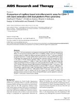
Porcine circoviruses (PCV) are small non‐enveloped viruses with a circular single‐
stranded DNA genome (1,700 nucleotides). The genome is packaged in an icosahedral capsid of
17 nm in diameter (Figure 1.1) and is considered the smallest virus infecting mammalian cells
(Tischer et al., 1982; Nawagitgul et al., 2000). PCV was serendipitously identified as a benign
picornavirus‐like contaminant persisting in a porcine kidney (PK‐15) cell line without causing
visible cytopathic changes (Tischer et al., 1982). Further study later grouped PCV as a member of
the Family Circoviridae and Genus Circovirus, and identified it as close relative to another animal
circovirus such as Psittacine Beak and Feather Disease Virus (BFDV) and other similar viruses
from the Geminiviridae Family such as Coconut Foliar Decay Virus (CFDV), and from Nanoviridae
Family such as Banana Bunchy Top Virus (BBTV) (Niagro et al., 1998; Pringle, 1998).
(A)
(B)
Figure 1.1 | PCV Ultrastructure and Genome Morphology. EM micrographs of (A) purified PCV
particles negatively stained with uranyl acetate. Scale bar = 100 nm; and (B) PCV single‐stranded
circular DNA genome (scale bar = 1µm). Source: (Tischer et al., 1982).
PCV was recently divided into two genotypes (PCV1 and PCV2) based on the
identification of PCV‐like entities in lesions derived from pigs affected with Post‐Weaning
Multisystemic Wasting Syndrome (PMWS), but displaying distinct antigenic properties from
Cell‐Based Screening Assay for Inhibitors of Porcine Circovirus Type 2 (PCV2) Replication
Page | 12
1. INTRODUCTION
currently known PCV1 (Allan et al., 1998; Ellis et al., 1998). PCV1 and PCV2 have the same
ambisense genomic organization, which is the main strategy employed by PCV2 to encode six
predicted overlapping Open Reading Frames (ORF), and three characterized proteins despite its
small genome (Meehan et al., 1998). ORF1, which is expressed on the plus (leading) strand of the
genome, encodes a replicase (Rep) protein essential for virus replication (Mankertz et al., 1998).
Rep has two splice variants, Rep’ and Rep, of which the former has a truncated C‐terminal
region; Rep’ and Rep both comprise the complex needed for initiation of replication. The other
two ORF are expressed from the minus (lagging) strand: ORF 2 encodes the major structural
protein (Cap) of the capsid (Nawagitgul et al., 2000), while ORF3 encodes an apoptosis‐inducing
protein (Liu et al., 2005; Liu et al., 2007).
Although phylogenetic analysis of genomic sequences of PCV1 and PCV2 found in
GenBank designate these viruses to different branches (Mankertz et al., 2004), they have similar
morphology, genomic organization, and display a high degree of sequence homology. PCV2
ORF1 (Rep) has shown 83% nucleotide identity and 86% predicted protein homology with ORF 1
of PCV1, while ORF 2 has shown 67% nucleotide identity and 65% predicted protein homology
between the two genotypes (Allan and Ellis, 2000). ORF 3 differs significantly between PCV 1 and
PCV 2 due to an internal stop codon in PCV2 resulting to a truncated protein; the predicted
amino acid identity between the two ORF3 is only 61.5% (Liu et al., 2005).
PCV is thought to undergo Rolling‐Circle Replication (RCR) because the stem‐loop
structure (Figure 1.2) characterizing the origin of replication is similar to plasmids, viruses, and
bacteriophages that replicate by the same manner (Finsterbusch and Mankertz, 2009). The
origin of PCV2 replication is located in the intergenic region between the transcriptional start
sites of ORF 1 and ORF 2. The loop has an AxTAxTAC sequence where replication is initiated,
while the stem has a 10‐nucleotide palindrome that serves as binding site for the Rep complex.
Downstream of the stem‐loop is a series of four hexanucleotide repeats (H1, H2, H3, and H4),
Cell‐Based Screening Assay for Inhibitors of Porcine Circovirus Type 2 (PCV2) Replication
Page | 13
1. INTRODUCTION
two of which (H1‐H2) function as Rep‐binding sites. The Rep complex comprised of Rep and Rep’
initiates replication by binding to the H1‐H2 hexamers and to the 10‐nucleotide palindrome on
the right side of the stem. Binding to H1‐H2 leads to effective silencing of the Rep promoter and
shuts down its transcription, in a negative feedback loop. Binding to the palindrome destabilizes
the origin of replication on the stem, thus making it a target for Rep‐catalyzed nicking leading to
a free 3’‐OH end that function as primer for the host‐encoded DNA polymerase II to catalyze
strand elongation. A free plus‐strand genome is generated at the end of this cycle (Faurez et al.,
2009; Mankertz et al., 2004), and it either participates in another round of replication or
packaged into the capsid. The exact mechanism of the lagging strand synthesis is currently
unknown, but at the end of the cycle a double‐stranded Replicative Form (RF) DNA is generated
from which transcription occurs in both strands.
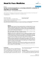
Figure 1.2 | Genome Organization of Porcine Circoviruses. ORF 1 (Rep) is encoded on the
positive (leading) strand, while ORF 2 (Cap) and ORF 3 are encoded on the negative (lagging)
strand of the PCV genome. ORF3‐encoded protein has a C‐terminal truncation compared with
the PCV1 homolog. The stem‐loop structure represents the origin of replication located in the
intergenic region between the 5’ ends of ORF 1 and ORF 2. The arrow indicates the nicking site
from where replication is initiated. Boxed regions represent hexanucleotide repeats, which act
as binding sites for Rep proteins. Source: (Finsterbusch and Mankertz, 2009).
Cell‐Based Screening Assay for Inhibitors of Porcine Circovirus Type 2 (PCV2) Replication
Page | 14
1. INTRODUCTION
1.1.2 Pathogenesis and Replication Cycle of PCV2
PCV2 is capable of infecting various cell types from various species (Liu et al., 2005;
Chaiyakul et al., 2010). Based on infection studies in 3D4/31 cells, a porcine‐derived monocytic
cell line, PCV2 utilizes the surface glycosaminoglycans (GAG) heparin, heparan sulfate and
chondroitin sulfate‐B on the host cell for attachment (Misinzo et al., 2006). The virus is
afterwards internalized by clathrin‐mediated endocytosis and passes through the endosome
pathway, where uncoating requires an acidic environment (Misinzo et al., 2005). In PK15 cells, a
porcine‐derived kidney cell line, inhibition of endosome acidification led to increased PCV2
disassembly (Misinzo et al., 2008) suggesting that acidification is unnecessary for virus
uncoating. This disparity in findings was speculated to be due to distinct serine proteases
catalyzing Cap protein cleavage found in the two cell lines.
Subsequent to capsid disassembly, the PCV2 genome is converted to a double‐stranded
RF intermediate, which is the template for both replication and transcription. The ORF1 is
transcribed to produce Rep protein, and viral genome replication begins. PCV2 replication is cell
cycle‐dependent and often slow because the virus DNA is “only able to enter the nucleus during
mitosis when nuclear material is being distributed to the daughter cells” (Tischer et al., 1987).
However, treatment with glucosamine enables the PCV DNA to “enter the nucleus directly”
through an unknown mechanism, hence circumventing the need for mitosis before replication
initiation (Tischer et al., 1987) and resulting to significantly higher infection rates.
Once the negative‐strand DNA has been produced, ORF 2 encoding the Cap protein is
expressed. The protein has a mass of 26 kilo Daltons (kDa) and is found to self‐assemble to form
the characteristic icosahedral structure of PCV2 capsid (Nawagitgul et al., 2000). The viral
genome is packaged into the capsid through still unknown mechanism, and the progeny virus
subsequently form paracrystalline arrays within inclusion bodies under the cell membrane of
infected cells, in preparation for release to the environment (Stevenson et al., 1999). At the end
Cell‐Based Screening Assay for Inhibitors of Porcine Circovirus Type 2 (PCV2) Replication
Page | 15
1. INTRODUCTION
of the cycle, the virus induces apoptosis of host cells through a pathway believed to be mediated
by ORF 3 protein (Liu et al., 2006; Liu et al., 2005), which was shown to interact with a porcine
homolog of human ubiquitin ligase E3 hPirH2 (human p53‐induced RING‐H2). ORF3 protein
binding destabilizes pPirH2 (porcine p53‐induced RING‐H2) and results to increased p53
expression and subsequent apoptosis (Liu et al., 2007; Karuppannan et al., 2010). Apoptosis
allows progeny virus to be disseminated into the environment and aids infection of neighboring
cells, resulting to increased viral load (Karuppannan and Kwang, 2010).
Although both PCV1 and PCV2 have been detected in and isolated from both healthy
and diseased pigs (Allan and Ellis, 2000; Chae, 2004; Harding, 2004) and despite the documented
high homology between the two virus genomes, only PCV2 had been associated with porcine
disease while PCV1 remained benign. Determinants hypothesized to cause the differential
pathogenic potential of these two closely related viruses are ORF2 and ORF3 proteins. ORF2
exhibits high sequence diversity between PCV1 and PCV2 (Fenaux et al., 2000; Larochelle et al.,
2002) and this translates to alterations in host cell tropism and virus‐host interactions (Chae,
2005). ORF3, which had been shown to induce p53 expression and subsequent cell death (Liu et
al., 2007; Karuppannan et al., 2010), is differentially expressed between PCV1 and PCV2, and
ORF3‐induced apoptosis was shown to play a key role in PCV2 pathogenesis by facilitating virus
exit from the host cell and consequently aiding its subsequent spread (Karuppannan and Kwang,
2010).
1.2 PCV2‐Associated Diseases (PCVAD)
Since its association with PMWS‐affected pigs, PCV2 has been implicated with a number
of swine diseases later categorized under the umbrella term “PCV‐Associated Diseases” (PCVAD).
PCVAD is divided into “clinical syndromes and diseases that have pre‐or post‐natal
manifestations” (Grau‐Roma et al., 2010) and include Postweaning Multisystemic Wasting
Cell‐Based Screening Assay for Inhibitors of Porcine Circovirus Type 2 (PCV2) Replication
Page | 16
1. INTRODUCTION
Syndrome (PMWS), Porcine Dermatitis and Nephropathy Syndrome (PDNS), Porcine Respiratory
Disease Complex (PRDC), Reproductive Failure, etc. PCV2 is a ubiquitous virus and has been
reported from all continents (Grau‐Roma et al., 2010). It is believed to be horizontally
transmitted by direct contact among pigs through the oronasal route, although it can also be
shed in bodily secretions such feces, saliva, urine, milk, and semen (Larochelle et al., 2000;
Shibata et al., 2003; Ha et al., 2009), which may contribute to viral transmission.
1.2.1 Postweaning Multisystemic Wasting Syndrome (PMWS)
PMWS is a well‐established porcine disease that has existed since 1962 (Jacobsen et al.,
2009) although it was first detected only in 1996 (Ellis et al., 1998). To date, it has been reported
in 5 continents (Figure 1.3) and primarily affects postweaned piglets aged 7‐15 weeks. It is
diagnosed by 6 clinical signs which are: wasting, shortness of breath or dyspnea, enlarged lymph
nodes, diarrhea, pallor, and jaundice or yellowing of the skin (Harding, 2004; Chae, 2005).
Isolation of microscopic lymphoid lesions with PCV2 antigen detected either through in situ
hybridization (ISH) or immunohistochemistry (IHC) is also necessary to diagnose PMWS (Chae,
2004). Although PCV2 is necessary to cause PMWS, it is not sufficient and it only causes the
disease in the presence of either immunomodulatory agents or other swine pathogens. The
most commonly found pathogens in PCV2 co‐infections resulting to PMWS include porcine
parvovirus (PPV), porcine reproductive and respiratory syndrome virus (PRRSV), swine influenza
virus (SIV), Streptococcus suis, and Mycoplasma hyopneumoniae. Noninfectious
immunomodulators leading to the disease include keyhole limpet hemocyanin in incomplete
Freund’s adjuvant (Kennedy et al., 2000; Tomás et al., 2008). One hypothesis put forward to
explain this phenomenon is that lymphoid depletion caused by PCV2 infection leads to an
immunocompromised state in pigs resulting to enhanced susceptibility to other swine
pathogens. This is supported by the observation of successful induction of PMWS in postweaned
Cell‐Based Screening Assay for Inhibitors of Porcine Circovirus Type 2 (PCV2) Replication
Page | 17
1. INTRODUCTION
pigs once maternal antibodies have waned, and following immunosuppression caused by
immunomodulators.
Figure 1.3 | Map of Worldwide Occurrence of PMWS. Countries around the world are mapped
along with the initial reports of PMWS. The disease has been reported from North and South
America, Europe, and Asia. Source: (Nawagitgul et al., 2000; Chae, 2004). PCV2 but not the
disease has been reported from Australia and New Zealand (Raye et al., 2005; Muhling et al.,
2006).
1.2.2 PDNS and other PCVAD
PDNS is a fatal but sporadic disease first recognized in 1993 and affects pigs 12‐14 weeks
of age (Chae, 2005). It has been reported in Asia and North America and is characterized by the
appearance of primary lesions in both the skin and kidney. High mortality rates are observed
upon onset of anorexia, weight loss and depression. Similar with PMWS, PCV2 is necessary but
insufficient to cause PDNS and the disease results only through co‐infection with other swine
Cell‐Based Screening Assay for Inhibitors of Porcine Circovirus Type 2 (PCV2) Replication
Page | 18
1. INTRODUCTION
pathogens such as Pasteurella multocida and PRRSV. In contrast with PMWS, PCV2‐filled lesions
in PDNS‐affected pigs are localized in kidneys and not in lymphoid tissues.
PRDC is another health problem observed in pigs 16‐22 weeks of age. It is also caused by
co‐infections of PCV2 with PRRSV, SIV, M. hyopneumoniae, and other swine pathogens. PRDC is
diagnosed by meeting criteria, which includes presence of respiratory signs such as prolonged
dyspnea, pulmonary microscopic lesions with PCV2 antigens, and absence of lymphoid lesions
characteristic of PMWS (Ellis et al., 2004).
PCV2 is also associated with high rates of abortion, stillbirths and fetal mummification.
PCV2 has been isolated from specimens with reproductive failure at different stages of gestation
(Sanchez et al., 2001), although characteristic lesions were absent in recovered fetuses. Aside
from the diseases and syndromes mentioned, PCV2 has been associated with other infectious
porcine diseases including necrotizing lymphadenitis, congenital tremors, and other hepatic,
enteric and renal diseases (Ellis et al., 2004; Chae, 2005).
1.2.3 Treatment of PCVAD
Agents that inactivate PCV2 could potentially aid in the treatment of PCVAD. PCV2 is a
non‐enveloped virus and therefore resistant to lipid‐dissolving disinfectants commonly used in
farms such as alcohol, chlorhexidine, and phenol. Moreover, PCV2 is highly thermostable and
cannot be attenuated successfully with heating and drying (O'Dea et al., 2008). However, PCV2
can be inactivated by alkaline disinfectants, oxidizing agents and quaternary ammonium
compounds (Martin et al., 2008), although the success rate has yet to be studied.
Several vaccines against PCV2, which express the Cap protein, had been developed and
are currently available in the market: Suvaxyn® (Fort Dodge Animal Health Inc) is a whole‐virus
chimeric
vaccine
derived
from
attenuated
PCV1
expressing
Cap
Cell‐Based Screening Assay for Inhibitors of Porcine Circovirus Type 2 (PCV2) Replication
from
PCV2
Page | 19
1. INTRODUCTION
TM
(FortDodgeAnimalHealth, 2009); Ingelvac® CircoFLEX (Boehringer Ingelheim Vetmedica Inc) is
a subunit vaccine containing purified Cap protein expressed from Baculovirus
(BoehringerIngelheimVetmedica, 2010); CircumventTM vaccine (Intervet Inc., Millsboro, Denver,
USA) is derived from killed baculovirus expressing purified PCV2 Cap antigen (Schering‐Plough,
2009) . Both CircoFLEXTM and Suvaxyn® are single‐dose vaccines, while CircumventTM is a 2‐dose
vaccine. All three vaccines are recommended to be given to healthy piglets prior to weaning and
had been tested in field conditions. Trial immunizations resulted to reduction of viremia in
unchallenged pigs (Kixmöller et al., 2008; Segalés et al., 2009) and development of long‐term
protective memory upon experimental triple challenge with PCV2‐PPV‐PRRSV (Shen et al., 2010).
Other PCV2 vaccines currently being developed include DNA vaccines, subunit vaccines,
recombinant virus vaccines, and chimeric PCV1‐PCV2 vaccines (Blanchard et al., 2003; Kamstrup
et al., 2004; Song et al., 2007; Wang et al., 2007).
Although commercially available vaccines lead to significant reduction in PCV2 viremia
and induce protective immunity against challenge with PCV2, these do not benefit pigs that are
already affected with PCVAD. Treatment of diseased pigs entails administration of antivirals that
limit PCV2 replication but unfortunately, no such drug exists to date.
1.3 Assay Development and Screening
Since PCV2 causes a wide array of swine diseases and vaccination, which is the only
antiviral strategy currently available, confers only limited protection, there is urgent need for
developing drugs that can cure diseased pigs. No such drugs exist to date and they have to be
discovered. The drug discovery process is usually divided into two phases: 1) Research and 2)
Development. The research phase deals with the identification of an array of suitable
compounds with desired effect against a selected target, while the development phase deals
with (1) optimization of the compound for greater potency and efficacy, and (2) formulation into
Cell‐Based Screening Assay for Inhibitors of Porcine Circovirus Type 2 (PCV2) Replication
Page | 20
1. INTRODUCTION
a marketable drug suitable for consumption. The research phase typically begins with
identification and selection of a target, for which an assay to determine target response has to
be developed. The research phase ends with the candidate selection process, where hits are
narrowed down to a few candidates and at which the development phase begins with the proof
of concept (POC) trials in animal models.
Figure 1.4 | Flow of a Typical Drug Discovery Process. Drug discovery is divided into two
phases: 1) Research and 2) Development. The research phase deals with identification of
compounds against a selected target, while the development phase deals with
candidate
optimizing the compound for human consumption and formulating as a suitable marketable
drug. Source: (Novartis, 2010)
1.3.1 Cell‐based and Cell‐free Assays
Generally, assays in drug discovery are classified as either cell‐based or target‐based
techniques. In the former, whole‐cell phenotypic responses are measured; while in the latter,
binding and kinetics of individual molecules (the target and candidate drug) are measured in cell‐
free systems. Cell‐based assays are further classified into three broad categories: (1) second
messenger assays, (2) reporter gene assays, and (3) cell proliferation assays. Second messenger
Cell‐Based Screening Assay for Inhibitors of Porcine Circovirus Type 2 (PCV2) Replication
Page | 21
1. INTRODUCTION
assays involve measurement of signal transduction resulting from activation of cell‐surface
receptors, such as in the discovery of ion channel activators and inhibitors (Gonzales et al.,
1999). Reporter gene assays monitor responses at the transcriptional and translational levels of
cells transfected with reporter genes depending on the expression of the reporter upon
activation of the target gene (Mankertz et al., 2003). These offer advantages such as availability
of multiple instrumentation platforms, relative low cost of reagents, and high amenability for
HTS (Johnston, 2002). Lastly, cell proliferation assays monitor the activated or stunted cell
growth upon activation of the target.
Cell‐free or biochemical assays, on the other hand, are divided into 1) enzyme assays, 2)
receptor‐mediated assays, and 3) immunoassays. Common drug targets include five main
protein families: G protein‐couples receptors (GPCRs), kinases, proteases, nuclear receptors, and
ion channels (Inglese et al., 2007). Although responses measured in cell‐free assays are generally
less complicated due to the absence of whole biological systems, hits obtained are often
confounded by nonspecific binding and background signals from the compounds being tested
(Feng et al., 2007; Feng et al., 2008).
1.3.2 Signal Detection Systems
Prior to the 21st century, majority of screening assays employed radiometric methods of
signal detection, often through Scintillation Proximity Assay (SPA; Amersham Pharmacia Biotech)
and FlashPlatesTM (NEN Life Science Products, Boston, MA). The technology is based on either
SPA microbeads or FlashPlateTM surfaces containing scintillant and coated with the target of
interest. Binding of a radiolabeled molecule to the target brings the radioisotope in close
proximity to the solid support, leading to energy transfer between the emitted beta particle and
the scintillant and subsequent release of photons. However, radiometric assays were slowly
Cell‐Based Screening Assay for Inhibitors of Porcine Circovirus Type 2 (PCV2) Replication
Page | 22
1. INTRODUCTION
phased out due to safety issues, limited reagent stability, and relatively long read times
(Hertzberg and Pope, 2000).
Fluorescence is now the most widely used detection system in screening assays due to
its intrinsic sensitivity, safety, short read times, the development of modified highly fluorescent
probes such as the Alexa Fluor (Molecular Probes, Invitrogen) and Cy family of dyes (Amersham
Life Sciences), and the advent of more sophisticated fluorescence microplate readers (Hertzberg
and Pope, 2000). Sensitivity in fluorescence‐based assays, however, is often limited by
background signal arising from assay reagents, containers, or compounds being tested, and this
could also lead to high amounts of false‐positive hits. This problem, however, can be overcome
by time‐resolved techniques such as Homogeneous Time‐Resolved Fluorescence (HTRF) using
lanthanide cryptates as dyes.
Another method to circumvent problems resulting from high background signal is
through luminescent detection systems (Inglese et al., 2007). Luminescence generates light
through catalytic reactions on a substrate, either through an enzyme (Bioluminescence) or
through decay of an unstable chemical intermediate (Chemiluminescence) (Schweitzer and
Abriola, 2002). It doesn’t depend on an external source of excitation energy, hence significantly
reducing background signals.
1.3.3 Assay development for HTS
Once the format and detection system have been chosen, the assay is developed as a
bench top method with low throughput, and later miniaturized to smaller‐volume formats
(typically using microtiter plates) amenable to high‐throughput screening (HTS). Factors to
consider when developing an assay include: (1) defined response to be measured, (2) clear
parameter‐response dependence, and (3) lag time between stimulation with the compounds
Cell‐Based Screening Assay for Inhibitors of Porcine Circovirus Type 2 (PCV2) Replication
Page | 23