Effects of plant polyphenols and mutational analysis of multidrug resistance protein 4 (MRP4 ABCC4) functions
Bạn đang xem bản rút gọn của tài liệu. Xem và tải ngay bản đầy đủ của tài liệu tại đây (887.38 KB, 134 trang )
EFFECTS OF PLANT POLYPHENOLS AND
MUTATIONAL ANALYSIS OF MULTIDRUG
RESISTANCE PROTEIN 4 (MRP4/ABCC4) FUNCTIONS
WU JUAN
NATIONAL UNIVERSITY OF SINGAPORE
2005
EFFECTS OF PLANT POLYPHENOLS AND
MUTATIONAL ANALYSIS OF MULTIDRUG
RESISTANCE PROTEIN 4 (MRP4/ABCC4) FUNCTIONS
WU JUAN
(B.M., Peking University)
A THESIS SUBMITTED
FOR THE DEGREE OF MASTER OF SCIENCE
DEPARTMENT OF BIOCHEMISTRY
NATIONAL UNIVERSITY OF SINGAPORE
2005
2
Acknowledgements
I would like to express my heartfelt thanks and appreciates to my advisor, Dr Theresa
Tan, Department of Biochemistry, National University of Singapore, for her keen
supervision, valuable suggestion and discussion, patient guidance and encouragement
during my study.
I deeply thank Ms Yang Shu and Mr. Li Yang for their technical support and kind
help. I also thank Mr. Wang Penghua, Mr. Zhang Shaochong, Miss Sherry Ngo, and
Mr. Bian Haosheng, who gave me valuable suggestions. I thank Dr Robert Yang for
use of the fluorescent microscope.
I am grateful to the members of my family for their understanding and great support,
especially to my dear parents, sister and husband, for their loving encouragement and
caring.
3
Table of Contents
Acknowledgements........................................................................................................3
Summary ........................................................................................................................6
List of Tables .................................................................................................................8
List of Figures ................................................................................................................9
List of Abbreviations ...................................................................................................11
1. Introduction...........................................................................................................14
1.1. Transporters ...................................................................................................14
1.2. ABC transporter .............................................................................................17
1.3. MRP family....................................................................................................19
1.3.1.
The role of MRPs in detoxification ......................................................23
1.3.2.
MRP1 ....................................................................................................26
1.3.3.
MRP2 ....................................................................................................29
1.3.4.
MRP3 ....................................................................................................30
1.3.5.
MRP4 ....................................................................................................31
1.3.6.
MRP5 ....................................................................................................35
1.3.7.
MRP6 ....................................................................................................36
1.3.8.
MRP7 ....................................................................................................37
1.3.9.
MRP8 ....................................................................................................38
1.3.10. MRP9 ....................................................................................................39
1.4. Flavonoids.....................................................................................................39
1.5. Identification of domains and amino acid residues for determining substrate
specificity of MRPs......................................................................................44
1.5.1. Substrate specific domains.......................................................................44
1.5.2. Identification of key amino acids.............................................................45
1.5.3. Single-nucleotide polymorphisms (SNPs) in transporters.......................47
2. Aims and overview of study .................................................................................50
3. Materials and Methods..........................................................................................52
3.1. Mammalian cell culture ..................................................................................52
3.1.1. Materials ................................................................................................52
3.1.2. Cell line and cell culture ........................................................................52
3.1.3. Initiating a new flask..............................................................................52
3.1.4. Passaging cells .......................................................................................53
3.1.5. Harvesting cells......................................................................................53
3.1.6. Freezing cells .........................................................................................53
3.2. Functional study of MRP4 protein.................................................................54
3.2.1.
Materials ...............................................................................................54
3.2.2.
Cytotoxic assay .....................................................................................54
3.2.3.
Export assay with MCB ........................................................................55
3.2.3.1. Detection and measurement of transport activity ...........................55
3.2.3.2. Effects of plant polyphenols on bimane-GS efflux.........................56
3.2.4.
Reduced glutathione efflux assay ........................................................56
3.2.4.1. Detection and measurement of transport activity ...........................56
3.2.4.2. Effects of plant polyphenols on GSH efflux...................................57
3.3. Cloning site-directed mutated MRP4 cDNA .................................................57
3.3.1.
Materials ...............................................................................................57
3.3.2.
Site-directed mutagenesis .....................................................................58
3.3.2.1. Primer design ..................................................................................58
3.3.2.2. Polymerase chain reaction (PCR) ...................................................58
3.3.2.3. Extraction and purification of DNA ...............................................62
4
3.3.3.
TA sub-cloning ....................................................................................62
3.3.3.1. Ligation of PCR products to a TA cloning vector ..........................62
3.3.3.2. Culture of bacterial cells .................................................................63
3.3.3.3. Preparation of competent cells........................................................64
3.3.3.4. Transformation................................................................................64
3.3.3.5. Selection and screening...................................................................65
3.3.3.6. DNA extraction: mini-prep ............................................................65
3.3.3.7. Restriction enzyme digestion..........................................................65
3.3.3.8. DNA extraction: midi-prep .............................................................66
3.3.3.9. DNA sequencing.............................................................................67
3.3.4.
Plasmid construction..........................................................................67
3.4. Transfection and expression of mutated MRP4.............................................69
3.4.1.
Materials ..............................................................................................69
3.4.2.
Transfection and selection ...................................................................69
3.4.3.
SDS-PAGE gel electrophoresis ...........................................................70
3.4.3.1.
Preparation of reagent and solution ...............................................70
3.4.3.2.
Preparation of sample ....................................................................71
3.4.3.3.
Procedure .......................................................................................71
3.4.4.
Western blotting....................................................................................72
3.4.5.
Immunostaining ...................................................................................73
3.5.
Functional study of mutated MRP4 protein.................................................74
3.5.1.
Cytotoxic assay .....................................................................................74
3.5.2. Export assays with MCB ......................................................................74
3.5.3. Export assays of GSH ...........................................................................74
4.
Results...............................................................................................................75
4.1. Functional study of MRP4 protein...............................................................75
4.1.1. Export of bimane-GS by MRP4/Hep G2 cells........................................75
4.1.2. Effects of plant polyphenols on bimane-GS efflux mediated by MRP4 78
4.1.3. Export of reduced glutathione by MRP4/Hep G2 cells ..........................84
4.1.4. Effects of plant polyphenols on GSH efflux mediated by MRP4...........87
4.2.
Cloning and expression of mutant MRP4....................................................93
4.2.1. PCR ........................................................................................................93
4.2.2. Cloning of mutant MRP4 into cloning vector........................................94
4.2.3. Construction of mutant full-length MPR4 expression plasmid .............96
4.2.4. Expression of mutant MRP4 protein in Hep G2 cells............................98
4.2.5. Localization of mutant MRP4 in Hep G2 cells....................................100
4.3. Functional study of mutant MRP4................................................................102
4.3.1. Cytotoxic assay ....................................................................................102
4.3.2. Export of bimane-GS of mutant MRP4/Hep G2 cells .........................103
4.3.3. Export of reduced GSH of mutant MRP4/Hep G2 cells......................104
5. Discussion ............................................................................................................106
6. Conclusions..........................................................................................................119
References..................................................................................................................120
5
Summary
Multidrug resistance protein 4 (MRP4/ABCC4) is a member of the ATP-binding
cassette transport superfamily. MRPs are able to transport structurally diverse
conjugated organic anions including glutathione-S-conjugates and function as efflux
pumps of therapeutic drugs and endogenous compounds. Previous studies had shown
that the substrates of MRP4 include methotrexate, cAMP and cGMP, metabolites of
chemotherapeutic agents, glutathione-conjugated and glucuronide-conjugated organic
anions.
Like MRP1-3, MRP4 can also perform the transport of glutathione-S-conjugates
despite the differences in the membrane topology and drug resistance profiles
between MRP4 and MRP1-3. MRP4 has only two transmembrane domains and two
ATP-binding domains with the absence of a third (N-terminal) membrane spanning
domain, which is present in MRP1-3. Using cells stably overexpressing MRP4, this
study confirmed that MRP4 can indeed facilitate the efflux of the glutathione
conjugate, bimane-glutathione. The efflux increased with time and > 72% of the
conjugate was exported after 20 minutes. A concentration-dependent inhibition of
bimane-glutathione efflux was observed with some common dietary plant
polyphenols including ellagic acid, curcumin, apigenin, luteolin and kaempferol. In
addition, MRP4 can facilitate the efflux of glutathione directly and the concentrationdependent inhibition of glutathione efflux was also observed with these plant
polyphenols including ellagic acid, curcumin, apigenin, kaempferol, luteolin,
genistein and quercetin.
6
As a step toward determining the substrate-binding sites of MRP4, site-directed
mutagenesis of highly conserved residues were carried out on the basis of the
alignment of the protein sequences of MRP family. We replaced three highly
conserved charged amino acids Arg165, Arg951 and Asp953 with conserved or nonconserved substitution. The single-nucleotide polymorphism (SNP) site Cys171Gly in
the transmembrane domain of MRP4 was also examined. All mutant clones were
transfected into human Hep G2 cells and the localization and the expression levels of
mutant MRP4 were comparable to that of wild-type MRP4. Our finding shows that
both R165N and C171G mutants lost their ability to confer resistance to purine
analogues 6-TG and 6-MP and to transport glutathione-S-conjugates (bimane-GS).
Only the R165N mutant is unable to transport glutathione. In brief, our present study
indicates that highly conserved charged amino acids Arg165 and the SNP site
Cys171Gly in the transmembrane domains of MRP4 are important determinants for
MRP4-mediated transport and drug resistance.
7
List of Tables
Table 1.1
The structures of twelve compounds used in the study ...................42
Table 3.1.
Primers for mutagenesis..................................................................61
Table 3.2
Composition of SDS-PAGE gel .....................................................72
Table 4.1
Effect of curcumin on bimane-GS efflux .......................................79
Table 4.2
Effect of ellagic acid on bimane-GS efflux ....................................79
Table 4.3
Effect of keampferol on bimane-GS efflux ....................................80
Table 4.4
Effect of luteolin on bimane-GS efflux ..........................................80
Table 4.5
Effect of apigenin on bimane-GS efflux.........................................81
Table 4.6
No effect of compounds on bimane-GS efflux...............................82
Table 4.7
Effect of curcumin on GSH efflux..................................................88
Table 4.8
Effect of ellagic acid on GSH efflux ..............................................88
Table 4.9
Effect of keampferol on GSH efflux...............................................89
Table 4.10 Effect of luteolin on GSH efflux.....................................................89
Table 4.11 Effect of apigenin on GSH efflux ...................................................90
Table 4.12 Effect of quercetin on GSH efflux..................................................90
Table 4.13 Effect of genistein on GSH efflux ..................................................91
Table 4.14
No effect of compounds on GSH efflux .........................................91
Table 4.15. IC50 of resistance to drugs of mutant MRP4/Hep G2 cells. ........102
Table 4.16
Bimane-GS synthesis of mutant MRP4 and controls over a 10-min
time course. ..................................................................................103
Table 4.17
Total GSH of mutant MRP4 and controls over a 10-min time course.
......................................................................................................105
8
List of Figures
Figure 1.1
Classification of the types of transporters .....................................18
Figure 1.2
Topology of MRP family members...............................................21
Figure 1.3
Subcellular localization of MRPs in polarized epithelial cell
surrounding a hypothetical lumen...............................................22
Figure 1.4
Model showing interrelation between multidrug resistanceassociated protein (MRP) and glutathione (GSH) .......................25
Figure 1.5
Involvement of glutathione in MRP1-mediated transport ...........28
Figure 1.6
Alignment of predicted TM segments in MRP4 and corresponding
TM segments in other human MRP family members..................49
Figure 2.1
Flow chart of the project...............................................................51
Figure 3.1
PCR-based overlapping extension to produce mutants ................60
Figure 3.2
The map of the pGEM-T vector ...................................................63
Figure 3.3
The map of pcDNA6/V5-His vector. ...........................................68
Figure 3.4
Schematic diagram of full-length MRP4 with restriction enzyme
sites. .............................................................................................69
Figure 4.1
Efflux of bimane-glutathione from control and MRP4
overexpressing cells. ....................................................................77
Figure 4.2
Effects of polyphenols on bimane-glutathione efflux. .................83
Figure 4.3
Efflux of GSH from control and MRP4 overexpressing cells......86
Figure 4.4
Effects of polyphenols on GSH efflux. ........................................92
Figure 4.5
Template for mutant MRP4 fragments.........................................93
Figure 4.6
Mutant MRP4 fragments ..............................................................93
Figure 4.7
Restriction enzyme digestion of R165K, R165N and C171G
clones by EcoRI and EcoRV in pGEM-T vector.........................94
Figure 4.8
Restriction enzyme digestion of R951M and D953Q clones by
HincΙΙ and XhoΙ. ..........................................................................95
Figure 4.9
Restriction enzyme digestion of pcDNA6-mutant MRP4 vector by
EcoRI and XhoI ...........................................................................96
9
Figure 4.10
DNA sequence results of mutant pcDNA6-MRP4.......................97
Figure 4.11
Western blot analysis of wild-type and mutant MRP4 expression
in Hep G2 cells.............................................................................99
Figure 4.12
Immunostaining of Hep G2 cells overexpressing wild-type and
mutant MRP4. ............................................................................101
Figure 4.13
Efflux of bimane-GS from mutant MRP4/Hep G2 cells and
controls at 10-min time point.....................................................104
Figure 4.14
Efflux of GSH from mutant MRP4/Hep G2 cells and controls at
10-min time point.......................................................................105
10
List of Abbreviations
6-MP
6-Mercaptopurine
6-TG
6-Thioguanine
ABC
ATP-binding Cassette
ALD
Adrenoleukodystrophy
AP
Ammonium persulfate
ATP
Adenosine triphosphate
Bimane-GS
Bimane-glutathione
Bp
base pair
BSA
bovine serum albumin
BSEP
bile salt export pump
cAMP
Cyclic AMP
CFTR
Cystic Fibrosis Transmembrane conductance Regulator
cGMP
Cyclic GMP
DMSO
Dimethyl sulfoxide
DMEM
Dulbecco’s Modified Eagle Medium
E217βG
estradiol 17-β-D-glucuronide
E.coli
Escherichia coli
EST
Expressed sequence Tag
FBS
Fetal bovine serum
GSH
Glutathione
GSSG
Glutathione disulphide
GST
Glutathione S-transferase
HBSS
Hanks Balanced Salt Solution
11
IC50
50% growth inhibitory concentration
IPTG
Isopropythio-beta-D-galactoside
Kb
Kirobase
LB
Luria Broth medium
LBA
Luria Broth medium with Ampicillin
LTC4
Leukotriene C4
MCB
Monochlorobimane
MDR
Multidrug Resistance
MOAT
Multispecific Organic Anion Transporter
MRP
Multidrug Resistance-associated Protein
MSD
Membrane spanning domain
MTS/PES
([3, (4,5-dimethylthiazol-2-yl)-5-(3-carboxymethoxyphenyl)-2(4-sulfophenyl)-2H tetrazolium] / phenazine ethosulfate
MTX
Methotrexate
NBD
Nucleotide Binding Domain
NUMI
National University Medical Institute
OATs
organic anion transporters
OATPs
organic anion-transporting polypeptides
OCTs
organic cation transporters
ORF
Open Reading Frame
PAH
p-Aminohippurate
pBS
pBlueScript SK ΙΙ(+) vector
PBS
phosphate-buffered Saline
pcDNA6
pcDNA6/V5-His
PCR
Polymerase Chain Reaction
12
Pgp
P-glycoprotein
SNPs
single-nucleotide polymorphisms
SDS
Sodium dodecyl sulfate
SDS-PAGE
Sodium dodecyl sulfate-polyacrylamide gel eletrophoresis
TAE
Tris-Acetate-EDTA
TBS-T
Tris-Buffered Saline/Tween 20
TEMED
N,N,N’,N’-Tetramethylethylenediamine
TMD
Transmembrane Domain
TMD0
Third NH2-terminal transmembrane domain
UV
Ultra-violet
X-gal
5-bromo-4-chloro-3-beta-D-galactoside
13
1. Introduction
1.1.
Transporters
Transporter-mediated processes play key roles in the absorption, distribution and
excretion (ADE) of many endogenous and xenobiotic compounds. Drugs ingested
into the body are transported through the plasma membrane several times. Most drugs
need transporters for their trans-membrane transport. These transporters are classified
into five groups by their difference in molecular structures, substrate specificities and
transport mechanisms. They are organic ion transporter superfamily, ATP-dependent
transporter superfamily, peptide transporter family, organic anion transporting
polypeptide family and amino acid-polyamine-choline transporter superfamily (Endou,
2000). The disposition of endogenous compounds, drugs and other xenobiotics are
performed by transporters in many organs. In the intestine, liver, kidney and brain,
transporters are important in the absorption, distribution and excretion of therapeutic
drugs. In the liver, transporters are involved in the uptake of drugs from blood to liver,
across the sinusoidal membrane and hepatobiliary distribution and excretion of drugs
and metabolites (Kim, 2000; Ayrton and Morgan, 2001). In the kidney, transporters at
the basolateral and luminal membranes are involved in renal secretion of drugs (Inui
et al., 2000; Ayrton and Morgan, 2001). In the intestine and brain, transporters, such
as P-glycoprotein, play a significant role in the extrusion of drugs from these organs
so that the drug absorption and brain penetration are attenuated (Suzuki and Sugiyama,
2000; Ayrton and Morgan, 2001). Drug efflux transporters, such as multi-drug
resistance protein 1 and 2 (MPR1 and MRP2), and the uptake transporters, such as
members of the organic anion-transporting polypeptides (OATPs) and organic anion
14
transporter families (OATs), can mediate the cellular efflux and uptake of a large
number of structurally divergent compounds, respectively, especially in organs such
as the intestine, liver and kidney (Marzolini et al., 2004).
Drug absorption mainly happens in the gastrointestinal tract. The intestine, primarily
regarded as an absorptive organ, is also able to eliminate certain organic acids. The
interactions of drugs with intestinal membrane transporters have an important impact
on the intestinal drug absorption and secretion (Kunta and Sinko, 2004). Some
transporters are involved in the active absorptive influx of compounds from the lumen
into the portal bloodstream (Tsuji and Tamai, 1996). Conversely, other transporters
are responsible for the active efflux of drugs and xenobiotics from gut epithelial cells
back into the lumen. Transporters present in the gut epithelial plasma membrane
include members of a number of transport protein families such as MDR, MRP,
OATP, OCT and OAT (Ayrton and Morgan, 2001). Certain organic solutes, such as
amino acid-mimetic drugs, monocarboxylic acid drugs, phosphonic acid drugs, bile
acids, are thought to be absorbed from the gastrointestinal tract through transportermediated mechanisms. By contrast, absorption of many lipophilic drugs is limited by
Pgp or other ATP-dependent active secretory mechanisms at the brush border
membranes of intestinal epithelial cells (Sai and Tsuji, 2004). For example, Pglycoprotein (MDR1), a member of MDR, is present on the villus tip of the apical
brush border membrane of gut enterocytes and is orientated to pump substrates from
inside the cells back into the lumen of the intestine (Wagner et al., 2001).
The hepatobiliary system and the kidneys are the main routes by which drugs and
their metabolites leave the body (van Montfoort et al., 2003). The liver plays a key
15
role in the clearance and excretion of many drugs and hepatobiliary excretion of drugs
involves passage from blood, through the hepatocyte, and into the bile. Many
transporters are present on the canalicular membrane to mediate this process. The
OATPs, which have been shown to be specifically located on the liver sinusoidal
membrane in rodents and humans, are mainly responsible for the hepatic uptake of
large amphipathic organic anions, organic cations and uncharged substrates, whereas
OCTs and OATs mediated the uptake of predominantly small organic cations and
anions in liver (van Montfoort et al., 2004; Ayrton and Morgan, 2001). Members of
ATP-binding cassette family of transporters are mainly involved in the active drug
secretion into bile. These transporters include P-glycoprotein encoded by multidrugresistance gene (MDR), the bile salt export pump (BSEP) and a distinct ATPdependent transport system referred to as cMOAT or MRP2 (Ayrton and Morgan,
2001). The kidney plays an important role in the elimination of many drugs which
include active tubular secretion in renal clearance. It has been demonstrated that an
increasing number of transporter families, such as OAT, OCT, OATP, MDR and
MRP, are present in the kidney. The first step in drug elimination in kidney is uptake
into proximal tubular cells, which is mainly mediated by OCTs and OATs. Various
transporters mediate the active secretion of drugs, and hydrophilic cations and anions
in the renal tubule (van Montfoort et al., 2004; Ayrton and Morgan, 2001).
16
1.2. ABC transporter
To date, more than fifty human ATP-binding cassette (ABC) genes have been
identified. High sequence homology in the nucleotide binding domains (NBDs)
allows identification and classification of members of the ABC transporter family.
The functional protein usually is comprised of two NBDs and two transmembrane
domains (TMDs). There are seven subfamilies, ABCA through ABCG. These are
expressed in both normal and malignant cells. They are involved in the transport of
many substances, including the excretion of toxins from the liver, kidneys, and
gastrointestinal tract, and they limit permeation of toxins to vital structures, such as
the brain, placenta, and testis. Mutations in the genes encoding these transporter
proteins can induce a multitude of defects, presenting as autosomal recessive
conditions (Leonard et al., 2003).
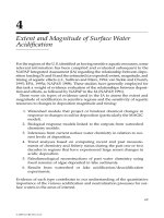
The ABC superfamily is one of the largest protein superfamilies and contributes to the
active transport of a wide variety of compounds across biological membranes (Klein
et al., 1999). Most ABC proteins are membrane transporters, which can translocate
various substrates to various compartments. There are four types of transporters on
the cell membrane: ion channel, passive transporter, primary active transporter and
secondary active transporter. ABC proteins belong to the primary active transporter
category (Figure 1.1).
17
Figure 1.1 Classification of the types of transporters.
ABC protein family is divided into four subfamilies: MRP/CFTR (ABCC, to-date:
thirteen members), MDR/TAP (ABCB, eleven members), ALD (ABCD, four
members) and ABC1 (ABCA, twelve members) and three smaller groups: white
(ABCG, six members), GCN20 (ABCF, three members) and the subgroup OABP
(ABCE) with only one ‘single member’. A large number of the known ABC proteins
are active pumps (Borst et al., 1999; Cole and Deeley, 1998; Hipfner et al., 1999;
Higgins, 1992; Klein et al., 1999).
As membrane transporters, the typical eukaryotic ABC protein contains four domains.
These include two hydrophobic, polytopic transmembrane domains (TMDs), also
called membrane spanning domains (MSDs), and two hydrophilic, cytosolic
nucleotide binding domains (NBDs). They are organized in pairs (TMD-NBD or
NBD-TMD) and expressed either as one continuous unit or two separate polypeptides
(Decottignies and Goffeau, 1997; Hipfner et al., 1999). In most ABC transporters, the
binding and subsequent hydrolysis of ATP at their NBDs provide energy for
transporting substrates across the membrane. The substrates include phospholipids,
ions, organic anions, amino acids, peptides, steroids, drugs and other xenobiotics.
18
1.3.
MRP family
MRPs are the members of the ATP binding cassette (ABC) superfamily of transport
proteins. MRPs are multispecific organic anion transporters, which can transport
negatively charged anionic molecules and neutral molecules conjugated to glutathione,
glucuronate or sulfate. The MRP family comprises nine related ABC transporters that
are able to transport structurally diverse lipophilic anions and function as efflux
pumps of therapeutic drugs and endogenous compounds (Kruh et al., 2003).
The amino acid sequence of MRP1 resembles P-glycoprotein encoded by human
MDR1 gene only to a modest extent (about 15%), and its structure is distinct as well.
MDR1, a member of the MDR/TAP subfamily, is the most extensively studied
transporter involved in multidrug resistance. The P-glycoprotein (MDR1) was the first
cloned human ABC protein (Roninson et al., 1986). It is located on the apical (or
luminal) surface of polarized epithelial cells. It is found at the pharmacological barrier
of the body and present on the brush border membrane of intestinal cell, on the biliary
canalicular membrane of hepatocytes, as well as on the luminal membrane in
proximal tubules of kidney (Bosch et al., 1996). The MDR1 transporter can extrude a
wide range of structurally unrelated hydrophobic toxic compounds. It is suggestive
that the physiological function of MDR1 is to protect cells against toxic compounds.
MDR1 is also expressed in tumor cells. At some stages of treatment with natural
product drugs, the expression level of MDR1 increases by 50% in all human tumors.
The failure of some tumors to respond to therapy is clearly related to the increase in
Pgp. Therefore, increased Pgp activity in tumor cells can lower the concentration of
cellular chemotherapeutic agents and this results in anti-cancer drug resistance.
19
However, the multidrug resistance mediated by MDR1 is not the only single factor in
the therapeutic outcome in human malignancies (Bosch et al., 1996). Experimental
studies in vitro also showed that Pgp is not the only cause of MDR. Many cells
selected for resistance do not contain increased levels of Pgp but nevertheless are
resistant to a broad range of natural product drugs. Several of these cell lines contain
raised levels of a second member of the ABC transporter proteins, the MDRassociated protein (MRP), which was discovered by Cole et al. (Cole et al., 1992).
The drug resistance phenotype of MRP protein overlaps with that of Pgp. It is
associated with resistance to anthracyclines, etoposide, and vinca alkaloids. However,
the spectrum of drug resistance of MRP and Pgp is not exactly the same. MRP does
not confer resistance to taxol, which is a clinically important agent and a part of the
Pgp resistance profile. Moreover, Pgp-mediated multidrug resistance is readily
reversed by verapamil and cyclosporin A (analogues), but that mediated by MRP is
not.
The MRP subfamily of ABC transporters from mammals consists of nine members,
six of which have been implicated in the transport of amphipathic anions. Based on
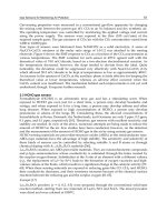
the structure, MRP1, MRP2, MRP3, MRP6 and MRP7 are termed as ‘long’ MPRs
because of an additional MSD0 at the N-terminal, while MRP4, MRP5, MRP8 and
MRP9 are ‘short’ MRPs (Figure 1.2). In polarized epithelial cells, MRP1, MRP3,
MRP5 and MRP6 are localized on the basolateral membranes. MRP2 is localized on
the apical membranes. MRP4 is localized on the basolateral membranes in human
prostatic glandular cells and on the apical membranes in rat kidney tubule cells. The
localizations of MRP7, MRP8 and MRP9 have not been determined (Figure 1.3).
20
Figure 1.2
Topology of MRP family members. (a). Schematic depicting the
organization of protein domains. Stripes, membrane spanning domain; open,
cytoplasmic loops located between MSD0 and MSD1, NBF1 and MSD2 and at the Cterminus; black, nucleotide binding folds. (b). Topological model of MRP1 (which
resembles MRP2, MRP3, MRP6 and MRP7) (top) and MRP4 (which resembles
MRP5, MRP8 and MRP9) (bottom) (Hopper et al., 2001).
21
Figure 1.3 Subcellular localization of MRPs in polarized epithelial cell
surrounding a hypothetical lumen (Kruh et al., 2003).
The structures of MRP1, MRP2, and MRP3 are very similar. They confer resistance
to a variety of natural products as well as methotrexate, and have the facility for
transporting glutathione and glucuronate conjugates. MRP1 is a ubiquitously
expressed efflux pump for the products of phase II xenobiotic detoxification. It is also
involved in immune responses involving cysteine leukotrienes. MRP2, whose
hereditary deficiency results in Dubin-Johnson syndrome, functions to extrude
organic anions into the bile. MRP3 is distinguished by its capacity to transport
glycocholate, a monoanionic bile constituent, and may function as a basolateral backup system for the detoxification of hepatocytes when the usual canalicular route is
impaired by cholestatic conditions. MRP4 and MRP5 resemble each other more
closely than they resemble MRPs 1-3 and confer resistance to purine and nucleotide
analogs which are either inherently anionic, as in the case of the anti-AIDS drug
PMEA, or are phosphorylated and converted to anionic amphiphiles in the cell, as in
the case of 6-MP. Given their capacity for transporting cyclic nucleotides, MRP4 and
MRP5 have also been implicated in a broad range of cellular signaling processes
involving cyclic GMP and cyclic AMP. The drug resistance activity and physiological
22
substrates of MRP6 are unknown. However, its hereditary deficiency results in
pseudoxanthoma elasticum, a multisystem disorder affecting skin, eyes, and blood
vessels. Hence, MRP6 may play a role in elastic tissue homeostasis. The
physiological functions of MRP7, MRP8 and MRP9 are still unknown. Some MRPs
can also transport neutral drugs if co-transported with glutathione. It is hoped that
elucidation of the resistance profiles and physiological functions of the different
members of the MRP subfamily will provide new insights into the molecular basis of
clinical drug resistance (Kruh and Belinsky, 2003; Hopper et al., 2001; Borst et al.,
1999).
1.3.1.
The role of MRPs in detoxification
Metabolism of toxicants
Lipophilic xenobiotics and endogeneous compounds are often metabolized before
being eliminated from the cell. The metabolism of these compounds can be grouped
into either phase I or phase II reactions.
In phase I, a function polar group including a hydroxyl, carboxyl, amino or thio group,
is introduced to the compounds. In phase II, the phase I metabolite is conjugated with
various endogenous substrates, such as sugars, amino acids, glutathione (GSH) and
sulfate, to form water soluble products that are readily excreted (Hodgson et al., 1998).
An important phase II reaction is the reaction catalyzed by glutathione-S-transferases.
Glutathione-S-transferases are anionic in nature and are transported out of cells
through an ATP-dependent process. A key feature of MRP proteins is the ability to
transport glutathione-S-conjugates.
23
GSH related transport
Glutathione (GSH), γ-Glu-Cys-Gly is a tripeptide, and is present in all cells at high
levels. GSH has many important roles in the protection of cells from oxidative stress.
It is responsible for the removal of toxic peroxides that form in the course of growth
and metabolism under aerobic conditions. The ratio of reduced GSH to the oxidized
form, glutathione disulphide (GSSG), is a reflection of cellular redox status.
Maintenance of low cellular GSSG concentrations and high GSH level is important
for cellular homeostasis. Some MRP proteins, MRP1 and MRP2, may be important in
maintaining cellular redox status as they can transport both GSH and GSSG.
Aiding detoxification is another important function of GSH. A variety of electrophilic
compounds, including anticancer drugs, such as chlorambucil and melphalan, can be
conjugated to GSH by glutathione S-transferase (GST) and are then transported out of
the cell by MRPs (Klein et al., 1999; Borst et al., 2000).
Glutathione conjugation reaction results in the removal of reactive electrophiles. This
helps to protect vital nucleophilic groups in macromolecules, such as proteins and
nucleic acids. The resulting glutathione-conjugate is further metabolized through a
series of reactions and finally into mercapturic acids that can be excreted either in the
bile or in the urine (Hodgson et al., 1997).
In other instances, GSH is not conjugated to compounds but is co-transported with the
drugs by MRP. In both cases, a constant supply of GSH is required (Figure 1.4).
24
Figure 1.4 Model showing interrelation between multidrug resistance-associated
protein (MRP) and glutathione (GSH). MRP1 transports oxidized glutathione (GSSG)
at a relatively high concentration. Reduced GSH is transported out of the cell with
very low affinity. However, some xenobiotics, such as the flavone apigenin and the
calcium channel blocker verapamil, can be conjugated to GSH by glutathione Stransferase (GST) and then transported by MRP; others are co-transported with GSH.
In both cases, drug transport is dependent on the continue supply of GSH (Leslie et al.,
2001a).
Elimination of xenobiotics by MRPs
MRP proteins are amphipathic anion transporters that can transport uncharged,
anionic or mildly cationic anticancer agents. Considering the structurally diverse
substrates transported by MRP proteins, it is complex to decipher the mechanism. The
current efflux model is that MRP1 contain a bipartite or multipartite binding site. One
side of the structure can bind to the hydrophobic or anionic conjugated compounds or
similarly to the unconjugated substrates while the other to GSH. Unconjugated
compounds may be co-transported with free GSH rather than converted into anions
inside of the cells (Loe et al., 1996a; Borst et al., 1999)
25