Detection of cancer specific peptides in prostate cancer using MHC tetramer technology
Bạn đang xem bản rút gọn của tài liệu. Xem và tải ngay bản đầy đủ của tài liệu tại đây (1.78 MB, 104 trang )
DETECTION OF CANCER-SPECIFIC PEPTIDES IN
PROSTATE CANCER USING
MHC TETRAMER TECHNOLOGY
DR CHONG KIAN TAI
MBBS (SINGAPORE), MRCS (EDINBURGH)
A THESIS SUBMITTED FOR THE DEGREE OF
MASTER OF SCIENCE
DEPARTMENT OF SURGERY
NATIONAL UNIVERSITY OF SINGAPORE
2007
2
ACKNOWLEDGEMENTS
I am grateful for the constant support, guidance and advice from John M. Corman, MD
(Virginia Mason Medical Centre, Seattle, Washington, USA), William W. Kwok, PhD
and Gerald T. Nepom, MD, PhD (Benaroya Research Institute at Virginia Mason,
Seattle, Washington, USA) for the fruitful and academically stimulating research in
USA.
I am also grateful to my graduate programme supervisor A/Prof Kesavan Esuvaranathan
and scientific advisor Dr Ratha Mahendran (Department of Surgery, National University
of Singapore) for their constant support and guidance in my research progress in
Singapore.
I am also thankful for the wonderful teamwork and outstanding contributions from our
clinical and laboratory colleagues without whom these studies will not be possible.
Work in this thesis has received publishing permission from William W. Kwok, PhD,
who has copyright ownership for the generation and use of MHC class II tetramers,
data of mice and human tetramer staining studies, and data on phenotypic
characteristics of antigen-specific CD4+ T cell clones.
Detecting cancer-specific peptides in prostate cancer
using MHC tetramer technology
3
TABLE OF CONTENTS
TOPICS
PAGE
1.
ACKNOWLEDGEMENT
2
2.
TABLE OF CONTENTS
3
3.
SUMMARY
6
4.
LIST OF TABLES
8
5.
LIST OF FIGURES
9
6.
LIST OF ABBREVIATIONS
7.
MAIN BODY OF THESIS
(1)
11
INTRODUCTION
•
Background of Prostate Cancer
12
•
T cells and Major Histocompatibility Complex
18
•
CD4+ T cells and Tumour Immunology
21
•
Tumour Antigens
25
•
Immunotherapy
27
•
Roles of MHC Class II Tetramers
31
•
Preliminary Data
36
•
Specific Research Goals
40
Detecting cancer-specific peptides in prostate cancer
using MHC tetramer technology
4
TOPICS
(2)
(3)
(4)
(5)
PAGE
METHODS
•
Study design
41
•
Modifications of Tetramer staining protocols
42
•
Processing and stimulating PBMCs
44
•
Design and use of MHC Class II Tetramers
47
•
Measuring cytokine secretions
52
•
Typing of T cell Receptors (TcR)
52
RESULTS
•
Tetramer staining outcome
53
•
Background staining for controls
64
•
CD4+ T cell clones
66
DISCUSSION
•
Development of tetramer staining protocol
74
•
MHC Class II tetramer staining
76
•
Uses of peptide epitopes and antigen-specific T cells
81
•
Analyses of CD4+ T cell clones
84
CONCLUSION AND FUTURE DIRECTIONS
Detecting cancer-specific peptides in prostate cancer
using MHC tetramer technology
87
5
TOPICS
8.
BIBLIOGRAPHY
9.
APPENDICES
PAGE
88
(1) Appendix A - Original Tetramer staining protocol
94
(2) Appendix B - Revised protocol for processing PBMC
96
(DRB1*0401 Tetramer Binding)
(3) Appendix C - MHC Class II tetramer staining protocol from
99
Benaroya Research Institute at Virginia Mason
(4) Appendix D - General T-cell Clone Growing protocol
102
(5) Appendix E - 3H-thymidine incorporation proliferation assay
104
Detecting cancer-specific peptides in prostate cancer
using MHC tetramer technology
6
SUMMARY
Prostate cancer is the 5th most common male cancer in Singapore and the most common
non-cutaneous malignancy in North American men. Serum prostate specific antigen
(PSA) is the most common serum marker used for prostate cancer diagnosis, prognosis
and disease monitoring after therapy. However, it is not prostate cancer-specific and
does not have high specificity or sensitivity.
The PSA protein has antigenic sequences that induce T-cell responses. Amongst Tlymphocytes, CD4+ T-cells are highly specific in recognition of peptide antigens
presented by major histocompatibility complexes (MHC) class II molecules and vital for
secondary expansion and activation of cytotoxic T cells. Hence CD4+ T-cells recognise
peptide-specific antigens and co-ordinate the immune repertoire to attack cells with
foreign antigens for eventual cellular lysis.
Unfortunately its MHC-peptide-T cell
receptor complexes are of low serum frequency and low binding avidity.
MHC tetramers are soluble recombinant human leucocyte antigen (HLA) molecules that
bind to specific peptide antigens.
They enable surrogate interactions with antigen-
specific T-cell receptors even in the absence of antigen-presenting cells. Highly specific
MHC class II-peptide antigen complexes can be analysed using these MHC class II
tetramers.
Detecting cancer-specific peptides in prostate cancer
using MHC tetramer technology
7
This study applied MHC class II tetramers to screen for new prostate specific CD4+ Tcell responsive epitopes in peripheral blood leucocytes of prostate cancer patients.
CD4+ T cells were isolated and stimulated in vitro with test peptides from DRB1*0401,
DRB1*0701 and DRB1*1501 prostate cancer patients and non-cancer controls.
Fluorophobe-labelled MHC class II tetramers loaded with specific test peptides assessed
the presence of antigen specific T cells by cell flow cytometry.
The results in DRB1*0401 volunteers showed that PAGE-113-25, a prostate associated
cancer-testis antigen, was identified in this highly specific interaction for both prostate
cancer patients and normal controls. Further study is needed to define its role in allelespecific prostate tumour vaccine development and to monitor outcomes of successful
vaccine trials.
Detecting cancer-specific peptides in prostate cancer
using MHC tetramer technology
8
LIST OF TABLES
TABLES
PAGE
1. Examples of tumour antigens in human
26
2. List of synthesised peptides tested in the study
48
3. HLA typing of study participants
53
4. Analysis of background staining in normal control volunteers and
prostate cancer patients
65
Detecting cancer-specific peptides in prostate cancer
using MHC tetramer technology
9
LIST OF FIGURES
FIGURES
PAGE
1. Top ten most frequent male cancers in Singapore (Singapore Cancer
Registry, 1998-2002)
12
2. Age-standardised rates between 1968-2002 (top) and age-specific rates
between 1968-2002 (bottom) for prostate cancer in Singapore
13
3. SEER Age-adjusted incidence rates by race for all prostate cancer (SEER
9 Registries for 1975-2003; Age-Adjusted to year 2000 USA Standard
Population)
15
4. MHC class II tetramers with 4 biotin molecules attached to central
Strepavidin (S) core, which is labelled with fluorochrome PE
34
5. DR0401 PSA 64-78 tetramer binding to DR0401 transgenic mice
immunised with HA 307-319 or PSA 64-78 peptides
37
6. DR0401 PSA 64-78 tetramer binds to CD4+ human T cells
39
7. Processing of peripheral blood mononuclear cells for peptide stimulation
46
And MHC class II tetramer staining for eventual analysis by flow cytometry
8. Sorting of T cells by MACS microbeads and FACS cell flow cytometry
55
9. A 64 year old prostate cancer patient with six-fold increase in positive
staining intensity of the DRB1*0401/PAGE-1 13-25 tetramer
57
10. A 65 year old prostate cancer patient also had positive staining of the
DRB1*0401/PAGE-1 tetramer and the DRB1*0401/PAP 22-34 tetramer
58
Detecting cancer-specific peptides in prostate cancer
using MHC tetramer technology
10
FIGURES
PAGE
11. A 77 year old prostate cancer patient with six-fold increase in positive
staining intensity of the DRB1*0401/ PSMA 459-473 tetramer and
DRB1*0401/ PAP 22-34 tetramer
59
12. PAGE-1 loaded MHC class II tetramer staining on Day 13 for a 64-year
old prostate cancer patient.
60
13. Positive HA control peptide-loaded MHC class II tetramer staining on
Day 13 for a 64-year old prostate cancer patient.
62
14. A 60 year old healthy normal volunteer also had a six-fold increase in
positive staining intensity of the DRB1*0401/PAGE-1 13-25 tetramer
63
15. CD4+CD25-CD45RA+ T cell test clones for 65 year old prostate cancer
patient
67
16. Phenotype of a CD4+ T cell clone
69
17. Cytokine production of CD4+ T cell clone that recognises PAGE-1 13-25
epitope
70
18. Clonal T cells, P005.RA.4B, respond to specific peptide stimulation
(PAGE-1) in a peptide concentration-dependent manner.
72
19. P315.RA.5A CD4+CD25-CD45RA+ naïve T cell clone.
73
Detecting cancer-specific peptides in prostate cancer
using MHC tetramer technology
11
LIST OF ABBREVIATIONS
APC
Antigen presenting cell
HLA
Human leucocyte antigen
MHC
Major histocompatibility complex
PAGE-1
Prostate associated gene
PAP
Prostatic acid phosphatase
PBMC
Peripheral blood mononuclear cell
PSA
Prostate specific antigen
PSMA
Prostate specific membrane antigen
TcR
T cell receptor
Detecting cancer-specific peptides in prostate cancer
using MHC tetramer technology
12
INTRODUCTION
BACKGROUND OF PROSTATE CANCER
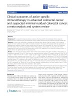
Prostate cancer is the 5th most common male cancer in Singapore, affecting 7.2% of all
male cancers between 1998 and 2002 (Figure 1). Its age-standardised rate had increased
four-fold from 4.2 per 100,000 per year between 1968 and 1972 to 17.4 per 100,000 per
year between 1998 and 2002. It occurs mostly in men after 50 years old and has the
highest average annual percent rate of increase for age-standardised incidence rate at
5.6% between 1968 and 2002 (Figure 2) [1].
Figure 1: Top ten most frequent male cancers in Singapore
(Singapore Cancer Registry, 1998-2002)
Lung
19.0
Colo-rectum
17.4
Liver
8.1
Stomach
7.7
Prostate
7.2
Percentage of all male cancers
Nasopharynx
5.7
Skin
4.3
Lymphomas
4.1
Bladder
3.3
Leukemias
3
0
5
10
Detecting cancer-specific peptides in prostate cancer
using MHC tetramer technology
15
20
13
Figure 2: Age-standardised rates between 1968-2002 (top) and age-specific rates
between 1968-2002 (bottom) for prostate cancer in Singapore
Detecting cancer-specific peptides in prostate cancer
using MHC tetramer technology
14
Prostate cancer is also the most common non-cutaneous male cancer in North America,
affecting one in every six men. The American Cancer Society estimates 234,460 new
prostate cancer cases in year 2006 with 27,350 prostate-cancer specific deaths [2].
The Surveillance, Epidemiology and End Results (SEER) programme from the National
Cancer Institute (NCI) in USA is a comprehensive cancer registry database to collect and
track cancer incidence and survival statistics in different population-based registries
throughout USA [3].
Although prostate cancer has maintained its lead in cancer
incidence in North America, the SEER database showed a gradual decrease in new
prostate cancer detection from the peak in early 1990’s recorded in 9 registries between
1975 and 2003 (Figure 3).
When compared to the USA, the trend in detecting new prostate cancers in Singapore
appears to be rising without any signs of reaching a plateau currently. It is vital for us to
work on better ways to detect and determine prognosis for these prostate cancer patients.
Detecting cancer-specific peptides in prostate cancer
using MHC tetramer technology
15
Figure 3:
SEER Age-adjusted incidence rates by race for all prostate cancer
(SEER 9 Registries for 1975-2003; Age-Adjusted to the year 2000
USA Standard Population)
Detecting cancer-specific peptides in prostate cancer
using MHC tetramer technology
16
Currently, clinicians have limited tools to test for prostate cancer. These tools include
clinical digital rectal examination (DRE) to palpate the prostate, serum prostate specific
antigen (PSA), and histological analysis of prostate tissues obtained by prostate needle
biopsies.
Prostate specific antigen is a serine protease from the kallikrein gene family [4]. It is
produced by the normal male prostatic epithelium and periurethral glands. However its
serum level is elevated in several prostatic diseases, which range from non-malignant
conditions including the benign prostatic hyperplasia, prostatic inflammation or
infections, to malignancies like prostate cancers. Hence, even though serum PSA is the
most common serum biomarker used for prostate cancer diagnosis, prognosis and disease
monitoring after therapy, it does not have high specificity or sensitivity because it is not
prostate cancer-specific.
The PSA era has led to increased diagnosis of early-staged prostate cancer, but stage-forstage cancer-specific mortality has unfortunately remained similar to decades ago.
Despite the widespread use of serum PSA, patients with apparently normal PSA values
may also have histologically-proven prostate cancer from transrectal prostatic needle
biopsies [5]. This problem is highlighted when up to 25% of men with prostate cancer
have PSA within the ‘normal range’ of less than 4.0 ng/ml [6].
Detecting cancer-specific peptides in prostate cancer
using MHC tetramer technology
17
In view of the limitations of PSA, population screening for prostate cancer has come
under scrutiny [7]. The exceptions for effective cancer screening may only apply to highrisk groups for prostate cancer, including African-Americans ethnicity, strong family
history of prostate cancer, and age of the patient.
Prostate cancer screening is further limited by the high false-negative first needle biopsy
rate. One-third of patients are not diagnosed from single-session needle biopsy due to
potential sampling errors. In addition, prostate needle biopsies are invasive procedures
with potential risks, including hemorrhage and infection.
Detecting cancer-specific peptides in prostate cancer
using MHC tetramer technology
18
T CELLS AND MAJOR HISTOCOMPATIBILITY COMPLEX (MHC)
T lymphocytes defend against intracellular micro-organisms and activate other cells like
B lymphocytes and macrophages. To achieve intercellular interactions, T cell receptors
(TcRs) recognise cell-associated antigens with high specificity through proteins encoded
by major histocompatibilty complex (MHC) locus. The TcR is highly specific in peptide
antigen binding before forming a complex with the MHC molecules on the target cell.
The MHC locus located on chromosome 6 at 6p21.3 is in one of the most gene-dense
regions of the human genome. It encodes some of the most polymorphic human proteins
in MHC class I and II molecules, which may contain over 200 allelic variants. With
complete gene sequencing by the MHC Sequencing Consortium in 1999, linkage
disequilibrium and genomic polymorphisms of the MHC genes are better understood for
future applications [8].
Segments of genes in the MHC locus encode cell surface-specific proteins in human,
known as the human leucocyte antigens (HLA). Traditionally, these refer to 3 main
MHC regions: the centromeric class II, the telomeric class I with the class III region in
between them. The HLA-A, HLA-B, HLA-C genes belong to MHC class I molecules,
while the HLA-DP, HLA-DQ and HLA-DR genes belong to MHC class II molecules.
Detecting cancer-specific peptides in prostate cancer
using MHC tetramer technology
19
The two main types of MHC gene products are the class I and class II molecules. The
MHC class I molecules are heterodimers consisting of a single transmembrane α-chain, a
β2-microglobulin and an antigenic peptide within the α1-α2 cleft needed for its stable
expression to present peptides to the CD8+ cytolytic T cells. These peptides are derived
from cytosolic proteins which have been degraded by proteasome from larger
intracellular proteins.
The MHC class II molecules are found on antigen presenting cells, like dendritic cells,
macrophages, activated T cells and B cells. They are heterodimers composed of two noncovalently associated homologous peptides, the α-chain and β-chain, which present
extracellular proteins to CD4+ helper T cells.
The MHC class III region contains genes that encode for complement components of
inflammation (e.g. C2, C4) and tumour necrosis factor (TNF) superfamily.
The nomenclature for HLA system had been updated regularly by the IMGT/HLA
Database, which is part of the international ImMunoGeneTics (IMGT) project that
operates as a high-quality resource centre for immunoglobulins, T cell receptors, major
histocompatibility
complex,
immunoglobulin
superfamily
(IgSF),
major
histocompatibility complex superfamily (MhcSF) and related proteins of the immune
system (RPI) of human and other vertebrate species [9]. It provides a specialist database
for updated sequences of the human major histocompatibility complex (HLA).
Detecting cancer-specific peptides in prostate cancer
using MHC tetramer technology
20
This database also includes the official sequences for the World Health Organisation
(WHO) Nomenclature Committee for Factors of the HLA System. As of Dec 2006, there
are 1,723 HLA class I alleles and 858 HLA class II alleles in the database.
Detecting cancer-specific peptides in prostate cancer
using MHC tetramer technology
21
CD4+ T-CELLS AND TUMOUR IMMUNOLOGY
In adaptive immunity, T cells play a key role in specific recognition of and response to
foreign peptide antigens, with the collaboration of MHC-restricted peptide bearing
antigen-presenting cells.
During cell-mediated immunity, CD4+ T cells activate
macrophages for phagocytosis while cytotoxic CD8+ T cells achieve targeted cell lysis.
For humoral immunity, CD4+ T cells stimulate proliferation and differentiation of B
lymphocytes.
The unique properties of T cells include recognising only specific amino acid sequences
of peptides and protein antigens. These antigen-specific T cells respond to foreign
peptides only if these antigens are attached to cell surfaces of antigen presenting cells
(APCs) of the particular individual. This process, known as self MHC restriction, affects
both the CD4+ and CD8+ T cells.
The formation of TcR-peptide-MHC complex is therefore highly regulated and this
allows for appropriate T cell activation and function. Hence, MHC Class II-restricted
CD4+ T cells recognise extracellular proteins that have been internalised into the vesicles
of APCs, with the help of co-stimulators like interferon-γ and CD40-CD40L interactions.
Similarly, CD8+ class I-restricted T cells recognise peptides specifically degraded from
cytosolic proteins that have undergone endogenous synthesis.
Detecting cancer-specific peptides in prostate cancer
using MHC tetramer technology
22
The T lymphocytes gain their unique functions from the lymphocytic maturation process.
Immature precursor cells in the bone marrow do not express antigen receptors until they
develop into mature lymphocytes in the peripheral lymphoid tissues. By the time they
mature, T cells have undergone sequential gene expression, generated diverse repertoire
of antigen receptor specificity and received functional and phenotypic characteristics that
are unique to their subtypes.
To ensure useful antigen receptor specificities are preserved in T cells, positive selection
of T cells whose receptors bind with weak and low avidity to self MHC molecules in the
thymus reach eventual T cell maturation. They are rescued from programmed cell death.
However, developing T cells with TcRs that do not recognise any thymic MHC
molecules are eliminated by apoptosis. Alternatively, if developing T cells with TcRs
that bind too strongly to self MHC antigens, they are also eliminated by apoptosis
through negative selection to maintain central tolerance, so as to prevent autoimmune self
destruction.
Detecting cancer-specific peptides in prostate cancer
using MHC tetramer technology
23
The end result of T cell maturation in the thymus is the formation of naïve mature T
lymphocytes. Once released to peripheral lymphoid organs, the T cells will be activated
if their TcRs recognise specific antigens found on peptide-MHC complexes carried by
antigen presenting cells. Upon activation in the presence of co-stimulators with cytokine
signals, these T cells proliferate and differentiate into memory and effector T cells.
For the CD4+ T cells, effector T cells like T-helper (TH) cells will secrete cytokines,
activate B cells and help macrophages in phagocytosis. Effector T cells differentiate into
various subsets from mature CD4+ T cells to perform different effector functions. The
TH1 cell lineage produces interferon-γ (IFN-γ) to combat microbials that activate
macrophages and natural killer cells, while the TH2 lineage secretes interleukin-4 (IL-4)
and interleukin-5 (IL-5) in the presence of helminthic worms and allergens.
It is
important that these activated T-cell responses are reduced when antigens have been
eliminated.
This is a normal process to ensure antigen-activated T cells undergo
apoptotic cell death and return the immune system to baseline homeostasis.
As for memory T cells, they survive long after the elimination of antigen stimulation and
are responsible for better and stronger secondary immune responses during future
exposures to the same antigen.
Unfortunately, the mechanisms, maintenance and
stimulation of CD4+ memory T cells are not well understood.
Detecting cancer-specific peptides in prostate cancer
using MHC tetramer technology
24
The idea of an immune system that seeks out and destroys developing cancer cells leads
to the concept of immune surveillance. In clinical practice, there is an increase in cancer
incidence of melanoma, Kaposi sarcoma and liver cancer in kidney transplanted patients
who received immuno-suppression treatment [10-11]. In patients with breast cancer and
melanoma, they have longer cancer-specific survival if histopathology analysis showed
that their tumour tissues are surrounded by infiltrates of T cells, NK cells and
macrophages [12].
Detecting cancer-specific peptides in prostate cancer
using MHC tetramer technology
25
TUMOUR ANTIGENS
Tumours antigens that are expressed exclusively on tumour cells and not on normal host
cells are known as tumour-specific antigens, while those that are also expressed on
normal cells are called tumour-associated antigens. These antigens may be products of
oncogenes or tumour suppressor genes, silent genes in normal tissues that had abnormal
expression, over-expressed genes or oncogenic viruses.
Some of them may be differentiation antigens normally found in its tissue of origin, like
the serum prostate specific antigen, while other antigens are oncofoetal proteins that are
absent in normal adults but present in both cancer and normal developing foetal tissues
(Table 1).
Detecting cancer-specific peptides in prostate cancer
using MHC tetramer technology