Evaluation of advanced paclitaxel drug delivery implants for controlled release post surgical treatment against glioblastoma multiforme in the brain
Bạn đang xem bản rút gọn của tài liệu. Xem và tải ngay bản đầy đủ của tài liệu tại đây (742.27 KB, 46 trang )
Ong Yung Sheng, Benjamin
EVALUATION OF ADVANCED PACLITAXEL DRUG DELIVERY
IMPLANTS FOR CONTROLLED RELEASE POST-SURGICAL
TREATMENT AGAINST GLIOBLASTOMA MULTIFORME IN THE
BRAIN
ONG YUNG SHENG, BENJAMIN
MSc, DIC, BEng (Hons)
A THESIS SUBMITTED
FOR THE DEGREE OF MASTER OF ENGINEERING
DEPARTMENT OF CHEMICAL & BIOMOLECULAR
ENGINEERING
NATIONAL UNIVERSITY OF SINGAPORE
Page 0
Ong Yung Sheng, Benjamin
CONTENT
I
Acknowledgements
2
II
Abstract
3
1.0
Introduction
4
1.1
5
2.0
Background & Literature Review
7
2.1
7
2.2
2.3
3.0
Motivation, Objectives & Organization
Development of Controlled Release Implants
for Chemotherapy to the Brain
Paclitaxel
Local Implants for Paclitaxel Delivery
8
9
Evaluation of Paclitaxel Foams for local implants
11
3.1
Materials and Methods
3.1.1 Paclitaxel Foam Formulations
3.1.2 In vitro release of Paclitaxel in PBS
3.1.3 Cell Culture Maintenances
3.1.4 Cell Growth, Viability and Apoptotic activity Studies
3.1.5 Animal Care
3.1.6 In vivo release of Paclitaxel
3.1.7 Intracranial Survivability Analysis
3.1.8 In vivo intracranial bio-distribution Studies
11
11
12
12
12
13
14
14
15
3.2
Results and Discussion
3.2.1 In vivo Release
3.2.2 In vitro Cell Proliferation & Apoptotic Activity
3.2.3 Intracranial Survivability Studies
3.2.4 In vivo Bio-distribution
16
16
20
23
23
3.3
Conclusions
25
4.0
Evaluation of EHDA Microparticles
4.1
Materials and Methods
4.2
In Vivo Release
4.3
Tumor Volume Response Study
26
26
27
30
5.0
Evaluation of Spray-Dried 0.8% Paclitaxel Loaded Discs
32
6.0
7.0
8.0
Conclusions and Recommendations
List of Figures
Reference
35
37
39
Appendix A: Raw Data from Experiments
42
Page 1
Ong Yung Sheng, Benjamin
ACKNOWLEDGEMENTS
The author would like to thank his group members in the drug delivery lab in particularly, Ms
Laiyeng Lee and Mr Jingwei Xie for providing the formulations and other technical support.
Thanks go to Mr Sudhir Hulikal Ranganath, Ms Dawn Ng, Ms Meijia Ng and Mr Junjie Huang for
laboratory assistance. Also thanks to Ms Fan Lu and A/Prof Lee How Sung, from Dept of
Pharmacology, NUS, for carrying out the LCMSMS analysis. To A/Prof Gavin Dawe, Ms Alice Ee
and Ms Han Siew Peng, Dept of Pharmacology, NUS, for technical training and advise in
intracranial surgery, to Ms Kho Jia Yen, NUMI histology Lab, NUS, for consultation on tissue
staining and preparations, and A/Prof Ong Wei Yi, Dept of Anatomy, NUS, for his valuable inputs
and time on experiment design and concept. Final thanks go to Prof Nick Sahinidis, UIUC, and
A/Prof Wang Chi-Hwa, NUS as thesis advisors.
Page 2
Ong Yung Sheng, Benjamin
ABSTRACT
In this thesis, evaluation of three different Paclitaxel controlled release biodegradable implants for
post-surgical implantation was carried out. The Poly-(DL-lactic-co-glycolic acid) (PLGA) based
implants were fabricated in the form of Pressure Quenched Foams, Electro-Hydrodynamic
Atomized microparticles and Spray-Dried Discs.
Two formulations of foams with different functions were evaluated. The formulations were (F1)
5% Paclitaxel loaded PLGA 85:15 foams as the slower but prolonged releasing implant and (F2)
10% Paclitaxel PLGA 50:50 foam for faster drug release. Experiments carried out were in vitro
cell cultures to compare controlled release from foams vs. acute Paclitaxel exposure over 24
hours in terms of cell proliferation response and apoptotic activity. We were able to show through
the biodistribution in brain tissue experiment that Paclitaxel levels were sustained at ~ 3 mm from
the site of implantation over a period of 28 days.
Electro-hydrodynamic microparticles were showed to agree with in vitro release within an in vivo
environment releasing Paclitaxel for up to 28 days after implantation. In an tumor response study,
the results suggests enhanced tumor suppression by prolonging time taken to reach max tumor
volume of 3,000mm3 by 7 days over the commercial Taxol® product.
The in vivo release of Sprayed Discs was carried out and the results show some correlation to the
published in Wang et al (2003) [18]. The results suggest that in an in vivo environment, sustained
release can be achieve for up to 42 days with a peak release into systemic circulation observable
at 21 days after implantation
Page 3
Ong Yung Sheng, Benjamin
CHAPTER 1:
INTRODUCTION
One of the main challenges of modern pharmacology has been the delivery of the therapeutic
agent to the site of action (where the agent is need) and to reach concentration levels high
enough to achieve the desired treatment response. Often in many cases, duration of exposure at
these levels over prolong periods of time are essential to prevent a relapse back into the
diseased state and to provide a sustainable environment for patient recovery. Moreover, control
of drug levels below toxicity limits are crucial to prevent/reduce side effects to an acceptable
level.
Many of these requirements and challenges are not unlike those encountered in the field of
chemical engineering. The encapsulation drugs in a biodegradable matrix from which the drug
can diffuse out from is analogues to chemical reactants diffusing into a catalyst pore. By changing
constituent block concentrations in the polymer matrix, control of the rate of polymer degradation
can be achieved thereby changing rate of release of the drug.
Our study focuses on developing controlled releases implants in the form of discs for Paclitaxel to
be surgically inserted to remove remaining tumor cells after a debulking surgery (to remove the
main tumor bulk) in the brain. The discs are inserted into the cavity (where the resected tumor
was removed from) and the wound is closed. Over time, the discs will release Paclitaxel into the
peripheral tissue up to the durations of more than a month. It is hoped that by applying this
strategy, tumor cells around the cavity would be eliminated and tumor remission avoided.
Page 4
Ong Yung Sheng, Benjamin
1.1 MOTIVATION, OBJECTIVES & ORGANIZATION
The long-term vision beyond the scope of this project would be to develop accurate
computational models that would aid in the analysis and design of controlled release implants.
Results from here can be extrapolated to consider synergetic treatments e.g. changes in
transport of the drug under environment of periodic irradiation therapy which is know to induce
interfering physiological changes. Irradiation is known to increase blood brain barrier (BBB)
permeability which affects drug transport from the implant through drug loss from interstial tissue
across BBB into cerebral spinal fluid (CSF) circulation besides drug diluting effects due to CSF
coming into the interstial space. Modeling such dynamics can help medical practitioners and
scientist explain causes for or lack of treatment efficacy of strategies undertaken.
To begin on this vision, the goal of this MEng project was to carry out preliminary in vivo
experiments to evaluate the treatment with novel Paclitaxel release foam developed by Ms Lai
Yeng, a fellow research group member, based on a high pressure quench and rapid solidification
of drug-polymer melt.
This thesis presents a step-by-step approach in the analysis of the use of foams for controlled
release of Paclitaxel as implants within the brain for the post-surgical treatment of glioblastoma
multiforme through combining cell culture and in vivo experiments to evaluate the efficacy of this
treatment within the body.
Key issues for evaluation of the foams carried out in this thesis involve
(i)
In vitro drug release in PBS to examine at the degradation rate of the polymer and hence
the drug release profile. This section was undertaken by Ms Lee Lai Yeng but is presented
in this thesis for completeness.
(ii)
In vivo release subcutaneously in mice to obtain release profiles within the body. This
experiment reconfirms the release profile in a physiologically lipophilic environment. This
Page 5
Ong Yung Sheng, Benjamin
was thought to be significant since degradation rate of PLGA is likely to change according
to proportions of hydrophilic and lipophilic blocks. Moreover, this step evaluates the safety
of the implants against bulk release of the drug resulting in systemic toxicity. Weights of the
animals were regularly to check this point.
(iii)
Level of toxicity response of tumor cells cultures to sustained release from the foams
through cell growth and relative caspase 3 activity levels. Design of controls compared the
recovery of the (a) cells to acute exposure (over 24 hrs) with commercial taxol and (b) the
experimental foams. Experimental design was on the basis of two times the Area Under the
Curve (AUC) levels. An AUC level is the area under the curve of a plot of axis between
Concentration of the drug vs. the duration of exposure to the drug and is used here to
provide consistency in the design of the control groups with commercial Taxol®. This study
attempts to show the value of sustained release on a cellular level.
(iv)
Intracranial biodistribution of the drug in the brain over time. This study presents the ability
of the foams to maintain therapeutic levels of paclitaxel at distances away from the implant
over a period of one month. This is important as it illustrates sustain release and
penetration distance of the drug from the site of implantation. It also serves as raw data for
computational model validation
(v)
Intracranial Survivability of tumor-laden rats treated with Paclitaxel laded and placebo
(blank PLGA polymer without Paclitaxel) PLGA implants. Prolong survivability of the
experimental groups over the placebo groups indicate enhanced treatment by the foams.
Besides the foams implants, evaluation of two other implant formulations was undertaken.
Namely, 16.8% Paclitaxel loaded EHDA (ElectroHydroDynamic Atomization) microparticles
where we analysed the in vivo release profile as well as tumor volume response and 0.8%
Paclitaxel loaded Spray-dried compressed discs in an in vivo release study.
Page 6
Ong Yung Sheng, Benjamin
CHAPTER 2:
BACKGROUND & LITERATURE REVIEW
This section provides a summary of research in the development of controlled release implants to
the brain. Section 2.1 provides an outline of the development and challenges to effective
treatment, Section 2.2 covers the background of Paclitaxel, which is the chemotherapeutic agent
to be delivered and Section 2.3 will give a review of the research to date specifically of controlled
release implants for Paclitaxel.
2.1 DEVELOPMENT OF CONTROLLED RELEASE IMPLANTS FOR CHEMOTHERAPY TO
THE BRAIN
Over the last three decades there has been a rise in brain cancers like glioblastoma multiforme
(GBM), oligodendroglioma, anaplastic astrocytoma, medulloblastoma, and mixed glioma has
been on the rise. Of these, GBM is the most frequent accounting for 16,797 cases out of 38,453
cases per year of malignant brain tumors between 1973 and 2001 in America alone [1].
The conventional clinical treatment for glioma is by surgical debulking of the accessible tumor
from the patient’s brain. The amount of tumor removed is often limited by proximity to critical
regions for brain function and this presents a risk of tumor re-growth from residual tumor. The
approach for limiting cancer remission is carried out by conventional systemic post-surgical
chemotherapy and radiotherapy courses. Unfortunately, these have resulted in limited clinical
effectiveness due to restricted transport of chemotherapy agent across the BBB (blood brain
barrier) and significant PgP (P-glycoprotein) mediated efflux barrier effects [2]. To overcome
barriers to effective drug transport, biodegradable controlled-release polymers implants could be
surgically located at the site of tumor removal during the debulking surgery. Commercial implants
Page 7
Ong Yung Sheng, Benjamin
®
like the Gliadel Wafer delivering BCNU (Carmustine) has enjoyed limited successes in improving
patient survival rates. Clinical trials with Gliadel® Wafer vs. placebo wafers have been shown to
prolong survival in people with newly diagnosed high-grade malignant gliomas (in addition to
surgery and radiation) from a median survival of 11.6 months to 13.9 months. With recurrent
glioblastoma multiforme in addition to surgery, median survival increased to 6.4 months from 4.6
months [3, 4]. Since only one third of GBM patients are responsive to BCNU [5] with other Gliadel
wafer associated complications like cerebal edema [6], several groups have been working on
controlled release for other drugs such as doxorubicin [1] and paclitaxel.
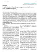
2.2 Paclitaxel
Paclitaxel (see Figure A for chemical structure), a chemotherapeutic drug originating from the
pacific yew Taxus brevifolia, and other members of the Taxaceae family [7] is commonly used as
a chemotherapeutic agent of for ovarian and breast cancer. Paclitaxel functions through
promotion of the assembly and stabilization of microtubules inhibiting cellular division. It also
prevents de-polymerization of the assembled microtubules and thereby halts mitosis or cell
division and binds to Bcl-2 [8, 9] which normally blocks the process of apoptosis, allowing
apoptosis to proceed. Unfortunately, Paclitaxel is highly hydrophobic and exhibits a fast plasma
clearance when administered by infusion [10]. Absorption across the BBB was also poor due to pglycoprotein (p-gp) efflux effects [11, 12, 13]. However, studies have over showed that prolong
exposure to Paclitaxel for more than 24 hrs can provide significant clinical efficacy [14].
Figure A: Chemical Structure of Paclitaxel
Page 8
Ong Yung Sheng, Benjamin
2.3 Local Implants for Paclitaxel Delivery
Several studies have been carried out using different materials to achieve controlled release of
Paclitaxel from surgical implants. Von Eckardstein et al. used a nitrosoureas liquid crystalline
cubic phase encapsulating carboplatin and paclitaxel and reported reduction in tumor sizes in F98
rat brains. Brain tissue concentration of Paclitaxel showed little or no drug in the vicinity of 3 mm
beyond 7 days. Clinical observations of the same formulations have suggested feasible and safe
usage if < 15 mg paclitaxel was used [15, 16].
Li et al. used implants based on polyphsophoester p(DAPG-EOP) polymer at 10% drug loading
into Polilactofate microspheres which were combined with PEG-100. Brain tissue concentrations
after 30 days showed drug concentrations above LD90 (Drug concentration need to kill 90% of
tumor cells) of a depth between 5 to 7 mm and enhanced survivability [17].
Wang et al. reported the in vitro release profiles of discs released from Poly (DL-lactic-co-glycolic)
acid 50:50 (MW 45,000- 75,000) fabricated by spray-drying followed by 2 ton compression, a
delay of 15 days before drug release was observed [18].
Elkharraz et al. fabricated injectable Poly (DL-lactic-co-glycolic) acid 50:50 based microparticles
from oil-in-water extraction/evaporation method and glycerol tripalmitate-based implants with 29
and 60% w/w and showed that release of 73 to 87 % of the encapsulate drug within 7 days in the
presence of N,N-Diethylnicotinamide (DENA), a hydrotropic agent for paclitaxel, significantly
increase the release of paclitaxel increased due to elevated hydrolysis rate of PLGA polymers
and the paclitaxel solubility [19, 20]. However, how DENA would be used in drug delivery seemed
to be in question since DENA affects the central nervous system expressed in seizures and
behavioral changes [21].
Ho et al., was able to show that a constant zero-order in vitro release of 0.92+/-0.03 pg/day
Paclitaxel over 5 days was achievable using Chitosan-egg phosphatidylcholine (chitosan-ePC)
Page 9
Ong Yung Sheng, Benjamin
films. Inhibition of SKOV-3 (human ovarian adenocarcinoma cell line) proliferation was shown
with an ED50 of 211 ng/ml from the films. A sustained, zero-order release of Paclitaxel was also
seen in vivo over a 2 week period in mice implanted with the films [22].
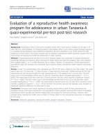
Ruan et al., developed paclitaxel loaded poly(lactic acid)-poly(ethylene glycol)-poly(lactic acid)
(PLA-PEG-PLA) microspheres and found faster release rates than conventional Poly (DL-lacticco-glycolic) acid (PLGA), besides being able to provide sustained release of 49.6% of the
encapsulated drug after one month [23]. In a latter work, paclitaxel was encapsulated with
Vitamin E TPGS-emulsified Poly(D,L-lactic-co-glycolic acid) (PLGA) nanoparticles, of an average
size of 240 nm, prepared by a modified solvent extraction/evaporation techniques with vitamin E
as an emulsifier.
Figure B: Chemical Structure for Poly (DL-lactic-co-glycolic) acid
Page 10
Ong Yung Sheng, Benjamin
CHAPTER 3:
EVALUATION OF PACLITAXEL FOAMS FOR LOCAL IMPLANTS
3.1 Materials and Methods
3.1.1 Paclitaxel Foam Formulations
Two main formulations were used in this work, the first was based on a 5%w/w Paclitaxel loaded
foam with Poly-(DL-lactic-co-glycolic acid) 85:15 (F1) and 10%w/w Paclitaxel loaded foam with
Poly-(DL-lactic-co-glycolic acid) 50:50 (F2).
Paclitaxel (Bristol-Myers Squibb, New Brunswick, NJ) was incorporated into the polymer matrix
(Poly (DL-lactic-co-glycolic acid 85:15 or Poly (DL-lactic-co-glycolic acid 50:50) (Mol. Wt. 50,000 –
75,000 and Mol. Wt. 40,000 – 75,000 respectively) by dissolution in Dichloromethane and spray
drying the solution to form microparticles using a Buchi Spray Drier with inlet air flow-rate of 700
L/min, inlet temperature at 70oC, aspirator setting of 100% and a pump rate of 30%. The
microparticles were collected and freeze dried for 72 hours to remove any residual solvents
before being subjected to a high pressure of 70 bar for 45-60 minutes under CO2 within a
chamber. The high pressure depresses the glass transition temperature of the polymer and
allowing dissolution of CO2 gas into the polymer melt. Upon rapid decompression at 15 bars per
minute, the glass transition temperature of the polymers rise forming gas pockets which escapes
leaving interconnecting uniform pores in the foam [24].
The foam was set as a 3 mm diameter disc with a 1 mm thickness by the mold holding the
polymer melt in the compression chamber. Discs were used for the in vitro, in vivo release
profiles and intracranial survivability studies while rods of dimensions of 7 mm length x 1 mm
diameter were used for intracranial bio-distribution studies. Blank placebo foams were fabricated
in the same way without Paclitaxel.
Page 11
Ong Yung Sheng, Benjamin
The foam discs and the rods packaged into 2 ml eppendorf tubes before sterilization by gamma
irradiation to a dose of 15 kGy. Total weight of the foam discs and rods were 3 mg and 2 mg
respectively.
3.1.2 In vitro release of Paclitaxel in PBS
The foams discs and rods were incubated in 5 ml of PBS (pH 7.4) at a temperature of 37 oC.
Paclitaxel in PBS was measured by extraction into Dichloromethane and dissolution into HPLC
Acetonitrile/water (50/50%) mobile phase. The PBS replaced with fresh PBS after every sampling
to ensure that the solubility limit of Paclitaxel in PBS is not reached.
3.1.3 Cell Culture Maintenances
The cell line used was a rat glioma cell line (ATCC® Number: CCL-107™), C6 cells, established
by Benda et al. [25] and reported to be derived from N-methyl-nitrosourea-transformed rat
astrocytes. The cells were grown in DMEM (Dulbecco’s Modified Eagle Medium, Sigma)
supplemented with 10% Bovine Fetal Serum (Gibco, Invitrogen) and 1% Penicillin/Streptomycin
(Gibco, Invitrogen) in a humidified incubator. After reaching confluence, the cells were prepared
by washing in PBS and detached from the T-flask with Trypsin-EDTA (Gibco, Invitrogen). The
cells were re-suspended to obtain a concentration of 3 x 10⁵ cells/2.5 µL before inoculation into a
75 cm3 T flask containing 15 ml of fresh media.
3.1.4 Cell Growth, Viability and Apoptotic activity Studies
Investigation of the cellular response to the Foams was carried out by comparison with three
control groups (namely Blanks, 5050_Taxol & 8515_Taxol – see following for explanation). All
groups (experimental and controls) were inoculated at a density of 1 x 10⁶ C6 Glioma cells into
175 cm3 T-Flask into 50 ml of DMEM culture medium on Day 0 and cell density was counted on
Days 4 and 8. The cells in all the flask were allowed 2 days to attach and grow in the flask before
the respective foams 5% Paclitaxel Loaded PLGA 85:15 (F1) and 10% Paclitaxel Loaded PLGA
50:50 (F2) were administered into the flask.
Page 12
Ong Yung Sheng, Benjamin
2 control flasks, 5050_Taxol and 8515_Taxol, received 49 ug and 7 ug of Paclitaxel respectively
administered in the form of the commercial Taxol®, obtained from Bristol-Myers Squibb, on day 2
(the two Paclitaxel concentrations were calculated to give an equivalent dosage of 2 x AUC over
24 hrs as the two foams implants would released over 6 days of exposure). After 24 hours, the
Paclitaxel laden medium was removed and the cells washed with two rounds of PBS washes
before replacement of Paclitaxel-free medium. These controls were used to simulate acute
exposure to paclitaxel to cells over 24 hours window and observing cellular recovery on Day 4
and 8.
Growth curves of the groups were as separate flasks carried out in triplicates and counted by
conventional Trypan Blue dye exclusion method in a hemocytometer to obtain cell densities and
cell viability on days 2, 4 and 8. 3 x 10⁶ cells were collected for caspase 3 activity level
measurement using a Caspase-3/CPP32 Fluorometric Assay Kit from Biovision.
Caspase 3 activities levels were determined by re-suspending cells in 50 µl of chilled cell lysis
buffer. The cells were then incubate cells on ice for 10 minutes before adding 50 µl of 2x
Reaction Buffer (containing 10 mM DTT) to each sample. 5 µl of the 1 mM DEVD-AFC substrate
were then added before incubating at 37oC for 2 hours before read samples in a fluorometer
equipped with a 405-nm excitation filter and 485-nm emission filter. The upon cleavage of the
substrate by CPP32 or related caspases, free AFC emits a yellow-green fluorescence (λmax = 505
nm). Absorbances were then compared with controls for comparison on caspase activity
response.
3.1.5 Animal Care
All experiments were carried out with approval from the National University of Singapore’s
Institutional Animal Care and Use Committee (IACUC) and housing and care of animals are
provided in accordance with the National Advisory Committee for Laboratory Animal Research
(NACLAR) Guidelines (Guidelines on the Care and Use of Animals for Scientific Purposes) in
Page 13
Ong Yung Sheng, Benjamin
facilities licensed by the Agri-Food and Veterinary Authority of Singapore (AVA). A total of 55
Wistar Rats (obtained from Centre for Animal Resources, CARE, Singapore) were used for the
work in this present study. The rats were housed in cages and given free access to standard
laboratory food and water.
The tranquilization, induction and maintaining agent for Wistar Rats was administered as 100 mg
Ketamine / kg body weight and 10 mg Xylazine/kg body weight for all surgeries. For BALB/c mice,
the dose was 75 mg Ketamine/kg body weight and 1 mg Medetomidine/kg body weight.
3.1.6 In vivo release of Paclitaxel
In vivo release of Paclitaxel for Foams F1 and F2 was carried out by implanting the foams
subcutaneously in BALB/c male mice (beginning at an average weight of 30 g). At fixed time
points Day 7, 14, 28, 42, 56 and 70, one experimental group is sacrificed (n = 3 mice) and the
foams recovered from the cadaver. The foam is re-dissolved in DCM and analyzed for residual
Paclitaxel by HPLC. The animals were anesthetized with ketamine (75 mg/kg) and medetomidine
(1 mg/kg), shaved and scrubbed down with 70% alcohol, dilute Hebis scrub (chlorhexidine)
followed with a final scrub with Betadine. A small 1 cm incision is made on the lateral flank of the
animal and the foam inserted. The wound was then sutured up and allowed to heal before
removing stitches after 1 week. Weights of the groups were taken weekly to check on Paclitaxel
related toxicity.
The objective of this study was to verify release profile in an in vivo environment and to check for
safety for use against implant failure.
3.1.7 Intracranial Survivability Analysis
5 x 10⁶ C6 Glioma Tumor cells were injected subcutaneously on the lateral flank of the Wistar rat
and allowed to grow. Once the tumor had reached the required volume, the animal was sacrificed
and the tumor resected from its back and cut into 2 mm pieces for intracranial implantation.
Page 14
Ong Yung Sheng, Benjamin
For tumor implantation, each rat was anaesthetized, shaved and scrub down with 70% alcohol,
dilute Hebis scrub (chlorhexidine) followed with a final scrub with Betadine. An incision is made to
the scarp of the rat and a 3 mm diameter burr hole was made in the skull 5 mm posterior and 3
mm to the right of the bregma for the animals undergoing intracranial surgery. The dura was
incised sharply, and the underlying cortex was resected with light suction. Hemostasis was
obtained by light compression using sterile gauze, and the wound was subsequently irrigated.
Dissected 2 mm pieces of C6 tumor tissue were implanted in the resection cavity, and the wound
was closed by suturing. On day 8, the animal was re-anesthetized, the wound reopened and the
foam was placed on top of the tumor implant. The wound was then re-closed by suturing.
A total of 30 rats were used for the survivability analysis, a control placebo (Control, n = 10)
received a 3 mm diameter blank PLGA discs and two experimental group were used , one for (i)
5%w/w Paclitaxel loaded PLGA 85:15 discs (F1, n = 10) and (ii) 10% w/w Paclitaxel loaded PLGA
50:50 discs (F2, n =10). The total weight of discs was 3 mg.
The rats were weighted once every two days and were sacrificed when they showed signs of
persistent anorexia or dehydration, body weight loss of 20%, inability to maintain an upright
position or to move, moribundity, lethargy or failure to respond to gentle stimuli, or bloodstained
or mucopurulent discharge from nose or eyes.
3.1.8 In vivo intracranial bio-distribution Studies
For biodistribution studies, only (i) 5% w/w Paclitaxel loaded PLGA 85:15 rods (F1, n = 5) and (ii)
10% w/w Paclitaxel loaded PLGA 50:50 rods (F2, n =5)) were used over 3 separate time points (
14, 21 and 28 days) The rods were ~ 1 mm diameter x 7 mm in length and weight 2 ± 0.2 mg
each. Total number of rats for bio-distribution studies was 60 rats.
Each Wistar rat was anaesthetized, shaved and scrub down with 70% alcohol, dilute Hebis scrub
(chlorhexidine) followed with a final scrub with Betadine. An incision is made to the scarp of the
Page 15
Ong Yung Sheng, Benjamin
rat and a 3 mm diameter burr hole was made at 1.5 mm posterior of the bregma and 2 mm left
from the midline under stereotaxic control. To create a path for inserting the rod, an 18 Gauge
needle was inserted to a depth of 7 mm from the brain surface and retracted. The foam rod was
then inserted completely into the incision created. The scarp of the rat was then closed by
suturing and the animal allowed to recover while the weights of the animals were monitored daily.
At the respective time points, the rats were sacrificed and their brain harvested. The brains were
immediately frozen, kept at - 80 oC before being sectioned coronally in a rat brain matrix with 1.0
mm thickness. Each slice was carefully weighted, homogenized and analyzed for Paclitaxel
concentration by Liquid Chromatography Mass Spectrometry (MS) method (LCMSMS) in Dept of
Pharmacology, NUS.
3.2 Discussion & Results
3.2.1 In Vivo Release Profile
The Paclitaxel release rate was evaluated in vivo to determine the actual rate of drug release
within the body’s environment. This was used to observe the rate of diffusion of Paclitaxel from
the pores of the foam in body fluids and to check for major bulk degradation of the polymer
matrix.
The 2 foam formulations were first evaluated in vitro in PBS (carried out by Ms Lee Lai Yeng, but
shown here for completeness).
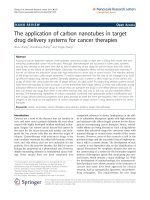
Figure 1 shows a comparison between the release between two copolymer (PLGA 50:50 & PLGA
85:15) used starting a common 5% Paclitaxel Loading. The data highlights the fact that the
release rate of PLGA 50:50 is much faster than the PLGA 85:15. This is because of the higher
hydrophilic poly-Lactate content in the copolymer which results in faster molecular weight drop in
aqueous PBS than the PLGA 85:15 which has a higher hydrophilic polymer content. This
understanding formed the basis for selecting these polymer proportions for PLGA. We wanted to
Page 16
Ong Yung Sheng, Benjamin
offer two options: a fast releasing foam to arrest fast growing tumors besides bringing up the
surrounding tissue to a therapeutic concentration and a slower releasing foam to maintain the
tissue’s Paclitaxel concentration over a prolong period of time. PLGA 50:50 Paclitaxel drug
loading was doubled to 10% to rise the absolute amount of Paclitaxel release while PLGA 85:15
drug loading was kept at 5% to meet this objective. It is hoped that these two options or the
combination of both would provide the surgeon with room for decision making based on the
specific circumstances in the operating theatre.
The in vitro release profile of the 10% Paclitaxel loaded PLGA 50:50 foams in the form of discs
and rod (rods will be used in biodistribution latter) are presented in Figure 2. This figure confirms
that the release variability between discs and rods are not significantly difference and allows the
use of rods for bio-distribution studies as a model (rods are used due to ease of insertion into the
brain and improves survivability from the surgery). No characteristic initial drug burst was
observed and the data shows a cumulative 8% drug release after 35 days in PBS. In terms of
total mass of Paclitaxel releases by day 35, 24.1 µg was released out of 305 µg encapsulated for
discs and 18.2 µg out of 230 µg encapsulated in rods.
Figure 5A & 5B present the in vivo cumulative % release of Paclitaxel in from 3 mm discs
implanted in BALB/c Mice (n = 3 mice) for 5% Drug Loaded PLGA 85:15 & 10% Drug Loaded
PLGA 50:50
over 3 weeks respectively for procedure as described in Section 3.1.6. This
experiment was carried out to evaluate the safety and drug release behavior of the implant in an
in vivo environment. It was observed that in the 5% Drug Loaded PLGA 85:15 foams, 22 wt% of
the Paclitaxel released after 28 days while for the 10% Drug Loaded PLGA 50:50 foams, 9 wt%
of the Paclitaxel were released. By normalizing both foams to an initial drug loading of 10%, we
get about 18 wt% drug release for PLGA 85:15 foams and this suggest no significant difference
for in vivo release profile between PLGA 50:50 and PLGA 85:15. Considering the safety in using
these implants, the weights of animal were consistently rising throughout the experiments and did
Page 17
Ong Yung Sheng, Benjamin
not show signs of Paclitaxel related toxicity, indicating no sign of sudden burst release over the 3
weeks window after implantation.
It was observed during the in vitro experiment that the PLGA 50:50 foams showed significant
swelling in PBS. This was not evident in the discs recovered from the mice. The measured
diameters of the discs in Figure 6 showed a varying diameter about 3 mm and a possible
explanation for the lack of the swelling phenomenon maybe the less hydrophilic environment of
the body as compared to PBS.
35
PLGA 50:50 (5% Paclitaxel Loading) 3 mm Diameter Disc
PLGA 85:15 (5% Paclitaxel Loading) 3mm Diameter Disc
% C um ula tiv e Pa clitaxe l R elea se
30
25
20
15
10
5
0
0
5
10
15
20
25
30
35
40
Time (Days)
Figure 1: Comparison of In vitro Paclitaxel release from PLGA 50:50 and PLGA 85:15 Foams (n =3) [24]
9
30
% C u m u lativ e Pac litaxel R ele as e
8
PLGA 50:50 (10% Paclitaxel Loading) rods
PLGA 50:50 (10% Paclitaxel Loading) 3mm disc
a
C u m ula tiv e Pa cl ita xe l R el ea se d f rom F o am [µ g
PLGA 50:50 (10% Paclitaxel Loading) 3mm disc
7
6
5
4
3
2
1
b
PLGA 50:50 (10% Paclitaxel Loading) rods
25
20
15
10
5
0
0
0
5
10
15
20
Time (Days)
25
30
35
40
0
5
10
15
20
25
30
35
40
Time (Days)
Figure 2: In vitro release Profile of 10% Paclitaxel Loaded PLGA 50:50 Foams in the form of 3 mm diameter
discs (Total Weight = 3 mg) and 1 x 1 x 7 mm rods (Total Weight = 2 mg) in terms of Cumulative % (a) and
mass Paclitaxel release (b) (n =3) [24]
Page 18
Ong Yung Sheng, Benjamin
16
16
12
10
8
6
4
2
0
0
5
10
15
20
Time (Days)
25
30
35
b
14
12
10
8
6
4
2
0
40
0
5
10
15
20
25
30
35
40
Time (Days)
Figure 3: In vitro release Profile of 5% Paclitaxel Loaded PLGA 85:15 Foams in the form of 3 mm diameter
discs (Total Weight = 3 mg) and 1 x 1 x 7 mm rods (Total Weight = 2 mg) in terms of Cumulative % (a) and
mass Paclitaxel release (b) (n =3) [24]
Figure 4: SEM image of pores structures in foams (Mean Pore Diameter = 396.7 um; S. D = 160.7um) [24]
35
20
y = 2.8650E+00x2 - 5.3430E+00x + 1.1374E+01
30
R2 = 1.0000E+00
A
18
% Drug Release (ug Drug/ug Foam)
% Drug Release (ug Drug/ug Foam)
% C um ulativ e Pa clita xe l Re le a s e
C u m u la tiv e P a c lita xe l R e le a s e d fro m F o a m [ µ g ]
a
14
25
20
15
10
5
0
Day 14
Day 21
Day 28
16
y = 1.5150E-01x2 + 1.6264E+00x + 2.6682E+00
R2 = 1.0000E+00
B
14
12
10
8
6
4
2
0
Day 14
Day 21
Day 28
Figure 5: In vivo release profile foam discs recovered from BALB/c Mice (n = 3 mice) over 3 weeks from 3
mm foam discs. Graph A presents the % cumulative release of Paclitaxel from 5% Paclitaxel loaded PLGA
85:15 foams, Graph B presents the % cumulative release of Paclitaxel from 10% Paclitaxel loaded PLGA
50:50 foams
Page 19
Ong Yung Sheng, Benjamin
10% Paclitaxel Loaded PLGA 50:50 Foam
4.00
5 % Paclitaxel Loaded PLGA 85:15 Foams
Diameter of Discs [mm]
3.50
3.00
2.50
2.00
1.50
1.00
0.50
0.00
Day 14
Day 21
Day 28
Day 42
Figure 6: Diameter of foams (F1 & F2) implanted in vivo over 6 weeks. No discs were recoverable on
day 42 for PLGA 50:50 foams
3.2.2
In Vitro Cell Proliferation & Apoptotic Activity
The cell proliferation response to the 5% Paclitaxel loaded PLGA 85:15 foams (F1) shows a
reduction of 14.6 % and 61.8 % on Day 4 and Day 8 respectively over the Blank control (without
treatment). The 8515_Taxol controls arrest cell proliferation and showed a marginal recovery
after the 24 hr acute exposure (Paclitaxel concentration to give 2 x AUC for 85:15 foam release
over 6 days) from day 2 onwards with a cell growth ratio (Day 8 / Day 4) of 1.12 which was
smaller than the ratio for F1 groups which proliferated by 1.52 times over 4 days. The apoptotic
activity analysis data suggests that between day 4 and 8, the activity drops indicating a recovery
trend while activity increase for the F1 group which highlights increasing toxicity.
1.20E+08
Cells per flask
1.00E+08
Blank
8515
8515_Taxol
8.00E+07
6.00E+07
4.00E+07
2.00E+07
0.00E+00
Day 2
Day 4
Day 8
Figure 7: Cell Proliferation Response for Blank controls, 5% Paclitaxel loaded PLGA 85:15 foams (F1)
and 85:15_Taxol, the acute 24 hr exposure control group (Paclitaxel concentration based on 2 x AUC
with 6 day release of PLGA 85:15 foams)
Page 20
Ong Yung Sheng, Benjamin
5000
Blank
4500
8515
Flouresence Intensity
(Correlated Apoptotic Activity)
4000
8515_Taxol
3500
3000
2500
2000
1500
1000
500
0
Day 4
Day 8
Figure 8: Apoptotic Activity Analysis for Blank controls, 5% Paclitaxel loaded PLGA 85:15 foams (F1)
and 85:15_Taxol, the acute 24 hr exposure control group (Paclitaxel concentration based on 2 x AUC
with 6 day release of PLGA 85:15 foams)
Experiments with 10% Paclitaxel Loaded PLGA 50:50 foams showed a reduction of 35.3% and a
78.9% reduction in cell densities over the blank control groups on Days 4 and 8. Cell growth ratio
Day 8 and Day 4 for 5050_Taxol control group was 1.47 compared to 1.11 in flask with the foam.
This indicated stronger proliferation suppression by the foam. This was expressed in an
increasing apoptotic activity in the flasks with foams while the activity dropped between Day 4
and 8 showing recovery from the acute treatment.
This study highlights the benefits of sustained release of the foams over acute Paclitaxel
exposure treatment as observed in conventional IV infusion chemotherapy treatment at over two
times a similar AUC as the foams over 8 days. The cell cultures with both foams showed tumor
growth suppression and an increasing tread of Caspase 3 activation over an experimental
window of 4 days while, the acute control treatment suggests evidences of cellular recovery.
Hence, sustenance of a high level of Paclitaxel within the vicinity of the tumor is critical to prevent
cellular recovery and to ensure a commitment to the apoptosis pathway.
Page 21
Ong Yung Sheng, Benjamin
1.20E+08
Blank
5050
1.00E+08
5050_Taxol
Cells per flask
8.00E+07
6.00E+07
4.00E+07
2.00E+07
0.00E+00
Day 2
Day 4
Day 8
Figure 9: Cell Proliferation Response for Blank controls, 10% Paclitaxel loaded PLGA 50:50
foams (F2) and 50:50_Taxol, the acute 24 hr exposure control group (Paclitaxel concentration
based on 2 x AUC with 6 day release of PLGA 50:50 foams)
4500
Flouresence Intensity
(Correlated Apoptotic Activity)
4000
Blank
5050
5050_Taxol
3500
3000
2500
2000
1500
1000
500
0
Day 4
Day 8
Figure 10: Apoptotic Activity Analysis for Blank controls, 10% Paclitaxel loaded PLGA 50:50
foams (F2) and 50:50_Taxol, the acute 24 hr exposure control group (Paclitaxel concentration
based on 2 x AUC with 6 day release of PLGA 50:50 foams)
Page 22
Ong Yung Sheng, Benjamin
Cell Density Grow th Ratio
(Day 8/Day 4)
1.800
1.600
1.400
1.200
1.000
0.800
0.600
5050
5050_Taxol
Figure 11: Cell Density Growth ratio for (Day 8 / Day 4) for 10% Paclitaxel loaded PLGA 50:50
foams (F2) and 50:50_Taxol, the acute 24 hr exposure control group (Paclitaxel concentration
based on 2 x AUC with 6 day release of PLGA 50:50 foams
3.2.3
Intracranial Survivability Studies
The intracranial survivability studies for both foam formulations were still in progress at the time of
this thesis writing. Full data results would be shown in the upcoming publication covering this
work.
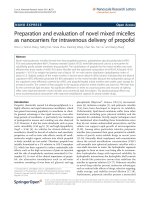
3.2.4
In vivo Bio-distribution
The bio-distribution of Paclitaxel in brain tissue was measured in vivo over 3 time points on day
14, 21 and 28. This was carried to evaluate the penetration and sustainability of Paclitaxel
concentration over time. The foams were cut into a 1 x 1 x 7 mm rod weighing 2 mg (Paclitaxel
concentration at 100 ug) and inserted vertically into the brain on day 0. At the specific time point,
the rat brain was harvested and sectioned coronally into slices of 1 mm thickness .
The results for the 5% Paclitaxel Loaded PLGA 85:15 foam suggest that paclitaxel concentrations
are still rising even up to 28 days after implantation. This indicates that the rate of drug
elimination is lower than the release of Paclitaxel from the foam. Also after 28 days, paclitaxel
levels beyond 3 mm from the site of implantation were observed to drop off by 10 times. No
Page 23
Ong Yung Sheng, Benjamin
visible signs of neurotic tissue were observed (dead tissue which may be a barrier to drug
transport). The LCMSMS is not able to differential between protein bound and free paclitaxel. It is
believed that the tissue-bound paclitaxel (which would account for a larger portion of the drug)
would serves as a depot for future continuous paclitaxel release. Over the duration of the
experimental groups, no sign of weight loss or indication of toxicity with Paclitaxel was observed.
While a low penetration depth of 3 mm is consistent with the LD90 depth reached by Khan et al. (5
to 7 mm) [17] due to the tissue-binding nature of Paclitaxel, this maybe an indication that
releasing free Paclitaxel alone from foams may not be sufficient to promote deep tissue
penetration. One strategy for penetration that maybe explored should include encapsulating
nanoparticles within the foam which would prevent pre-mature tissue binding and allow the drug
to penetrate deeper before being released as free paclitaxel.
100000
Day 14
Day 21
Day 28
ng Paclitaxel/100 mg tissue
10000
1000
100
10
1
5
4
3
2
1
0
-1
Distance from Im plante d Foam [m m ]
-2
-3
-4
Figure 12: Biodistribution of Paclitaxel in the brain from the site of implantation for 5% Paclitaxel loaded PLGA 85:15
Foams (n= 5 rats)
Biodistribution for 10% Paclitaxel loaded PLGA 50:50 foams were also carried out, however, due
to equipment relocation from the Department of Pharmacology, the data was not in time for this
thesis.
Page 24