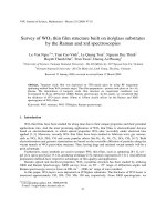
Multimodal tumor imaging by iron oxides and quantum dots formulated in poly (lactic acid) d alpha tocopheryl polyethylene glycol 1000 succinate nanoparticles
Bạn đang xem bản rút gọn của tài liệu. Xem và tải ngay bản đầy đủ của tài liệu tại đây (20.2 MB, 104 trang )
MULTIMODAL TUMOR IMAGING BY IRON OXIDES AND
QUANTUM DOTS FORMULATED IN POLY (LACTIC ACID)-DALPHA-TOCOPHERYL POLYETHYLENE GLYCOL 1000
SUCCINATE NANOPARTICLES
TAN YANG FEI
NATIONAL UNIVERSITY OF SINGAPORE
2010
MULTIMODAL TUMOR IMAGING BY IRON OXIDES AND
QUANTUM DOTS FORMULATED IN POLY (LACTIC ACID)-DALPHA-TOCOPHERYL POLYETHYLENE GLYCOL 1000
SUCCINATE NANOPARTICLES
TAN YANG FEI
(B.Eng. (Hons.), NUS)
A THESIS SUBMITTED
FOR THE DEGREE OF MASTER OF ENGINEERING
DEPARTMENT OF CHEMICAL AND BIOMOLECULAR ENGINEERING
NATIONAL UNIVERSITY OF SINGAPORE
2010
ACKNOWLEDGEMENTS
First of all, I would like to express my deep appreciation and gratitude towards the
following people who have helped me to complete the thesis.
A big thank you to my research project supervisor, Professor Feng Si-Shen, for
offering me an opportunity to be part of his Chemotherapeutic Engineering research
group. I want to thank him for his invaluable support, both physically and morally,
and all the guidance throughout the course of study.
All the professional officers and lab technologists, Mr. Chia Phai Ann, Dr. Yuan Ze
Liang, Mr. Boey Kok Hong, Ms. Lee Chai Keng, Ms. Chew Su Mei, Ms. Samantha
Fam, Ms. Alyssa Tay, Ms. Dinah Tan, Ms. Li Xiang, Mdm. Priya, Mdm. Li Fengmei,
and many other staff from Laboratory Animal Centre (LAC) who have
unconditionally helped in various kinds of administrative works as well as
experiments and have willingly shared their knowledge and expertise to further
enhance my learning process.
My dear colleagues, Mr. Prashant, Dr. Sneha Kulkarni, Mr. Liu Yutao, Mr. Phyo Wai
Min, Ms. Chaw Su Yin, Mr. Mi Yu, Ms. Zhao Jing and all the final year students for
all their kind assistances and supports they provided especially Ms. Wang Sui.
i
PUBLICATION
A journal with the same title as this thesis was published based on this work in
Elsevier under Biomaterials. I am the first author of the published journal. Below is
the relevant article information:
Multimodal tumor imaging by iron oxides and quantum dots formulated in poly(lactic
acid)-D-alpha-tocopheryl polyethylene glycol 1000 succinate nanoparticles.
Biomaterials. 32;2011:2969-2978
Authors
: Yang Fei Tan, Prashant Chandrasekharan, Dipak Maity, Cai Xian
Yong, Kai-Hsiang Chuang,Ying Zhao, Shu Wang, Jun Ding and Si-Shen Feng
Received
: 10 Dec 2010
Accepted
: 31 Dec 2010
Available online : 22 Jan 2011
ii
TABLE OF CONTENTS
ACKNOWLEDGEMENTS .............................................................. i
PUBLICATION ............................................................................. ii
TABLE OF CONTENTS ............................................................... iii
SUMMARY ................................................................................... v
LIST OF TABLES ......................................................................... x
LIST OF FIGURES ...................................................................... xi
LIST OF ABBREVIATIONS ........................................................ xv
CHAPTER 1: INTRODUCTION .................................................... 1
1.1 Background ...........................................................................................................1
1.2 Objectives and Scope ............................................................................................3
CHAPTER 2: LITERATURE REVIEW .......................................... 4
2.1 Cancer Facts ..........................................................................................................4
2.2 Causes of Cancer...................................................................................................5
2.3 Molecular Imaging ................................................................................................7
2.4 How Molecular Imaging Works............................................................................8
2.5 Molecular Imagers in Radiotherapy (RT) .............................................................9
2.6 Current Imaging Techniques...............................................................................10
2.7 Magnetic Resonance Imaging (MRI)..................................................................11
2.8 MRI Contrast Agents ..........................................................................................16
2.9 Superparamagnetic Iron Oxide (IO)....................................................................17
2.10 Fluorescence Imaging .........................................................................................18
2.11 Fluorescence Imaging Principle ..........................................................................19
2.12 Quantum Dots (QDs) ..........................................................................................21
2.13 Optical Properties of Quantum Dots (QDs) ........................................................21
2.14 Applications of Quantum Dots (QDs).................................................................22
2.15 Limitations of Quantum Dots (QDs)...................................................................24
2.16 Challenges of QDs and IO application in Imaging .............................................25
2.16.1 Insufficient Probes at Imaging Site .............................................................25
2.16.2 Cytotoxicity ..................................................................................................30
2.17 Nanotechnology in Molecular Imaging...............................................................33
2.18 Multi-modality ....................................................................................................34
CHAPTER 3: MATERIALS & METHODS ................................... 41
3.1 Materials..............................................................................................................41
3.2 Synthesis Methods...............................................................................................42
3.2.1 Flocculation of QDs .....................................................................................42
3.2.2 Formulation of QDs and IOs-loaded NPs ....................................................42
3.3 Characterization of QDs and IOs-loaded NPs: ...................................................43
3.3.1 Particle Size and Size Distribution .................................................................43
3.3.2 Surface Charge .............................................................................................43
3.3.3 TEM Analysis...............................................................................................43
3.3.4 QDs and IOs Encapsulation Efficiency ........................................................43
3.3.5 XPS...............................................................................................................44
3.4 Cell Line Experiment ..........................................................................................45
3.4.1 Cell Cultures .................................................................................................45
iii
3.4.2 In vitro cellular uptake of NPs......................................................................45
3.4.3 In vitro Cytotoxicity .....................................................................................46
3.5 Animal Study.......................................................................................................47
3.5.1 Tumor imaging (MRI) ..................................................................................47
3.5.2 Tumor Imaging (Fluorescent Imaging) ........................................................48
3.5.3 Biodistribution ..............................................................................................49
CHAPTER 4: RESULTS & DISCUSSIONS .................................. 50
4.1 Characterization of QDs and IOs-loaded nanoparticles ......................................50
4.1.1 Size and Size Distribution ............................................................................50
4.1.2 Surface Charge .............................................................................................50
4.1.3 TEM Analysis...............................................................................................51
4.1.4 QDs and IO Encapsulation Efficiency .........................................................52
4.1.5 XPS...............................................................................................................52
4.2 Cell Line Experiment ..........................................................................................58
4.2.1 In vitro cellular uptake of NPs.....................................................................58
4.2.2 In vitro Cytotoxicity .....................................................................................62
4.3 Animal Study.......................................................................................................64
CHAPTER 5: OUTLOOK ............................................................ 72
CHAPTER 6: CONCLUSION ...................................................... 74
CHAPTER 7: REFERENCES ....................................................... 80
CHAPTER 8: APPENDIX ............................................................ 86
iv
SUMMARY
Cancer has become the top killer of Man in recent decades. Thus, effective cancer
detection is crucial as cancer can be easily tackled at its early stages. Molecular
imaging enables the detection of a disease in its earliest stage. Three medical imaging
techniques often used in the current clinical practice are the X-ray computed
tomography (CT), positron emission tomography (PET) and magnetic resonance
imagery (MRI). CT and PET scans involve radiation exposures. Hence, the noninvasive MRI is preferred.
To
provide
a
better
contrast
in
MRI,
contrast
agents
are
introduced.
Superparamagnetic iron oxide (IO) is widely used as a contrast agent for MRI. It
exhibits excellent magnetic properties and acceptable biocompatibility. IO can vastly
enhance imaging due to its exceptional penetration depth. Furthermore, it has zero
retained magnetism after the removal of magnetic field. Another probe used for
amplification strategy is quantum dots (QDs) as luminescence probes in fluorescence
imaging. Advantages of fluorescence imaging includes high sensitive detection,
multicolor detection, probe stability, low hazard and low cost. Contrast agents such as
organic fluorescent dyes and Quantum Dots (QDs) are often used to promote
fluorescence imaging. Quantum dots (QDs) are composed of atoms from groups II-VI
or III-V of the periodic table. Their advantages include in vivo longevity and tunable
emission from visible to infrared wavelength by changing the size and composition of
QDs. QDs also have broad excitation spectra with high absorption coefficients, high
quantum yield of fluorescence, strong brightness, high resistance to photobleaching
and good sensitivity.
Although necessary, amplification strategies are not enough to produce high quality
images. Sufficient concentrations of probes must be gathered at the intended imaging
v
area for an adequate period in vivo. Nevertheless, the agent dose is limited by the side
effects of the agent and the rapid removal of probes from the blood system due to the
body’s mononuclear phagocyte system (MPS) interactions after opsonization. A
method to cloak nanoparticles from MPS recognition is the surface modification of
the probes to prevent opsonin proteins in the blood from being attached to the
particles surfaces. Generally, hydrophilic particles opsonize slower than hydrophobic
particles and neutrally charged particles opsonize slower than charged particles. Till
date, the most effective and most commonly used polymers as shielding groups are
the PEG-containing copolymers. One important example of such a copolymer is poly
(lactic acid)-D-alpha-tocopheryl polyethylene glycol 1000 succinate (PLA-TPGS)
that is gaining popularity in the research scene today.
Certain probes may have very good affinity with certain targets of imaging interest
however they may pose to be toxic to the body. To use such probes, encapsulation via
PEGylation may be needed to reduce cytotoxicity. Another method to decrease
cytotoxicity is by targeted delivery. Targeting is divided into passive and active
targeting. In passive targeting, nanoparticles accumulate at the tumor through the
enhanced permeability and retention (EPR) effect. The vascular structures of tumors
are defective and lack effective lymphatic drainage system, causing particles to
accumulate in them. Passive targeting is the prime objective for our probe system to
achieve.
Molecular imaging requires high affinity probes with reasonable pharmacodynamics.
Such probes are usually nanoparticles. Synthesizing imaging probes into
nanoparticles not only aids in escaping MPS detection but also increases cellular
uptake. Thus, the formulation of imaging probes such as IOs and QDs in
vi
nanoparticles of biodegradable polymers may provide an ideal solution to reduce
toxicity as well as enhance cellular uptake, hence improving imaging effects.
IO and QD probes are effective probes for amplification in molecular imaging.
However, individual imaging probes have their advantages and disadvantages. For
instance, IO probes provide high spatial resolution and unlimited depth penetration
but their sensitivity in imaging fails in comparison to optical fluorescence imaging
probes such as QDs. QDs, in turn; have excellent imaging effects and long half-life,
but their ability for tissue penetration is limited due to the refraction and adsorption of
light in the living organism. Therefore, it is very important to find an imaging method
that can fulfill the requirements in medical applications as much as possible, and this
can be achieved by applying multi-modal imaging.
Multi-modal imaging means applying two or more imaging modalities concurrently.
Multimodal imaging can be developed to make use of the advantages and overcome
the limitations, which can be realized by co-encapsulation of QDs and IOs in ligandconjugated nanoparticles of biodegradable polymers. To achieve a thorough analysis
of one multi-modal imaging system, in vivo, ex vivo and in vitro analyses should be
done and cross-referenced. Most studies in the research field are related to either ex
vivo or in vitro analysis, lacking in in vivo analysis. In addition, some imaging
modalities such as CT imaging have significant side effects on human health. Both
fluorescence imaging and MRI will not cause radiation injury. On top of that, QDs
and IO as contrast agents have been widely studied in biomedical applications.
Therefore, encapsulating both QDs and IO in PLA-TPGS copolymers, as multi-modal
imaging probes should provide high quality images. This probe should have high
sensitivity and depth penetration.
vii
This thesis illustrates a multimodal imaging system developed by co-encapsulating
superparamagnetic iron oxides (IOs) and quantum dots (QDs) in the nanoparticles
(NPs) of poly (lactic acid) - d-α-tocopheryl polyethylene glycol 1000 succinate (PLATPGS) for use in both magnetic resonance imaging (MRI) and fluorescence imaging.
This multimodal imaging system not only combines the advantages of both MRI and
fluorescence imaging, but also overcomes their disadvantages. This imaging system
also promotes sustained and controlled imaging with passive targeting effects to the
diseased cells. The QDs and IOs-loaded PLA-TPGS NPs were prepared by a modified
nanoprecipitation method, which were then characterized for their size and size
distribution, zeta-potential and the imaging agent encapsulation efficiency. The
transmission electron microscopy (TEM) images showed direct evidence for the welldispersed distribution of the QDs and IOs within the PLA-TPGS NPs. The cellular
uptake and the cytotoxicity of the PLA-TPGS NPs formulation of QDs and IOs were
investigated in vitro with MCF-7 breast cancer cells, which were conducted in close
comparison with the free QDs and IOs at the same agent dose. To investigate the
biodistribution of the QDs and IOs-loaded PLA-TPGS NPs among the various organs,
animal studies were conducted where mice cultivated with MCF-7 breast cancer
tumors were injected with the developed NPs. The results showed greatly enhanced
tumor imaging due to the passively targeting effects of the NPs to the tumor. Images
of tumors were acquired in vivo by a 7T MRI scanner. Further ex vivo images of the
tumors were obtained via confocal laser scanning microscopy. Such a multimodal
imaging system shows great advantages of both contrast agents making the resultant
probe highly sensitive with good depth penetration. A subject administered with the
developed NPs can undergo both MRI and fluorescence imaging. Any imagery
feature detected in one imaging picture which may suggest any disease or tumor
viii
growth, can be further compared and confirmed with the imaging picture taken by the
other imaging technique.
ix
LIST OF TABLES
Table
Description
Page no.
4.1
Characteristics of the QDs and IOs-loaded PLA-TPGS
nanoparticles including particle size and polydispersity (PDI),
zeta potential (ZP) and encapsulation efficiency percentage
(EE%).
51
x
LIST OF FIGURES
Figure
Description
Page no.
2.1
Cancer formation through mutations.
5
2.2
Causes of cancer.
7
2.3
CT imager.
10
2.4
PET imager.
11
2.5
MRI.
12
2.6
Zeeman effect.
13
2.7
(A): A collection of H nuclei in the absence of an externally
applied magnetic field. (B): An external magnetic field B0 is
applied which causes the nuclei to align themselves in one of
two orientations with respect to B0 (denoted parallel and antiparallel).
14
2.8
At Larmor frequency, the net magnetization flips 90°and the
spins are whipped to precess in phase.
15
2.9
Axial T1 weighted (A) and T2 weighted (B) images of the
brain magnetic resonance imaging (MRI) demonstrating a
lacunar infarction (arrow).
17
2.10
IVIS Fluorescence imager.
19
2.11
Jablonski diagram illustrating the processes involved in
creating an excited electronic singlet state by optical
absorption and subsequent emission of fluorescence.
➀:Excitation; ➁:Vibrational relaxation; ➂:Emission.
20
2.12
Excited quantum dots arranged according to size.
22
2.13
QDs applications.
23
2.14
CdSe QDs release of toxic Cd2+ ions by photolysis under UV
illumination.
24
2.15
Opsonization and Phagocytosis of a bacteria.
26
2.16
In vitro MRI of commercial IO (Resovist) and IO-loaded
PLGA-mPEG nanoparticles suspended in water (TE=7ms).
29
2.17
Passive and active tumor targeting.
32
xi
2.18
Schematic illustration of the multi-functional HSA-IONPs.
The pyrolysis-derived IONPs were incubated with dopamine,
after which the particles became moderately hydrophilic and
could be doped into HSA matrices in a way similar to drug
loading.
35
2.19
Synthesis of hybrid silica nanoparticles.
37
2.20
Schematic illustration of MFR-AS1411 synthesis. MF
particles had carboxyl group and Fmoc-protected amine
moiety, which was coupled with amine terminated AS1411
aptamer using EDC (MF-AS1411). After reaction of
MFAS1411 with p-SCN-bn-NOTA, particles were reacted
with 67Ga-citrate to form MFR-AS1411.
38
4.1
TEM Images of A: the IOs-loaded PLA-TPGS NPs, B: the
QDs-loaded PLA-TPGS NPs and C: the QDs and IOs-loaded
PLA-TPGS NPs (scale bar = 200 nm).
51
4.2
Particle XPS result for Cd showing no peaks (absence of Cd).
53
4.3
Grinded particle XPS for Cd showing 2 peaks (presence of
Cd).
54
4.4
Particle XPS result for Se showing no peaks (absence of Se).
54
4.5
Grinded particle XPS for Se showing 1 peak (presence of Se).
55
4.6
Particle XPS result for Zn showing no peaks (absence of Zn).
55
4.7
Grinded particle XPS for Zn showing 2 peaks (presence of
Zn).
56
4.8
Particle XPS result for Fe showing no peaks (absence of Fe).
57
4.9
Grinded particle XPS for Fe showing 2 peaks (presence of Fe).
57
4.10
CLSM images of MCF-7 cells treated with the QDs and IOsloaded PLA-TPGS NPs in vitro (scale bar = 10 µm). A: Bright
field image of cells. B: Blue coded DAPI stained nuclei. C:
Red coded QD from NPs in cytoplasm. D: Complete
overlapped image.
59
4.11
Cellular uptake efficiency of the MCF-7 cancer cells after 1, 2
and 4 h treatment with 100 µL of the QDs and IO-loaded
PLA-TPGS NPs of concentrations containing 1 µg/mL Cd, 0.5
µg/mL Cd and 0.25 µg/mL Cd respectively dispersed in
medium.
61
xii
4.12
In vitro viability of MCF-7 cells after 24 and 48 hour
treatment with the free IO, the free QDs (containing 1.42
µg/mL Cd), the free IO (containing 5.73 µg/mL Fe), and the
QDs and IOs-loaded PLA-TPGS NPs (containing 1.42 µg/mL
Cd and 5.73 µg/mL Fe) respectively dispersed in the medium.
63
4.13
Axial MRI image sections of the MCF-7 grafted tumor
bearing mice. Images A and B show the part of the tumor
(shown by the arrow) before and after 6 hours of
administration of the QDs and IOs-loaded PLA-TPGS NPs
into the mice. Images C and D show the kidney (K) and liver
(L) part of the mice before and 6 hours after the administration
of the PLA-TPGS NPs formulation of QDS and IOs (dosage:
1.5 mg of Cd/kg of body weight or equivalent of 6.0 mg of
Fe/kg body weight). The decrease in intensity in the regions of
the tumor and liver can be noticed in comparison with the
color scale shown aside.
64
4.14
Fluorescent Images of the various organs. Upper row: control.
Lower row: Organs of the mouse treated with the QDs and
IOs-loaded PLA-TPGS NPs (dosage: 1.5 mg of Cd/kg of body
weight or equivalent of 6.0 mg of Fe/kg body weight).
66
4.15
Fluorescence intensity increase percentage for the various
organs of the mice treated with the QDs and IOs-loaded PLATPGS NPs (dosage: 1.5 mg of Cd/kg of body weight or
equivalent of 6.0 mg of Fe/kg body weight).
67
4.16
Confocal laser scanning microscopy sections of the mouse
liver (scale bar = 60 µm). Images A, B and C show the liver
sections of the control with no treatment. A: Blue coded DAPI
stained nuclei. B: Red channel detection showing no signal
due to absence of QDs. C: Complete overlapped image of A
and B. Images D, E and F show the liver sections of the mouse
treated with the QDs and IOs loaded PLA-TPGS NPs. D: Blue
coded DAPI stained nuclei. E: Red coded QD from NPs in
cytoplasm. F: Complete overlapped image.
68
4.17
Confocal laser scanning microscopy sections of the mouse
kidney sections (scale bar = 60 µm). Images A, B and C show
the kidney sections of the control with no treatment. A: Blue
coded DAPI stained nuclei. B: Red channel detection showing
no signal due to absence of QDs. C: Complete overlapped
image of A and B. Images D, E and F show the kidney
sections of the mouse treated with the QDs and IOs loaded
PLA-TPGS NPs. D: Blue coded DAPI stained nuclei. E: Red
coded QD from NPs in cytoplasm. F: Complete overlapped
image.
69
xiii
4.18
Confocal laser scanning microscopy sections of the mouse
tumor sections. Images A, B and C (scale bar = 30 µm) show
the tumor sections of the control with no treatment. A: Blue
coded DAPI stained nuclei. B: Red channel detection showing
no signal due to absence of QDs. C: Complete overlapped
image of A and B. Images D, E and F (scale bar = 20 µm)
show the tumor sections of the mouse treated with the QDs
and IOs loaded PLA-TPGS NPs. D: Blue coded DAPI stained
nuclei. E: Red coded QD from NPs in cytoplasm. F: Complete
overlapped image.
xiv
70
LIST OF ABBREVIATIONS
Abbreviation
ADME
As
Cd
CLSM
cps
CT
Cu
DAPI
DI
DMSO
DNA
EDTA
EE
Er
EPR
F
FBS
FDA
Fe
Ga
Gd
HA
HLB
ICP-MS
In
InC
InS
IO
LLS
MDR
Mn
mPEG
MPS
MRI
ms
MTT
Description
Absorption, distribution, metabolism and excretion
Arsenic
Cadmium
Confocal laser-scanning microscope
Counts per second
X-ray computed tomography
Copper
4,6-Diamidino-2-phenylindole dihydrochloride
Deionized
Dimethyl sulfoxide
Deoxyribonucleic acid
Ethylenediaminetetraacetic acid
Encapsulation efficiency
Erbium
Enhanced permeability and retention
Florine
Fetal bovine serum
Food and drug administration
Iron
Gallium
Gadolinium
Hydroxyapatite
Hydrophile lipophile balace
Inductively coupled plasma mass spectrophotometer
Indium
Fluorescence intensity of cells in control wells
Fluorescence intensity of cells in sample wells
Iron oxide
Laser light scattering
Multiple Drug Resistance
Number averaged molecular weight
Methyl polyethylene glycol
Mononuclear phagocyte system
Magnetic resonance imagery
Milli second
Methylthiazolyldiphenyl-tetrazolium bromide
xv
Mz
N
Na
NIRF
NMR
NP
O
PBS
PDI
PEG
PET
PLA
PLA-TPGS
PLEA
PLGA
QD
RES
RF
ROI
RT
Ru
S
SCID
Se
Si
SWNT
T1
T2
Te
TE
TEM
THF
Tm
TR
UV
XPS
Yb
ZP
Zn
Net magnetization
Nitrogen
Sodium
Near-infrared imaging
Nuclear magnetic resonance spectroscopy
Nanoparticle
Oxygen
Phosphate buffered saline
Poly Dispersity Index
Polyethylene glycol
Positron emission tomography
Poly (lactic acid)
Poly (lactic acid)-D-alpha-tocopheryl polyethylene glycol
1000 succinate
Poly (lactic acid)-poly (ethylene glycol)
Poly (lactic–co-glycolic acid)
Quantum dot
Reticuloendothelial system
Radio frequency
Region of interest
Radiotherapy
Ruthenium
Sulphur
Severe combined immunodeficiency
Selenium
Silica
Single walled carbon nano tube
Longitudinal relaxation time
Transverse relaxation time
Tellurium
Echo delay time
Transmission electron microscope
Tetrahydrofuran
Thulium
Repetition time
Ultra violet
X-ray photoelectron spectroscopy
Ytterbium
Zeta potential
Zinc
xvi
CHAPTER 1: INTRODUCTION
1.1 Background
Cancer is the result of the uncontrolled growth and spreading of abnormal cells (Feng
SS and Chien S, 2003). Cancer cells can spread in the body through the blood and
lymph systems ( Cancer is the
leading cause of death in various developed countries. In the United States, there were
about 1,529,560 new cases of cancers reported in 2010. On top of that, cancer
associated death cases amounted to an alarming 569,490 in the very year
( Therefore, it is evidently
important to find efficient ways to combat cancer.
Massive advancements have actually been made in cancer treatments as compared to
the last decade. However, developments in molecular imaging systems to detect
cancer witnessed rather sluggish progress. Molecular imaging is an in vivo
characterization and measurement of the disease process at the cellular and molecular
level, which aims at investigating cellular functions without disturbance. In actual
fact, in order to effectively overcome cancer, it is of paramount importance to first
efficiently detect them. This is because, just like any other diseases, cancers can be
easily and effectively treated in their early stages especially before tumors
metastasize. Developing an advanced imaging system to detect cancer can realize this.
In recent years, researchers have finally realized the importance of advancing imaging
techniques resulting in great interests in advanced cancer imaging systems. Scientists
expected that by using efficient cancer imaging techniques, the stage and precise
locations of cancer could be determined efficiently. Apart from that, cancer imaging
can also aid cancer treatment especially during operations and help monitor the
1
treatment
effects
( />
Thus, an effective cancer imaging system is highly in demand.
In order to enhance molecular imaging, contrast agents are utilized as imaging probes.
Contrast agents make molecular imaging possible and effective by enhancing the
image contrast between healthy and abnormal tissues. Thus, they are needed for many
imaging techniques. However, most contrast agents have some toxicity issues and are
thus not biocompatible. Besides causing some sides effects in the human body due to
the toxicity, some contrast agents may have cell uptake limitation and could not be
efficiently delivered into cells. On top of that, human immune system detection of
these foreign contrast agents may also cause circulation limitations. Therefore, it is
crucial to find a better way to control deliver the contrast agents into human cells
while decreasing their cytotoxicity. Researchers found that by modifying contrast
agents into nanoparticles, advantages such as the desired control delivery system, long
vascular half-life and fewer side effects on human body can be achieved. In doing so,
the imaging quality can be increased and it will be easier for doctors to find the
accurate position of cancer in the body, locate the extent of cancer spreading, identify
specified cancer treatment and monitor the effect of the treatment.
Although contrast agents could enhance molecular imaging, every individual contrast
agents have its advantages and limitations. Therefore, by only using one contrast
agent and utilizing one mode of imaging may result in certain features within organs
suggesting the onset of a particular disease to be overlooked. Therefore, the idea of
dual modality was born which involves combining two contrast agents into a single
probe. One dosage of this probe enables the patient to undergo two modes of imaging
techniques. The results of the imaging can then be analyzed concurrently. This acts as
2
a more effective imaging practice to ensure no diseases get overlooked and left to
develop into tricky late stages where treatment may be complicated.
1.2 Objectives and Scope
The main objective of this project is to encapsulate both quantum dots (QDs) and
superparamagnetic iron oxide (IO) in biodegradable copolymer PLA-TPGS. Basic
characterization studies will be conducted on the nanoparticles to investigate the
particle size, polydispersity, surface charge and encapsulation efficiency. Cell line
work will be conducted using the nanoparticles. Cell studies include cell uptake and
cell toxicity experiments. On top of that, bio distribution experiments will be
conducted on treated cancer induced animals. Finally, molecular imaging will also be
used on animals treated with the particles.
3
CHAPTER 2: LITERATURE REVIEW
2.1 Cancer Facts
Cancer is currently the leading cause of death globally. According to the US National
Cancer Institute, cancer is defined as a category of affiliated diseases whereby
abnormal cells go through uncontrolled transformation (or mitosis) and have the
ability to spread to other parts of the body via the blood circulation and lymphatic
systems (metastasis).
In the normal state, cells grow and replicate to form new cells according to the needs
of the body. Whenever cells grow old and die, new cells replace them. However at
times, this ideal orderly process goes wrong in which new cells form when the body
does not need them, and old cells do not die when they should. The resultant extra
cells gather to form a mass of tissue. This mass is known as a tumor. Tumors can be
either benign (non cancerous) or malignant (cancerous). Benign tumors are localized
and do not spread to other parts of the body. They are rarely life threatening.
Malignant tumors, on the other hand, can spread (metastasize) and may be life
threatening ( />
4
Figure 2.1: Cancer formation through mutations.
(Adapted from />
A projection from statistics revealed that for every three people, one would be
diagnosed with cancer in his lifetime. On top of that, occurrences rate of cancer are
increasing at a rate of 1% per year ( />Till today, more than 200 different types of cancer have been discovered. The
probability of getting cancer is distinct in different types of tissues or organs, even
within the same individual.
2.2 Causes of Cancer
There are various causes for cancer. These causes can basically be subdivided into
two categories, namely the intrinsic and extrinsic factors. Intrinsic factors mainly
include the genetic make up of the body and the individuals cannot control this. It
implies that once a person is born, the genetic make up has already been coded to
determine the number of genetic mutations he or she will experience in the lifetime.
5
Some of these mutations may ultimately lead to cancer. The causes of such mutations
include inheritance from previous generations, abnormal fertilization or improper
fetal developments during pregnancy. Mutations may not always result in cancer.
However, inheritance of certain harmful gene mutations may increase the risk of
cancer development. For instance, research has shown that women who inherited
harmful BRCA1 and BRCA2 gene mutations can have a very higher risk of
developing breast cancer in their lifetime as compared to those who did not inherit
such gene mutations ( />In general, extrinsic factors play a bigger role in determining the development of
cancer. Extrinsic factors encompass a wide variety of causes, ranging from
environmental factors to the individual’s personal daily lifestyle. Daily lifestyle
practices such as diet directly influences the risk of getting cancer. Preservatives such
as nitrosamine, nitrosamide, sulphites as well as colorings, which are usually added
during food processing, can potentially accumulate in the body over an extended
period of time and cause cancer ( /> />
Genetically-modified
food
(staples such as rice and potatoes included) as well as food rich in methyl donors has
been reported to be able to potentially trigger genetic mutations, stimulating tumor
growth
(Watters,
2006;
/>
wellbeing/health-news/suppressed-report-shows-cancer-link-to-gm-potatoes436673.html). Besides dietary habits, harmful habits such as smoking and drinking
are also major factors causing cancers. For instance, more than 38,000 people are
diagnosed with lung cancer every year. Of these deaths, almost 90% is tobacco related
( />As the average human life span increases with groundbreaking discoveries in the
6
medical arena, mutations in cells and tissues are given enough time to develop into
cancer. On top of that, industrializations globally, increased radiation due to ozone
damage, extensive production of processed food and various failing personal lifestyle
has raised the risk of various cancers in the present human population. Therefore, it is
important to guard against cancer and the first step in doing so would be to do
molecular imaging periodically to detect any preliminary onset symptoms of cancer.
Figure 2.2: Causes of cancer.
(Adapted from />
2.3 Molecular Imaging
Early stage diagnosis plays a key role in determining the prognosis for diseases,
especially for fatal ailments such as cancer and cardiovascular diseases. Molecular
imaging provides critical information necessary to diagnose a disease in its earliest
stage, which is an in vivo characterization and measurement of the disease process at
the cellular and molecular level. Its objective is to investigate molecular basis and
diagnose abnormalities of cellular functions as well as follow up molecular processes
7