Clinicopathological study of biomarkers in prostrate cancer
Bạn đang xem bản rút gọn của tài liệu. Xem và tải ngay bản đầy đủ của tài liệu tại đây (5.96 MB, 131 trang )
CLINICOPATHOLOGICAL STUDY OF
BIOMARKERS IN PROSTATE CANCER
TAN YEN
(BSc, University of Melbourne)
A THESIS IS SUBMITTED FOR THE DEGREE
MASTER OF SCIENCE
DEPARTMENT OF ANATOMY
NATIONAL UNIVERSITY OF SINGAPORE
2011
ACKNOWLEDGEMENTS
First and foremost, I will like to express my heartfelt gratitude to my supervisor and Head
of Pathology, Singapore General Hospital (SGH), Assoc Prof Tan Puay Hoon, for
providing the opportunity to pursue my MSc degree. Her encouragement, support and
guidance played an important part in making the project a successful one. Next, I will
like to thank my co-supervisor, Prof Bay Boon Huat, Head of Department of Anatomy,
Yong Loo Lin School of Medicine, National University of Singapore (NUS), for
providing me an opportunity to pursue my MSc degree with the Department of Anatomy.
His encouragement and advices have allowed me to continue the degree with much ease.
I am very grateful to the Pathologists and my fellow colleagues at Histopathology Section,
Department of Pathology, SGH, for their assistance in the completion of the project.
Special thanks to Ms Maryam Hazly Hilmy and Ms Cheok Poh Yian for their guidance
on immunohistochemical staining and tissue microarray (TMA) construction respectively;
Ms Melissa Ng for scanning the H&E and TMA slides; Dr Aye for her guidance and
verification on all the immunostaining performed on the TMA sections and statistical
analysis; Dr Richie for patiently answering all my queries on prostate cancer. Special
thanks also goes out to Ms Bay Song Ling, Department of Anatomy, Yong Loo Lin
School of Medicine, NUS, for her wonderful re-illustration of the functional zones of the
prostate. Many thanks to Ms Yvonne Teng Huifang, for her help on the data collection
for this project, invaluable advices and friendship.
i
Apologies to those whom I have not mentioned by name, I will always be grateful to
them for helping me throughout the study.
I will also like to thank my family and friends for the tremendous support given to me
during this period.
Once again, I will like to thank Department of Anatomy, NUS for giving me the
opportunity to pursue my MSc.
ii
TABLE OF CONTENTS
Acknowledgement………………………………………………………………... i
Summary………………………………………………………………………….. v
List of Abbreviations……………………………………………………………... vii
List of Figures…………………………………………………………………….. viii
List of Tables……………………………………………………………………… ix
CHAPTER 1: INTRODUCTION………………………………………………...1
1.1 Epidemiology………………………………………………………………… 2
1.2 Anatomy of Prostate…………………………………………………………. 3
1.3 Risk Factors………………………………………………………………….. 7
1.4 Radical Prostatectomy……………………………………………………….. 10
1.5 Management of Prostate Cancer……………………………………………... 11
1.6 Gleason Grading System…………………………………………………….. 13
1.7 Prostate Specific Antigen (PSA) Screening and Pitfall……………………… 15
1.8 Androgen Receptor (AR).................................................................................. 17
1.9 Her-2/neu...........................................................................................................19
1.10 Ki-67…………………………………………………………………………20
1.11 p-53…………………………………………………………………………. 22
1.12 Neuroendocrine Expression and Markers…………………………………....24
1.13 CD56 ………………………………………………………………………...25
1.14 Chromogranin A……………………………………………………………...25
1.15 Synaptophysin (Syn)…………………………………………………………26
1.16 Tissue Microarray (TMA) Technology……………………………………....27
1.17 Scope of Study………………………………………………………………. 28
CHAPTER 2: MATERIALS AND METHODS………………………………… 30
2.1 Patients……………………………………………………………………….. .31
2.2 Gross Assessment of Radical Prostatectomy specimen and Preparation of Whole
Mounts for Histological Evaluation…………………………………………….... .32
2.3 Microscopic Evaluation of the Radical Prostatectomy Hematoxylin and Eosin
stained sections…………………………………………………………………... .33
2.4 Tissue Microarray (TMA) Construction……………………………………... .34
2.5 Immunohistochemistry…………………………………………………….......35
2.6 Scoring of immunohistochemically stained sections………………………… .38
2.7 Statistical Analysis………………………………………………………….... .41
CHAPTER 3: RESULTS…………………………………………………………..42
3.1 Clinicopathological Parameters Studied……………………………………….43
3.2 Immunoreactivity of the Biomarkers…………………………………………..48
3.3 Correlation of Androgen Receptor Expression and
Clinicopathological Parameters………………………………………………….. .54
3.4 Correlation of p53 protein with clinicopathological parameters…………….. .58
3.5 Correlation of Ki-67 antigen immunohistochemical expression with
clinicopathological parameters…………………………………………………... .61
3.6 Correlation of Clinicopathological Parameters and Biomarkers……………....65
3.7 Correlation of Androgen Receptor (AR) and p53, AR and Ki-67,
p53 and Ki-67……………………………………………………………………. 67
iii
3.8 Immunoreactivity of Neuroendocrine Biomarkers…………………………....69
3.9 Correlation of CD56 Expression and Clinicopathological Parameters………. 73
3.10 Correlation of Chromogranin A (Chr A) Expression and
Clinicopathological Parameters………………………………………………….. 76
3.11 Correlation of Synaptophysin (Syn) Expression and
Clinicopathological Parameters………………………………………………….. 81
3.12 Correlation of Neuroendocrine Differentiation (NED) and
Clinicopathological Parameters………………………………………………….. 85
3.13 Correlation of Neuroendocrine Differentiation (NED) and AR,
Ki-67 and p53 Immunohistochemical Expression………………………………. 88
CHAPTER 4: DISCUSSION……………………………………………………...90
4.1General Discussion…………………………………………………………… 91
4.2 Clinicopathological Features of Prostate Cancer…………………………….. 92
4.3 Correlation of Her-2/neu Expression with
Clinicopathological Parameters………………………………………………….. 96
4.4 Correlation of Androgen Receptor (AR) Expression with
Clinicopathological Parameters………………………………………………….. 100
4.5 Correlation of p53 Expression with Clinicopathological Parameters………... 102
4.6 Correlation of Ki-67 Expression with Clinicopathological Parameters……... 103
4.7 Correlation of Androgen Receptor (AR) and p53, AR and Ki-67,
p53 and Ki-67……………………………………………………………………. 104
4.8 Correlation of Neuroendocrine Markers with
Clinicopathological Parameters………………………………………………….. 105
4.9 Correlation of Neuroendocrine Differentiation (NED) with
Clinicopathological Parameters………………………………………………….. 107
4.10 Correlation of Neuroendocrine differentiation (NED) and AR,
Ki-67 and p53 Immunohistochemical Expression……………………………….. 108
4.11 Conclusion………………………………………………………………….. 109
4.12 Future Studies………………………………………………………………. 111
References…………………………………………………………………………. 113
iv
SUMMARY
Prostate cancer is currently the third commonest malignancy among Singaporean men.
The increase can be attributed to an aging population, serum PSA screening and the
application of transrectal ultrasound needle biopsy, which is the current gold standard for
diagnosis of prostate cancer.
Radical prostatectomy is currently the treatment given to male patients with localized
prostate cancer, and who are likely to benefit from the procedure. While serum PSA is
used to monitor the risk of biochemical recurrence after radical prostatectomy,
controversies surrounding the usefulness of serum PSA has led to a need for biomarkers
which can prognosticate disease aggressiveness and predict treatment outcome, and also,
better understanding of the pathogenesis.
For this project, 170 cases of prostatic acinar adenocarcinoma of male patients who
underwent radical prostatectomy at Singapore General Hospital from 2002 to 2005 were
being studied. Tissue microarrays (TMA) were constructed from whole mount
preparations of these radical prostatectomy specimens. Clinical details such as age,
ethnicity, pre- and post-operative serum PSA, histopathological parameters such as
histological types, Gleason score, size of tumour, location of tumour, extent of tumour,
presence or absence of perineural invasion, vascular/ lymphatic invasion, associated high
grade prostatic intraepithelial neoplasia as well as involvement of surgical margins were
used to study if these parameters had any correlation with the markers which were being
investigated.
v
Immunohistochemical staining was performed on TMA sections using androgen receptor
(AR), p53, Ki-67 Her-2/neu and neuroendocrine markers – synaptophysin (Syn),
chromogranin A (Chr A) and CD56 antibodies. These sections were then scored and
statistical analysis was performed using SPSS, student t test and Chi Square Test.
This study found Ki-67 and p53 to be associated with adverse pathological variables –
Ki-67 was positively correlated to PSA level as indicated by a p value of 0.041 while p53
intensity was positively correlated to Gleason score as indicated by p value of 0.018. Ki67 and p53 were also observed to be positively correlated with AR. Ki-67
immunoreactive score (IRS) was positively correlated to AR IRS as indicated by p value
of 0.055; p53 intensity-percentage score (IPS), p53 IRS were positively correlated to AR
IPS and AR IRS as indicated by p value of 0.041 and 0.015 respectively.
Correlations between Her-2/neu, neuroendocrine markers and clinicopathological
parameters did not yield any significant statistical p values. This could be due to the
relatively small case numbers that were positive for the various markers.
Further studies may include enlarging the patient cohort and widening the panels of
antibodies to be evaluated, in order to glean knowledge on the role of biological markers
in prostate cancer.
vi
LIST OF ABBREVIATIONS
ACT
AF
AR
AREs
BPH
Chr A
DAB
DBD
DFC
DNA
FDA
FFPE
FHA
HER2
H&E
HGPIN
HRP
IPS
IRS
LBD
NCAM
NE
NED
NK
NTD
PSA
Syn
TRUS
TURP
TMA
TZ
Antichymotrypsin
Activation factor
Androgen receptor
Androgen-responsive elements
Benign prostate hyperplasia
Chromogranin A
Diaminobenzidine
DNA binding domain
Dense fibrillary component
Deoxyribonucleic acid
Food and drug administration
Formalin-fixed-paraffin-embedded
Forkhead associated
Human epithelial growth factor 2
Heamatoxylin and eosin
High-grade prostatic intraepithelial neoplasia
Horseradish peroxidase
Intensity-percentage score
Immunoreactive score
Ligand binding domain
Neural cell adhesion molecular
Neuroendocrine
Neuroendocrine differentiation
Natural killer
N-terminal domain
Prostate-specific antigen
Synaptophysin
Transrectal ultrasound guided prostate needle
biopsy
Transurethral resection of the prostate
Tissue Microarray
Transition zone
vii
LIST OF FIGURES
Figure 1 Functional zones of prostate gland.
Figure 2 Immunohistochemical expression of androgen receptor (AR), p53, Ki-67, CD56,
chromogranin A (Chr A) and synaptophysin (Synap).
Figure 3 H&E stained whole mount sections of radical prostatectomy specimens.
Figure 4 H&E stained whole mount sections of radical prostatectomy specimens.
Figure 5 Immunohistochemical nuclear expression of androgen receptor in normal and
cancerous prostate tissues.
Figure 6 Immunohistochemical expression of p53 and cerb-B2 in benign and prostate
cancer tissue.
Figure 7 Immunohistochemical expression of nuclear Ki-67 in benign prostate tissue and
prostate cancer tissue.
Figure 8 Immunohistochemical expression of CD56 (NCAM) in prostate cancer tissue
Figure 9 Immunohistochemical expression of Chromogranin A (Chr A) in prostate
cancer tissue.
Figure 10 Immunohistochemical cytoplasmic expression of Synaptopysin (Syn) in
benign prostate tissue and prostate cancer tissue.
viii
LIST OF TABLES
Table 1. Ten Commonest Cancers Diagnosed in Singaporean Males from 2003 – 2007
Table 2. Details of antibodies and dilutions
Table 3. Clinicopathologic features of prostate cancer ( N=170 )
Table 4. Immunoreactivity of biomarkers in 170 cases of prostate cancer
Table 5. Correlation of androgen receptor (AR) expression with clinicopathological
parameters
Table 6. Correlation of p53 gene immunoreactivity with clinicopathological
parameters
Table 7. Correlation of Ki-67 antigen immunoexpression with clinicopathological
Parameters
Table 8. Correlation of Clinicopathological Parameters and Biomakers
Table 9. Correlation of androgen receptor (AR) and p53, AR and ki-67, p53 and ki-67
Table 10. Immunoreactivity of neuroendocrine markers in 170 cases of prostate
cancer
Table 11. Correlation of CD56 expression with clinicopathological
parameters
Table 12. Correlation of Chromogranin A (Chr A) expression with
clinicopathological parameters
Table 13. Correlation of Synaptophysin (Syn) expression with clinicopathological
parameters
Table 14. Correlation of neuroendocrine differentiation (NED) with clinicopathological
parameters
Table 15. Correlation of neuroendocrine differentiation (NED) and AR, Ki-67 and p53
immunohistochemical expression
ix
Introduction
CHAPTER 1
INTRODUCTION
1
Introduction
1 INTRODUCTION
1.1 Epidemiology
According to the “Trends in Cancer Incidence in Singapore 1968-2007”, Singapore
Cancer Registry Report Number 7, published by the Singapore Cancer Registry, prostate
cancer is currently the third most common cancer among Singaporean males, accounting
for 9.8% of all cancers in local men (Lee et al, 2008). Prostate cancer is also the third
most prevalent cancer among Chinese, Malay and Indian males in Singapore. The
increase in incidence can be attributed to an aging population, the advent of serum
prostate specific antigen (PSA) screening and the ready availability of transrectal
ultrasound guided prostatic core biopsies.
Serum PSA is currently the screening modality for early detection of prostate cancer. The
absolute serum level of PSA can also predict potential aggressiveness of a prostate cancer.
In Singapore, a PSA value more than 4ng/ml will generally indicate the possibility of
prostate cancer in an adult man, for which a subsequent transrectal ultrasound guided
prostate needle biopsy, which is the gold standard for confirmation of prostate cancer,
may be conducted. Radical prostatectomy and/or radiation therapy may be the treatment
modalities depending on Gleason scores and quantum of the cancer discovered on the
core biopsies, the preoperative serum PSA levels and patient age as well as preference.
2
Introduction
Table 1. Ten Commonest Cancers Diagnosed in Singaporean Males from 2003 –
2007
Rank Site
1
2
3
4
5
6
7
8
9
10
Colo-rectum
Lung
Prostate
Liver
Stomach
Lymphoid Neoplasm
Nasopharynx
Skin, including melanoma
Bladder
Kidney and other urinary
Number of Males
Diagnosed
3902
3828
2169
1700
1375
1309
1198
973
675
620
Percentage
(%)
18.4
17.6
9.8
7.9
6.6
5.8
5.5
4.3
2.9
2.7
Adapted from Trends in Cancer Incidence in Singapore 1968-2007, Singapore Cancer Registry Report No.
7, Singapore Cancer Registry, Page 31, 33
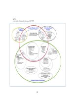
1.2 Anatomy of Prostate
The prostate gland, about the size of the walnut, weighs about 20gm upon maturity (Lee
et al, 1994). The gland is found low in the pelvis minor, surrounds the bladder neck
(Young and Heath, 2002) and the first part of the urethra, and is posterior to the
symphysis pubis. The gland lies ventral to the ampulla of the rectum, where the posterior
portion of the prostate can be easily palpated (Lee et al, 1994).
The prostate is made up of branched tubulo-acinar glands and fibromuscular tissues,
enclosed in a fascial sheath (Lee et al, 1994). A partial capsule encloses the posterior and
lateral aspects of the prostate while the anterior and apical surfaces are bounded by the
anterior fibromuscular stroma. (Young and Heath, 2002). The anterior fibromuscular
stroma is about a third of the entire bulk of the prostate, does not contain glands, covers
3
Introduction
the anterior part of the prostate and merges with the sphincter at the bladder neck
(Kissane, 1997).
The prostate consists of 4 zones, namely the transition zone, central zone, peripheral zone
and the anterior fibro-muscular stroma. The transition zone, which surrounds the
proximal prostatic urethra, consists of about 5% of the gland and is found at the junction
of the proximal and distal segments of the urethra. The transition zone is important in that
it can undergo hyperplasia, resulting in the formation of nodules known as benign
prostatic hyperplasia which may lead to clinical symptoms of prostatism such as urinary
frequency, hesitancy and dribbling.
The central zone, making up 20% of the glandular tissue, surrounds the ejaculatory ducts,
extending out from the verumontanum in a wedge-shaped fashion. The largest zone,
which is the peripheral zone, completes the remaining 70% of the prostate, and is also
where carcinoma of the prostate predominantly occurs (Lee et al, 1994).
4
Introduction
Figure 1 Functional zones of prostate gland.
(Figure is redrawn from Ross and Pawlina, Histology A Text and Atlas, 2011)
Three basic cell types, namely the secretory, basal and neuroendocrine can be found in
the prostate glands. The secretory cells, forming a continuous layer facing the glandular
lumen, extend throughout the ducts and acini. Secretory cells may produce substances
such as lactoferrin and neutral mucin. These cells also contain low-molecular weight
cytokeratins and receptors for androgen, estrogen, progesterone and lectin. More notably,
secretory cells are important in that they produce and store PSA and an isoenzyme of
prostatic acid phosphatase (PAP) (Kissane, 1997).
Basal cells, forming a discontinuous layer at the antiluminal surface of the prostatic
epithelium, are more commonly seen in the peripheral rather than in the central glands.
These cells do not secrete any substances, although they are immunoreactive to many
high-molecular cytokeratins – the presence of basal cells can be demonstrated by
immunohistochemistry. The presence of basal cells has diagnostic significance in that
5
Introduction
their identification precludes prostate cancer, since basal cells are absent in prostate
adenocarcinoma.
Basal cells are insufficiently differentiated to be considered
myoepithelium.
Neuroendocrine cells are often demonstrated by argentaffin-argyophil histochemical
stains. With advances in immunohistochemistry, neuroendocrine expression can now be
detected by various neuroendocrine markers, notably chromogranin A and synaptophysin.
Collagen, smooth muscle fibers, elastic fibres, lymphatic and blood vessels, nerves,
scattered foci of lymphocytes, make up the prostatic stroma. Fibroblasts and collagen
fibers occur in parallel arrangement; the discovery of nerve fibers circumferentially
surrounded by acini is a feature of prostatic carcinoma (Kissane, 1997, Epstein and
Murphy, 1997).
Prostate development arises from the urogenital sinus and is dependent on androgen and
5-α dihydrotestosterone (5α-DHT) which is a metabolite of fetal testosterone. It has also
been suggested that primitive prostatic mesenchyme is the target tissue for 5-α
dihydrotestosterone and not the epithelium lining the urogenital sinus (Kissane, 1997,
Epstein and Murphy, 1997).
5α-DHT is the most potent natural male hormone as well as natural ligand for androgen
with a Kd = 10-11M (Penning et al, 2008, Penning et al, 2007, Bauman et al, 2006). 5αDHT is essential for growth, development, differentiation, maintenance and secretory
6
Introduction
functions of the human prostate (Bauman et al, 2006, Rizner et al, 2003, Davies and
Eaton, 1991). 5α-DHT is formed when testosterone from Leydig cells of the testis is
reduced by the action of 5α-reductase type 2 in the prostate (Penning et al, 2008,
Penning et al, 2007, Bauman et al, 2006, Rizner et al, 2003). Regulation of 5α-DHT is
regulated by 3α/3β-hydroxysteroid dehydrogenase (3α-HSD) (Penning et al, 2008, Rizner
et al, 2003).
Overproduction of 5α-DHT can lead to either benign prostatic hyperplasia (BPH) or
prostate cancer (Rizner et al, 2003). Excess 5α-DHT production may be caused by
elevated 5α-DHT synthesis as a result of increased expression of 5α-reductase type 2 or
increased expression of the oxidative 3α-HSD isoforms which convert 3α-diol to 5α-DHT
or decreased inactivation of 5α-DHT due to the downregulation of 3-ketosteroid
reductase (Rizner et al, 2003).
1.3 Risk Factors
The risk factors which may contribute to the development of prostate cancer remain
relatively unknown although several efforts have been made to study whether age,
familial inheritance, racial groups, diet and even exposure to certain chemicals as a result
of occupational hazards, play a role in the development of prostate cancer. In addition,
there are a number of studies reporting on ethnicity as a risk factor, with black men
reportedly at highest risk (Boyle and Severi, 2003, Hoffman, 2006, Pomerantz et al, 2007,
Von Eschenbach, 1981).
7
Introduction
It is difficult to study the risk factors associated with prostate cancer, be it case-control
studies or prospective cohort studies, as reported by Boyle and Severi (Boyle and Severi,
2003) and Wolk (Wolk, 2005). Boyle and Severi also suggested that the obstacles faced
when designing studies to evaluate various risk factors associated with prostate cancer
were probably due to paucity of information relating to disease specificity, the
heterogeneous nature of phenotypes and genotypes of prostate cancer, which make
pathogenesis challenging to elucidate (Boyle and Severi, 2003).
Neverthless, Boyle and Severi and Wolk, had reported age being an established risk
factor associated with prostate cancer. This could be due to the fact that prostate cancer is
not frequent before the age of 50. In fact, Obek et al even suggested that age could be an
independent parameter in administration of treatment due to its direct impact on mortality
(Obek et al, 1999). However, Obek et al also reported that the studies available were not
conclusive enough to determine age as a prognostic factor for biochemical recurrence
after radical prostatectomy (Obek et al, 1999).
Family history and genetics as risk factors for prostate cancer had been well studied
(Boyle and Severi, 2003, Lichtenstein et al, 2000, Wolk, 2005). Boyle and Severi
reported that prostate cancer presented familial aggregation, which was similar to breast
and colon cancers. Linkage analyses, polymorphism studies were carried out to better
understand the relationship of gene mutation and risk of prostate cancer (Boyle and
Severi, 2003, Lichtenstein et al, 2000, Wolk, 2005).
8
Introduction
Hormones and growth factors were also being suggested to be risk factors associated with
prostate cancer (Boyle and Severi, 2003). The normal prostate epithelium growth and
maintenance were regulated by the androgen and vitamin D pathways – androgen
stimulates prostate cell proliferation while vitamin D inhibits proliferation, hence, it was
believed that high levels of vitamin D was therefore associated with lower risk of prostate
cancer (Boyle and Severi, 2003).
Besides genetic factors, growth factors and hormones reported to be risk factors
associated with prostate cancer, environmental factors such as diet, lifestyle, and type of
occupation, were also being studied to evaluate the association between these factors and
risk of prostate cancer.
Boyle, Severi, Chan and Wolk, (Boyle and Severi, 2003, Chan et al, 1998, Liang and
Liao, 1992, Wolk, 2005) reportedly found a positive association between prostate cancer
and consumption of meat and dairy products. Boyle, Severi and Wolk suggested that
when meat was cooked at high temperatures, such as grilling, carcinogenic substances
such as heterocyclic amines and polycyclic aromatic hydrocarbons, were produced. Wolk
also reported that consumption of dairy products was associated with increasing risk of
prostate cancer, yet the mechanisms involved in prostate cancer tumorigenesis were not
well studied. Kolonel (Kolonel, 2001) also reported inconsistent findings of association
of different types of fats and risk of prostate cancer.
9
Introduction
Previous studies also reported a weak association between body mass index (BMI) and
risk of prostate cancer (Lund Nielsen et al, 2000, Severson et al, 1998 ).
Types of occupation were also thought to be risk factors associated with prostate cancer,
but there was also a lack of significant findings to support a specific relationship (Boyle
and Severi, 2003, Lee et al, 1994).
While these risk factors might not be direct risk factors associated with prostate cancer,
the observations associated with these factors could be useful as measures to be
considered when managing prostate cancer in male patients.
1.4 Radical Prostatectomy
Radical prostatectomy is the treatment for localized prostate cancer and is performed
when there is a high likelihood of cure, reportedly with 10- and 15- year disease-free
survival rates (Lee et al, 1994). Radical perineal prostatectomy was introduced in 1904
by Hugh Hampton Young who suggested this method will allow better understanding of
the disease (Von Eschenbach, 1981). By 1979, radical anatomic prostatectomy which was
developed by Walsh and Partin was easier to perform, with less blood loss and fewer side
effects (Walsh and Partin, 1994). Walsh then introduced radical nerve-sparing
prostatectomy in 1982 (Walsh and Partin, 1994). By 1981, Von Eschenbach (Von
Eschenbach, 1981) reported that the retropubic radical prostatectomy was preferred by
many surgeons instead of the perineal approach as it allowed access to and surgical
staging of regional lymph nodes.
10
Introduction
Radical prostatectomy involves complete removal of the prostate, seminal vesicles and
adjacent tissues. Margins such as apex and base were also removed for histological
investigation (Srigley, 2006). Pelvic lymph nodes were also occasionally sampled. After
the procedure, the radical prostatectomy specimen is fixed in 10% buffered formalin
overnight prior to assessment by pathologist.
Pathologic evaluation of the radical prostatectomy specimen will give an insight on the
prognosis, which will aid in further management of the patient. Gleason score, tumour
size (in terms of volume), pathological stage, location and multifocality of tumour,
lymphovascular and perineural invasion can be determined on the radical prostatectomy
specimen.
One of the main disadvantages of radical prostatectomy is that it is a major operation and
could result in damage to structures around the prostate gland (Brawley et al, 2007).
However, with advances made to the procedure including laparoscopic and robotic
approaches, operative risks including postoperative morbidities such as urinary
incontinence and impotence are remarkably reduced.
1.5 Management of Prostate Cancer
Patients with localized prostate cancer are usually offered radical prostatectomy. There
are, of course, patients with localized prostate cancer who are also treated by
radiotherapy. Conventional radiotherapy is usually used to treat elderly patients who have
11
Introduction
co-morbidities. However, the true benefit of this treatment has been reported to be
difficult to evaluate (Lee et al, 1994, Moul, 2006).
Androgen ablation is another form of therapy for prostate cancer patients. As androgen
receptors are found to be involved with prostate cancer progression, it is thought that
administering antiandrogens to patients, especially those with metastatic disease, will be
beneficial. Flutamide, bicalutamide, nilutamide, are some antiandrogens approved by
Food and Drug Administration (FDA) for use on humans. Bicalutamide has been
extensively studied and has been found to improve the quality of life, and probably
survival. Flutamide, however, is reportedly not as promising as bicalutamide due to
inconclusive findings (Moul, 2006).
5-alpha-reductase inhibitor – finasteride, is administered at 10mg daily to patients who
have PSA-only recurrence after radical prostatectomy However, this approach was not
reported to be accompanied by a drop in serum PSA level, which is often an indication
that the cancer has been controlled (Moul, 2006).
Combination therapy has been reported to have a better outcome than monotherapy in
high grade prostate cancer. However, longer follow-up periods are required to better
understand the benefits of combination therapy. Also, administering bicalutamide with 5alpha-reductase inhibitors have not been studied (Brawley et al, 2007).
12
Introduction
1.6 Gleason Grading System
The Gleason grading system used for prostate cancer was developed in 1966 by Donald
F.Gleason, whereby the cancer was graded based on the morphology of the tumour.
Gleason score is reported as a sum of the primary (most predominant as determined by
area of involvement) and second most predominant patterns (Patel et al, 2007, Srigley,
2006). There are 5 grades assigned to each primary and secondary pattern. Each grade
describes a glandular pattern, with grade 1 being the best differentiated and grade 5
representing the worst or least differentiated pattern (Srigley, 2006, Epstein and Murphy,
1997).
The Gleason sum or score is calculated by adding the most predominant or
primary cancer pattern to the secondary or second most predominant pattern. The primary
and secondary grades of the most predominant and the second most predominant patterns
are then summed up to give a final Gleason score. At times, when the tertiary pattern is
also significant, especially in radical prostatectomy specimens, the tertiary Gleason grade
will be reported but not included in the final Gleason score.
Gleason scores range from 2 to 10, with the 5-8 range being the most common. Srigley
(Srigley, 2006) reported that low Gleason grade tumours were usually located in the
transition zone of the prostate gland, and higher Gleason scores, for instance 7, were
often found in the peripheral zone and associated with worse prognosis. Gleason scores
are determined in all radical prostatectomy specimens, as the score also aids in predicting
patient prognosis and outcome.
13
Introduction
Gleason grade 1 tumours consist of circumscribed nodules of uniform, single glands
which are closely packed. The glands in Gleason grades 1 and 2 are also larger than those
of higher Gleason grades. Gleason grade 1 and 2 patterns are associated with cells with
abundant and pale cytoplasm (Brawley et al, 2007).
Gleason grade 2 tumours are rather well circumscribed but tend to infiltrate beyond the
lobular margins into the nearby non-neoplastic gland. The glands are loosely arranged
and less uniform that those in grade 1.
Gleason grade 3 tumours infiltrate within non-neoplastic prostatic lobules. The sizes and
shapes of the glands are more variable. The glands can be large and cribriform, and are
considered Gleason grade 3 as long as the glands are not coalescent and still maintain
their rounded contours.
Gleason grade 4 glands, on the other hand, are coalescent and fused, with some abortive
glandular profiles. Cribriform patterns can be seen but the contours are now irregular and
the glandular outlines larger. These cells may have pale to clear cytoplasm.
Gleason grade 5 glands are made up of sheets, cords, single cells or solid nests. The
glands have sparse or no lumina. Comedonecrosis is also seen in Gleason grade 5
(Epstein and Murphy, 1997).
14
Introduction
The critical importance of pathologic assessment of prostate cancer to therapy and
prognostication calls for reproducibility, consistency and consensus on Gleason grading.
With PSA screening becoming more common and use of multiple needle biopsies to
detect prostate cancer, immunohistochemistry as an adjunct to prostate cancer diagnosis
has become more frequently used. Variants of prostate carcinoma that have implications
for treatment and prognosis need to be accurately identified and communicated to the
managing clinicians.
1.7 Prostate Specific Antigen (PSA) Screening and Pitfalls
For many years now, serum PSA has been the method of choice for screening as well as
detecting biochemical recurrence post-treatment of prostate cancer. And it has also been
widely accepted that a serum PSA level of more than 4ng/ml is usually indicative of
probable prostate cancer (Shariat et al, 2007).While serum PSA still retains an important
role in the prognostication of prostate cancer, there are additional markers that are
currently being investigated to assess the aggressiveness of the cancer.
PSA is a single chain, serine protease glycoprotein, with a molecular weight of 34,000
daltons and is produced by the epithelial cells of the prostate gland, even when the gland
is hyperplastic or cancerous. Its level is one million fold higher in prostatic fluid than in
serum. PSA functions in liquefying the seminal coagulum, and is contained within the
prostatic ducts, of which, some can be absorbed into the blood stream, binding to
antichymotrypsin (ACT) and alpha2-macroglobulin. The intraductal fluid within the
glandular lumen is divided from the capillary and lymphatic drainage by the secretory
15