A novel multiplex suspension array for rapid subgenogrouping of enterovirus 71 (EV71) strains from the 2008 epidemic of hand
Bạn đang xem bản rút gọn của tài liệu. Xem và tải ngay bản đầy đủ của tài liệu tại đây (4.19 MB, 228 trang )
A NOVEL MULTIPLEX SUSPENSION ARRAY FOR
RAPID SUBGENOGROUPING OF ENTEROVIRUS 71
(EV71) STRAINS FROM THE 2008 EPIDEMIC OF HAND,
FOOT AND MOUTH DISEASE, AND
SEROEPIDEMIOLOGY OF EV71 INFECTION IN A
PEDIATRIC COHORT IN SINGAPORE
WU YAN
(B.Sc.(Hons.), NUS)
A THESIS SUBMITTED
FOR THE DEGREE OF MASTER OF SCIENCE
DEPARTMENT OF MICROBIOLOGY
NATIONAL UNIVERSITY OF SINGAPORE
2010
ACKNOWLEDGEMENTS
I would like to express my heartfelt gratitude to my supervisors –A/Prof Vincent
Chow, A/Prof Poh Chit Laa and A/Prof Quak Seng Hock for giving me this
opportunity to study my master and work on this project. Without their invaluable
guidance, support and understanding, I would not have been able to finish this
project on my own. I would like to thank them for their encouragement and
willingness to share with me their research experiences.
I would like to thank Mrs Phoon Meng Chee for her technical advice in virus
isolation from clinical samples, cell culture work and plaque assays. I would also
like to thank Dr. Koo Seok Hwee from Department of Pharmacology for her
professional advice on development of multiplex suspension array. I sincerely
thank Dr. Andrea Yeo from Department of Pediatrics and other doctors and
nurses working in NUH for providing me with clinical specimens. I also thank Dr.
Tan Eng Lee from Singapore Polytechnic for guiding me in planning of this
project and giving constructive advice. I thank Dr. H Nishimura from Sendai
Medical Center, Japan for providing strain Y97-1188 and 10 more other EV71
strains, Dr. KP Chan from Singapore General Hospital for providing strain
3437/Sin/06 and Dr. MJ Cardosa from University of Sarawak for providing strain
MY104-9-SAR-97 and S10862-SAR-98. I am also grateful to the NUS Academic
Research Fund committee providing financial support for this project.
Special thanks to my friends and family for their companionships, support and
encouragement throughout my courses.
Lastly, I would like to thank my labmates, Audrey-Ann, Hui Xian, Mei Lan for
their help and understanding.
i
TABLE OF CONTENTS
Acknowledgements
i
Table of contents
ii
List of Tables
vii
List of Figures
ix
Abbreviations
xiii
Summary
xiv
CHAPTER 1
LITERATURE REVIEW
1.1
Enteroviruses
1
1.2
Enterovirus 71
4
1.2.1
Genomic structure for EV71
4
1.2.1.1
5’ untranslated region (5’UTR)
6
1.2.1.2
Structural proteins
9
1.2.1.3
Non-structural proteins
11
1.2.1.4
3’untranslated region (3’UTR)
12
1.2.2
Clinical diseases caused by EV71
16
1.2.3
Epidemiology of EV71
21
1.2.4
Molecular epidemiology of EV71
24
1.2.5
Putative EV71 receptors
32
1.3
1.3.1
Diagnosis of EV 71
Cell culture isolation and neutralization
33
33
ii
1.3.2
1.3.3
1.4
Serological approach
34
1.3.2.1
Enzyme linked immunosorbent assay
34
1.3.2.2
Indirect immunofluorescence assay
36
Viral nucleic acid approach
37
1.3.3.1
RT-PCR microwell detection
38
1.3.3.2
Conventional RT-PCR
39
1.3.3.3
Real-time RT-PCR
40
1.3.3.4
Microarray
42
1.3.3.5
Image-based approach
43
Management of EV71 infection
44
1.4.1
Treatment for EV71 infection
44
1.4.2
Prevention of EV71 infection
47
Beads based suspension array
48
1.5
1.5.1
Luminex Technology
48
1.5.2
Advantages of suspension array
50
1.5.3
Assay format
51
1.5.4
1.5.3.1
Direct DNA hybridization
51
1.5.3.2
Competitive DNA hybridization
54
1.5.3.3
Enzymatic methods
56
Applications
CHAPTER 2
2.1
2.1.1
59
MATERIALS AND METHODS
Development of multiplex suspension array for EV71 genogrouping
62
Virus strains, plasmid clones and clinical samples
62
iii
2.1.2
xTAG microspheres
65
2.1.3
Primers and probes design and production
65
2.1.4
Principle of the multiplex assay
67
2.1.5
Conventional PCR
69
2.1.6
Multiplex allele specific primer extension (ASPE)
70
2.1.7
Hybridization assay
70
2.1.8
Plaque assay
71
2.1.9
Sensitivity test for multiplex suspension array assay
71
2.1.10
2.2
Cutoff value
Clinical sample processing and virus identification
72
72
2.2.1
Clinical sample processing and storage
72
2.2.2
Virus isolation
73
2.2.3
RNA extraction
74
2.2.4
Reverse Transcription Real-time PCR hybridization assay
74
2.2.5
Reverse transcription PCR
75
2.2.6
Enterovirus identification PCR
75
2.2.7
Sequencing
77
2.2.8
VP1 Sequences of EV71 from GenBank
77
2.2.9
Nucleotide sequence analysis
83
2.2.10 Phylogenetic analysis
2.3
Neutralization test
83
83
2.3.1
Patient sera
83
2.3.2
EV71 neutralization test
84
iv
CHAPTER 3
DEVELOPMENT OF MULTIPLEX SUSPENSION
ARRAY FOR RAPID ENTEROVIRUS 71 GENOGROUPING
3.1
Introduction
86
3.2
Results
88
3.2.1
Amplification of the VP1 region using consensus primers
88
3.2.2
Design of subgenogroup-specific probes
91
3.2.3
Selection of xTAG microsphere sets
92
3.2.4
Specificity of probes designed for EV71 genogrouping
99
3.2.5
Detection and genogrouping of EV71from viral isolates
106
3.2.6
Detection limit
108
3.2.7
3.3
Detection and genogrouping of EV71 from clinical samples
113
Discussion
115
CHAPTER 4
THE LARGEST OUTBREAK OF HAND, FOOT
AND MOUTH DISEASE IN SINGAPORE 2008: THE ROLE OF
ENTEROVIRUS 71 AND COXSACKIE A STRAINS
4.1
Introduction
121
4.2
Results
117
4.2.1 Clinical features of patients with EV71 versus non-EV71 infections
121
4.2.2 Pan-Enterovirus RT-PCR, direct sequencing and virus isolation elucidate
the distribution of enterovirus types and the involvement of EV71 in HFMD
patients
127
4.2.3 Molecular epidemiology of EV71 outbreak strains identifies two major
subgenogroups
132
4.2.4 VP1 sequence comparison reveals interesting disparities between current
outbreak and known virulent strains
134
v
4.2.5 Amino acid differences are detectedwithin non-structural regions
140
4.2.6 Comparative analysis of 5′ UTR nucleotide sequences
140
4.3
Discussion
CHAPTER 5
144
SEROEPIDEMIOLOGY OF EV71 INFECTION IN
A PEDIATRIC COHORT IN THE SINGAPORE POPULATION
5.1
Introduction
150
5.2
Results
151
5.2.1
Analysis of age specific seroprevalence of EV71
151
5.2.2
Analysis of seroprevalence of EV71 based on age group
154
5.3
Discussion
REFERENCES
158
162
APPENDICES
LIST OF PUBLICATIONS
vi
List of Tables
Table 1.1:
Clinical manifestations of enterovirus serotypes.
Table 2.1:
Viral isolates, plasmid clone or genomic RNAs used for
EV71 genogrouping assay.
64
Table 2.2:
Consensus primers’ and specific probes’ sequences
used in genogrouping assay.
66
Table 2.3:
Primers used in enteroviruses’ identification.
79
Table 2.4:
VP1 gene sequences of 10 Singapore outbreak EV71
strains compared with selected enterovirus isolates for
81
3
phylogenetic analysis and dendrogram construction.
Table 3.1: Sequences, nucleotide composition and melting
temperature of probes used in genogrouping assay.
98
Red letter indicate the SNP site.
Table 3.2: Readings of EV71 subgenogroup-specific probes
to 11 reference strains at 53oC.
102
Table 3.3: Readings of EV71 subgenogroup-specific probes
to 11 reference strains at 58 oC.
103
Table 3.4: Readings of EV71 subgenogroup-specific probes
to 11 reference strains at 55 oC.
104
Table 3.5: Average readings of EV71 subgenogroup-specific
probes to 11 reference strains in genogrouping assay.
105
Table 3.6:
107
Specificity of EV71 subgenogroup-specific probes
to 11 viral isolates in genogrouping assay.
vii
Table 3.7:
Detection limit of EV71 genogroup-specific probes
to reference strains using either plaque forming units
or number of plasmid copies.
112
Table 3.8:
Detection of EV71 using genogrouping methods for
EV71 positive clinical samples.
114
Table 4.1:
Clinical information available for 42 patients in the study.
124
Table 4.2:
Identification of enteroviruses by classical and real-time
RT-PCR and virus isolation from different clinical
specimens.
129
Table 4.3:
Distribution of enterovirus types detected in 51 clinical
specimens.
130
viii
List of Figures
Figure 1.1:
Genome structure of EV71.
5
Figure 1.2:
Organization of the enterovirus 5’UTR.
8
Figure 1.3:
Capsid Structure of bovine enterovirus (BEV).
10
Figure 1.4:
Proteolytic processing of enterovirus polyprotein. 13
14
Figure 1.5:
Schematic representation of the spatial organization
of the 3-UTRs of PV1 (-) RNA strands.
15
Figure 1.6:
Vesicles on the palm of a child with hand,
foot and mouth disease (HFMD).
19
Figure 1.7:
Clinical syndromes associated with enterovirus 71 infection.
20
Figure 1.8:
Classification of 113 EV71 strains into genogroups
based on the VP1 gene (position 2442 to 3332).
28
Figure 1.9:
Phylogenetic tree showing classification of 25 EV71
field isolates into subgenogroups based on alignment
of the complete VP1 sequence
(nucleotide positions 2442–3332).
29
Figure 1.10: Phylogenetic classification of reference EV71 strains
based on the complete (891-nucleotide) VP1 sequence.
30
Figure 1.11: Dendrogram constructed by using the neighbor-joining
method showing the genetic relationships between 23
human enterovirus 71 (HEV71) strains isolated in
southern Vietnam during 2005.
31
ix
Figure 1.12:
Diagram of the microsphere-based direct hybridization
assay format.
53
Figure 1.13:
Diagram of the microsphere-based competitive
hybridization assay format.
55
Figure 1.14:
Diagram of ASPE, OLA and SBCE procedures used
for microsphere capture assays.
58
Figure 2.1:
Schematic view of multiplex suspension array for EV71
genogrouping.
68
Figure 2.2:
Flowchart depicting the processing of clinical specimens
from suspected HFMD patients during the 2008
Singapore epidemic.
80
Figure 3.1:
Electrophoretic analysis of amplicons generated from
consensus primers for viral RNA.
90
Figure 3.2:
Electrophoretic analysis of amplicons generated from
consensus primers for plasmid clones.
90
Figure 3.3:
Alignment results of VP1 region of 31 EV71 strains.
97
Figure 3.4a: Gel electrophoresis of PCR products by using
consensus primers for viral RNA.
110
Figure 3.4b: Gel electrophoresis of PCR products by using
consensus primers for viral RNA.
110
Figure 3.5:
Gel electrophoresis of PCR products by using
consensus primers for plasmid clones.
111
Figure 4.1:
Age distribution of HFMD patients infected by EV71
and enteroviruses other than EV71.
125
x
Figure 4.2:
Clinical characteristics of HFMD patients infected by
EV71 and enteroviruses other than EV71.
126
Figure 4.3:
Distribution of enteroviruses identified in clinical
specimens.
130
Figure 4.4:
Sequence alignment of 10 outbreak EV71 strains
against the hybridization acceptor probe for real-time
131
RT-PCR.
Figure 4.5:
Dendrogram constructed based on the complete VP1
gene sequences of 10 outbreak EV71 strains and selected
133
known strains.
Figure 4.6:
Alignment of VP1 nucleotides of 8 EV71 strains
belonging to subgenogroup B5 according to the time
of specimen receipt.
137
Figure 4.7:
Amino acid sequence variations within the VP1
neutralizing antibody epitopes SP12, SP55 and SP70
of 2008 outbreak EV71 strains.
138
Figure 4.8:
Comparison of VP1 amino acid sequence between
EV71/Fuyang.Anhui.PRC/17.08/3, 5865/Sin/000009
and 10 isolates of 2008 non-fatal strains.
139
Figure 4.9:
Mutations of fatal strains 5865/Sin/0009,
EV71/Fuyang.Anhui.PRC/17.08 and B5 strain
NUH0083/SIN/08, C2 strain NUH0075/SIN/08 at
position 73 and 362 of 3D polymerase region.
142
Figure 4.10:
Figure 5.1:
Nucleotide sequence alignment of 5’untranslated region
Internal Ribosome Entry Site.
143
Age specific seroprevalence of neutralizing antibodies to
Enterovirus 71.
153
xi
Figure 5.2:
Age group seroprevalence of neutralizing antibodies to
Enterovirus 71.
155
Figure 5.3:
Neutralizing antibody titer distribution of EV71 antibody
positive samples based on age group.
156
Figure 5.4:
Geometric mean titer of EV71 neutralizing antibody
for different age-group.
157
xii
Abbreviations
EV71
Enterovirus 71
CA16
Coxsackievirus A16
HFMD
Hand, foot and mouth disease
AFP
Acute flaccid paralysis
RD
Human Rhabdomyosarcoma cell line
Tm
Melting temperature
UTR
Untranslated region
RNA
Ribonucleic acid
cDNA
Complementary deoxyribonucleic acid
VP1
Viral capsid protein 1
RT-PCR
Reverse Transcription Polymerase Chain Reaction
ASPE
Allele specific primer extension
PFU
Plaque forming unit
xiii
Summary
Enterovirus 71 (EV71) belongs to the Picornaviridae family and is a singlestranded RNA virus with a linear genome. EV71 infections can cause various
clinical syndromes. This agent is the most common cause for hand, foot and
mouth disease (HFMD). High fatality rate has been associated with EV71
infections during large scale HFMD outbreaks in the Asia-Pacific region and it
has been found to cause neurological complication in patients. EV71 has been
classified into 3 genogroups A, B and C. Genogroups B and C are
subgenogrouped into B1 to B5 and C1 to C5. Subgenogroups C2, B4 and C4 have
caused high fatality rates in HFMD outbreaks in Taiwan, Singapore and China,
respectively. However, no association has been established between virulence and
genogroups of EV71.
Different approaches have been studied for enterovirus’ detection and
identification. Molecular methods are gradually replacing virus isolation and
neutralization test due to their rapidity, high specificity and sensitivity. PCR and
real-time PCR specific for EV71 detection have been developed and shown to be
very sensitive even for clinical samples. So far genogrouping of EV71 only relies
on direct DNA sequencing and phylogenetic analysis. An additional fact is that no
xiv
antiviral drugs or vaccines are available for treatment of EV71 infections.
Research groups are actively studying on the treatment EV71 infection. Synthetic
or natural compounds and monoclonal antibodies are all found be to potential
candidates. In terms of prevention, different types of vaccines have been explored
and some of them seem promising .
In order to develop a rapid and high-throughput method for EV71
genogrouping, the xMAP® technology was applied. This technology utilizes up to
100 sets of microspheres which can be differentiated by their fluorescence. The
method may adopt different assay formats and has been applied in various fields
such as human antibody and cytokine detection, virus and bacteria identification.
Genogrouping of EV71 is based on the sequence of the VP1 region, therefore
consensus primers and subgenogroup-specific probes were designed by aligning
the VP1 sequences of different EV71 strains. Due to the single nucleotide
differences observed among subgenogroups, allele specific primer extension
(ASPE) assay was chosen for multiplex suspension array development. Reference
strains of all EV71 subgenogroups were used for developing this novle array.
Reference strains were successfully identified and genogrouped. Viral isolates
from other sources were also tested and results were consistent with their
xv
documented identity. Sensitivity tests were carried out to find out how many virus
particles or number of plasmid copies is required for detection. As low as 5
plaque forming units (pfu) can be detected for 9 of the subgenogroups. The
subgenogroups B4 and C4, it required 100 pfu and 50 pfu respectively. In the
case of plasmid detection, at least 100 plasmid copies were required. Tests with
clinical samples gave 100% sensitivity and specificity. The result was consistent
with those obtained by RT-PCR and direct DNA sequencing.
Almost 30,000 children were affected during the largest HFMD outbreak that
occurred in Singapore in 2008. Clinical samples collected from National
University Hospital showed that 5 different enterovirus types were co-circulating
in the outbreak. CA6 and CA10 accounted for 50% of the enterovirus positive
samples, while EV71 alone accounted for 30% of enterovirus positive samples.
Two subgenogroups of EV71 were found to be responsible for the outbreak. The
predominant subgenogroups were B5 (found in 80% of EV71 positive samples)
and C2 (found in 20% of EV71 positive samples). Mutations were found in
different strains of subgenogroup B5 but not in the C2 strains. Mutations in the
VP1 region may explain the high incidence of cases. Sequence analysis of the
5’UTR and 3D regions showed that current strains may possess a low virulence.
xvi
HFMD incidence was high in Singapore since the year 2000; therefore
seroepidemiological study may help in disease control and management. A
national wide seroprevalence study was carried out in collaboration with Ministry
of Health. Serum samples from children under age 17 were collected for
measuring neutralizing antibodies to EV71. Neutralizing antibodies were detected
in 30% of investigated children. There was an increasing prevalence in older
children. High prevalence in older children indicated that natural exposure to
EV71 was common. Antibody titer analysis showed that infection occurred most
frequently in children younger than 7.
xvii
CHAPTER 1
LITERATURE REVIEW
1.1 Enteroviruses
Enteroviruses belong to the genus Enterovirus, family Picornaviridae and are
associated with different human diseases. Enteroviruses are initially classified
based on neutralization by antisera pools (Melnick, 1977). 89 serotypes are
identified and 64 serotypes are found to be infectious to humans (King, 2000;
Lindberg and Johansson, 2002). There are both human and non-human species
under genus Enteroviruses. The human enteroviruses are originally grouped on
the basis of human disease manifestations (poliovirus), replication and
pathogenesis in newborn mice (coxsackieviruses A and B), as well as growth in
cell culture without causing disease in mice (echoviruses) (Melnick, 1996a).
Based on their molecular properties, enteroviruses are reclassified into
Polioviruses and human enteroviruses of the A, B, C and D species (King, 2000).
In 2009. the enterovirus genus was newly classified into 10 species, including
Bovine enterovirus, Human enterovirus A, B, C and D, Human rhinovirus A, B
and C, Porcine enterovirus B and Simian enterovirus A (Internatioanl Committee
1
of taxonomy of viruses, 2010). Coxsackievirus A and enterovirus 71 are both
grouped under the human enterovirus A species. Enteroviruses are isolated using
cell culture methods. Various cell lines such as human Rhabdomyosarcoma (RD),
HeLa, Vero, Primary Monkey Kidney and human diploid lung (WI-38, MRC-5) may
be suitable for enteroviruses’ isolation (Schnurr, 1999).
All enteroviruses have a positive single-stranded RNA linear genome of
approximately 7.5 kb length (Li, 2005). After entering the host cell, the open
reading frame of the genome is translated into a single polyprotein, which is
subsequently cleaved by virus-encoded proteases into 4 capsid proteins and
several nonstructural proteins (Merkle, 2002). The stability of enteroviruses in
acidic enviroment allows them to be ingested and to reach the intestinal tract of
animals and humans (Levy, 1994). Although most enterovirus infections are mild
and asymptomatic, various fatal diseases such as aseptic meningitis, respiratory
illness, myocarditis, encephalitis and acute flaccid paralysis may occur (Rotbart,
2002). Table 1.1 summarizes the clinical manifestations produced by different
enterovirus serotypes .
2
Table 1.1: Clinical manifestations of enterovirus serotypes.
Clinical Manifestations
Enterovirus Serotypes
Paralysis and encephalitic disease
Poliovirus 1-3; Coxsackievirus A4, A7,
A9, A10, B1-5; Echovirus 1,2 4, 6, 7,
9, 11, 14-16, 18, 22, 30
Aseptic Meningitis and
meningoencephalitis
Poliovirus 1-3; Coxsackievirus A1, A2,
A4, A7, A9, A10, A14, A16, A22, B16; Echovirus 1-11, 13-23, 25, 27, 28,
30, 31; Enterovirus 71
Hand, foot and mouth disease (HFMD)
Coxsackievirus A5, A10, A16,
Echovirus 19, Enterovirus 71
Herpangina
Coxsackievirus A2-6, A8, A10, A12
Acute hemorrhagic conjunctivitis
Coxsackievirus A24, Enterovirus 70
Pericarditis, myocarditis
Coxsackievirus B1-5; Echovirus 1, 6,
9, 19, 22
Hepatitis
Coxsackievirus A4, A9, B5; Echovirus
4, 9; Enterovirus 72
Pleurodynia
Coxsackievirus B1-5
(Adapted from Melnick 1996b and Yin-Murphy 1996).
3
1.2 Enterovirus 71
1.2.1 Genomic structure for enterovirus 71
Enterovirus is a non-enveloped positive single-stranded RNA virus and has a
linear genome of approximately 7.5 kb in length. The genome is comprised of a
single open reading frame (ORF) which is flanked by untranslated regions (UTR)
at the 5’ and 3’ end. The 3’UTR is followed by a variable length of poly-A tract.
The single ORF is divided into 3 regions P1 to P3 and encodes a single
polyprotein of 2194 amino acids. The polyprotein is processed by proteases to
produce structural and non-structural proteins. The P1 region encodes for
structural proteins VP1 to VP4. Sixty identical units, each consisting of 4 capsid
proteins, form an icosahedral structure of 28 nm (Crowell and Landau, 1997)
known as the viral capsid. The P2 and P3 regions encode for non-structural
proteins including 2A to 2C and 3A to 3D. They are the viral proteases as well as
RNA polymerases which help in virus replication and formation. Figure 1.1 is the
schematic view of the genomic structure for enterovirus 71.
4
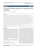
Figure 1.1: Genome structure of EV71. The single ORF is flanked by UTRs at
the 5' and 3' ends, a variable length poly-A tail is found at the 3' UTR. The ORF is
divided into three regions: the P1 region encodes four structural proteins VP1–
VP4, the P2 and P3 regions encode seven non-structural proteins 2A–2C and 3A–
3D, respectively. (Adapted from Brown and Pallansch, 1995)
5
1.2.1.1 5’ untranslated region (5’UTR)
Like other picornaviruses, enterovirus 71 has a long 5’ untranslated region
upstream of the start codon of about 750 bp. The 5’UTR is covalently linked to a
viral protein Vpg (Lee, 1977; Flanegan, 1977) and has multiple stem-loop
structures (Yang, 1997). Since the 5’cap is replaced by Vpg, enteroviruses use an
alternative, cap-independent, internal pathway for initiation of translation. The
secondary structure within the 5’UTR serves as an internal ribosome entry site
(IRES) for recruitment of ribosomes (Jang, 1988; Pelletier and Sonenberg, 1988).
The stem-loop structures were found to be important in both cap-independent
translation initiation and RNA replication. Stem-loop I is at the very beginning of
5’UTR and is a highly conserved cloverleaf-like structure. This structure is
involved in negative strand RNA synthesis (Andino, 1990). Stem-loops II to VI
serve as IRES and are required for cap-independent translation (Pelletier and
Sonenberg, 1988) (Figure 1.2). There is a pyrimidine tract found to be located
about 10–15 bases upstream of an AUG that is not recognized as an initiator
codon by the translation machinery; the sequence encompassing this silent AUG
of the enterovirus genome is termed box B (Pilipenko, 1992a and 1992b). Studies
demonstrated that the cellular protein, heterogeneous nuclear ribonucleoprotein K
6
(hnRNP K), interacts with stem-loops I-II and IV in the 5' UTR of enteroviruses.
Viral yields and RNA synthesis were significantly compromised in hnRNP K
knockdown cells (Lin JY, 2008). The sequence of 5’UTR was found to be quite
conserved among enteroviruses, and thus it has been widely utilized for the
detection of enteroviruses (Rotbart, 1990).
7