Identification and characterization of short vegetative phase (SVP) target genes
Bạn đang xem bản rút gọn của tài liệu. Xem và tải ngay bản đầy đủ của tài liệu tại đây (7.75 MB, 106 trang )
IDENTIFICATION AND CHARACTERIZATION OF
SHORT VEGETATIVE PHASE (SVP) TARGET
GENES
WU YANG
(B. Sc.)
A THESIS SUBMITTED
FOR THE DEGREE OF MASTER OF SCIENCE
DEPARTMENT OF BIOLOGICAL SCIENCES
NATIONAL UNIVERSITY OF SINGAPORE
2009
TABLE OF CONTENT
ACKNOWLEDGMENTS IV
CHEMICALS AND REAGENTS ..................................................................................................V
UNITS AND MEASUREMENTS ................................................................................................ VI
OTHERS ............................................................................................................................... VII
LIST OF TABLES
VIII
LIST OF FIGURES IX
SUMMARY
XI
CHAPTER 1 LITERATURE REVIEW 1
1.1 INTRODUCTION ................................................................................................................. 1
1.2 BIOLOGY OF ARABIDOPSIS ................................................................................................. 4
1.3 SHOOT APICAL MERISTEM (SAM) ORGANIZATION.......................................................... 5
1.4 STEM CELL MAINTENANCE AT SAM................................................................................ 7
1.5 MAJOR FLORAL PATHWAYS AND INTEGRATORS .............................................................. 9
1.6 SHORT VEGETATIVE PHASE (SVP)............................................................................ 14
1.7 MADS-BOX GENE FAMILY ............................................................................................ 17
1.8 ARABIDOPSIS PROTEIN INTERACTING WITH NIMA-1 (ATPIN1)......................................... 18
1.9 KIP-RELATED PROTEINS (KRPS) ..................................................................................... 19
1.10 CONCLUSION ................................................................................................................ 21
I
CHAPTER 2 MATERIALS AND METHODS
23
2.1 PLANT MATERIALS AND GROWTH CONDITIONS ............................................................. 23
2.2 RNA EXTRACTION ......................................................................................................... 23
2.3 REVERSE TRANSCRIPTION FOR CDNA SYNTHESIS ......................................................... 25
2.4 EXPRESSION ANALYSIS .................................................................................................. 26
2.4.1 Quantitative Real-time PCR .................................................................................. 26
2.4.2 Semi-quantitative RT-PCR..................................................................................... 26
2.5 NON-RADIOACTIVE IN SITU HYBRIDIZATION ................................................................... 27
2.5.1 RNA Probe Synthesis ............................................................................................. 27
2.5.2 Material Fixation ................................................................................................... 29
2.5.3 Dehydration and Embedding ................................................................................. 30
2.5.4 Sectioning............................................................................................................... 31
2.5.5 Pre-treatment of in situ Sections............................................................................ 32
2.5.6 In Situ Hybridization.............................................................................................. 33
2.5.7 In Situ Post-hybridization ...................................................................................... 34
2.6 CHROMATIN IMMUNOPRECIPITATION (CHIP) ASSAYS ................................................... 36
2.7 MICROARRAY EXPERIMENTS .......................................................................................... 37
2.8 GENOMIC DNA EXTRACTION ......................................................................................... 38
2.8.1 Rapid Extraction of Genomic DNA ....................................................................... 38
2.8.2 Kit-facilitated Extraction of Genomic DNA........................................................... 39
2.9 COMPETENT CELL PREPARATION ................................................................................... 41
2.10 TRANSFORMATION OF E. COLI COMPETENT CELLS ....................................................... 42
II
2.10.1 Heat Shock ........................................................................................................... 42
2.10.2 Verification of Constructs by Colony PCR .......................................................... 43
2.10.3 Plasmid DNA Extraction ..................................................................................... 43
2.10.4 Verification of Constructs by Sequencing............................................................ 45
2.11 TRANSFORMATION OF A. TUMEFACIENS COMPETENT CELLS ........................................ 46
2.12 PLANT TRANSFORMATION ............................................................................................ 47
CHAPTER 3 RESULTS 48
3.1 INTRODUCTION ............................................................................................................... 48
3.2 PHENOTYPIC ANALYSIS OF SVP-41 MUTANTS ................................................................. 49
3.3 EXPRESSION ANALYSIS OF SVP-41 MUTANTS .................................................................. 49
3.4 PHENOTYPIC ANALYSIS OF MUTANTS AND TRANSGENIC LINES .................................... 53
3.5 PHENOTYPES OF ATPIN1 KNOCKDOWN AND OVEREXPRESSION LINES ............................ 55
3.6 GENETIC CROSS ANALYSIS OF ATPIN1 .......................................................................... 59
3.7 CHIP ASSAYS OF ATPIN1 PROMOTERS ........................................................................... 63
3.8 FLOWERING PATHWAY ANALYSIS OF ATPIN1 ............................................................... 66
3.9 ATPIN1 EXPRESSION PATTERN ANALYSIS ..................................................................... 66
3.10 SEQUENCE ALIGNMENT OF ATPIN1 WITH ITS HOMOLOGS .......................................... 69
3.11 EXPRESSION ANALYSIS OF KRP1 AND KRP2 ............................................................... 71
CHAPTER 4 DISCUSSION
77
CHAPTER 5 CONCLUSION 83
REFERENCE 85
III
Acknowledgments
This thesis was written as a final report of my research for completing my
Master Degree. Taking this opportunity, I would like to express my gratitude
to all the people who have been so helpful and supportive during the period of
my study at NUS.
Specifically, I would like to thank my supervisor, Dr. Yu Hao, for his
guidance and support on my research project, and his help and encouragement
in my life in Singapore.
I would also like to thank all the lab members in the Plant Functional
Genomics Group for their generous help, support, and encouragement.
Lastly, I would like to say thank you to my parents and my fiancée, who have
always supported me with their love and trust.
Wu Yang
April 2009
IV
List of Abbreviations
Chemicals and Reagents
DEPC
diethylpyrocarbonate
dNTP
deoxynucleoside triphosphate
EDTA
ethylene-diamine-tetra-acetate
Gly
glycine
HCl
hydrochloric acid
KPO4
potassium phosphate
LB broth
Luria-Bertani broth
LiCl
lithium chloride
MgCl2
magnesium chloride
NaCl
sodium chloride
Na2HPO4
disodium phosphate
NaH2PO4
sodium phosphate
PBS
phosphate buffered saline
PMSF
phenylmehtylsulfonylfluoride
PVA
polyvinyl alcohol
SDS
sodium dodecylsulphate
Tris
tris-(hydroxymethyl)aminomethane
V
Units and Measurements
bp
base pair(s)
g
gram(s)
hr
hour(s)
kb
kilo base-pair(s)
kDa
kilo Dalton(s)
M
molar
min
minute(s)
ml
mililitre(s)
mM
milimolar
ng
nanogram(s)
OD600nm
absorbance at wavelength 600 nm
rpm
revolutions per minute
sec
second(s)
U
unit(s)
v/v
volume per volume
w/v
weight per volume
°C
degree Celsius
µg
microgram(s)
µl
microlitre(s)
µM
micromolar
VI
Others
amiRNA
artificial micro ribonucleic acid
BLAST
Basic Local Alignment Search Tool
cDNA
complementary deoxyribonucleic acid
ChIP
chromatin immunoprecipitation
cRNA
complementary ribonucleic acid
CDK
cyclin-dependent kinase
Col
Columbia
DNA
deoxyribonucleic acid
et al.
et alter (and others)
GA
gibberellin, or gibberellic acid
i.e.
that is
LD
long day
mRNA
messenger ribonucleic acid
PCR
polymerase chain reaction
RNA
ribonucleic acid
RT-PCR
reverse transcription polymerase
chain
reaction
SAM
shoot apical meristem
SD
short day
VII
List of Tables
Table 1. List of primer pairs used for real-time PCR analysis
51
Table 2. List of primers used for AtPIN1
58
Table 3. List of primers used for ChIP assays
65
VIII
List of Figures
Fig. 1 Schematic representation of major genetic flowering
10
pathways and floral pathway integrators
Fig. 2 Phylogenetic tree of StMADS11 clade
16
Fig. 3 Scanning electron microscopy analysis of adaxial
50
rosette leaves in svp-41 and wild-type plants
Fig. 4 Comparison of gene expression in svp-41 and wild-
54
type plants
Fig. 5 Phenotypes of AtPIN1 antisense and 35S:AtPIN1
56
plants
Fig. 6 Flowering time of AtPIN1 transgenic lines and expre-
57
-ssion of AtPIN1 in these lines
Fig. 7 Infertility phenotype of an AtPIN1 knockdown line
60
using amiRNA
Fig. 8 Genetic cross analysis of AtPIN1 transgenic lines
61
Fig. 9 Relationship of AtPIN1 with SOC1 and AGL24
62
Fig. 10 ChIP analysis of AtPIN1 promoter
64
Fig. 11 Flowering pathway analysis of AtPIN1
67
Fig. 12 AtPIN1 expression patterns in wild-type and svp-41
68
plants
Fig. 13 Sequence alignment of AtPIN1 and its homologs
70
IX
Fig. 14 Analysis of KRP1 and KRP2 expression in various
72
flowering mutants
Fig. 15 Analysis of KRP1 and KRP2 expression in long days
74
and short days
Fig. 16 Analysis of KRP1 and KRP2 expression under GA
75
treatment
Fig. 17 Analysis of KRP1 and KRP2 expression under
76
vernalization treatment
Fig. 18 Ser/Thr-Pro motifs in MADS-box transcription factors
80
X
Summary
Flowering plants undergo floral transitions from vegetative phase to
reproductive phase in response to multiple endogenous and environmental
signals. In Arabidopsis, SHORT VEGETATIVE PHASE (SVP) has been
suggested as a central regulator of flowering time. Recent findings have
indicated that SVP functions by interacting with FLC to control the
transcription of two floral pathway integrators, SUPPRESSOR OF
OVEREXPRESSION OF CONSTANS 1 (SOC1) and FLOWERING LOCUS T
(FT). In a search for novel target genes of SVP that mediate its function in
flowering regulation, we identified that AtPIN1 was transcriptionally regulated
by SVP and that it promoted flowering under both long days and short days.
AtPIN1 responds to both photoperiod and vernalization, and its function as a
flowering promoter depending on the activity of SOC1 and AGL24 was
revealed by genetic cross analysis. In addition, this interaction between
AtPIN1 and SOC1/AGL24 occurred at post-transcriptional level. Our data
suggest that, as an enzyme that catalyzes cis/trans conformation change,
AtPIN1 may bind to SOC1 and AGL24 and facilitates their conformational
change, leading to the accumulation of specific conformations of these two
proteins to promote flowering.
XI
Chapter 1 Literature Review
1.1 Introduction
Flowering plants, also known as angiosperms, are the most successfully
evolved and predominant group of land plants, characterized by their most
remarkable feature, i.e. flowers. They represent the most widespread group of
land plants and one of the only two extant groups of seed plants on the planet
earth (Magallón et al., 1999). They are easily distinguished from other seed
plants by their extremely diversified flower morphologies. Flowering plants
serve as the major basis for agriculture through livestock feed, and offer other
economic resources as well, including wood, paper, fiber, and medicines, etc.
Estimation of their number of species has been made to be in the range of
250,000 to 400,000 (Govaerts, 2001; Govaerts, 2003; Scotland and Wortley,
2003; Thorne, 2002). The reproductive successes of flowering plants depend
heavily on the correct timing to switch from vegetative to reproductive phase,
which allow plants to flower under desirable conditions for optimal seed
setting and synchronously for out-breeding species (Bernier, 1988). This
major developmental transition is tightly controlled by an integrated network
of pathways that respond to both environmental and endogenous signals and
distinct strategies for reproduction have been evolved in different plant species
(Simpson and Dean, 2002).
1
The last 20 years have seen an explosion of knowledge on the molecular and
genetic mechanisms underlying floral induction, patterning and organ identity.
Three dicot species, Antirrhinum majus, Arabidopsis thaliana, and Petunia
hybrida have been the primary sources from which the basic mechanisms are
elucidated. Among these three model plants, Arabidopsis thaliana is most
contributive in giving detailed and comprehensive knowledge about the
fundamental molecular mechanisms of flower development (Jack, 2004).
Arabidopsis thaliana is a small weed in the mustard family under the genus
Brassica and is native to Europe, Asia, and Northwestern Africa. Its adoption
as a genetic model organism was first proposed by Laibach in 1943 based on
his findings of the short generation time, fecundity, ease of crosses, and the
possibility of mutagenesis for Arabidopsis (Laibach, 1943). It was later
studied in detail by Rédei in the United States whose instrumental reviews
helped introduce the model to the scientific community (Rédei, 1975). Further
momentum for the use of Arabidopsis as a model organism came from the
release of the first complete and detailed genetic linkage map of Arabidopsis
(Koornneef et al., 1983), the summarization of the value of Arabidopsis as a
model system for research in plant biology, the demonstration that its small
genome is amenable to detailed molecular analysis (Meyerowitz and Pruitt,
1985), and the significant technical advances leading to the establishment of
transformation protocols (An et al., 1986; Feldmann and Marks, 1987; Lloyd
2
et al., 1986).
The increased enthusiasm for Arabidopsis led to the drafting of a vision
statement in 1990, which outlined the long-term objectives for the Arabidopsis
community, and the establishment of the Arabidopsis Genome Initiative in
1996 to coordinate the multinational endeavor of the large-scale sequencing of
Arabidopsis thaliana genome (Meinke et al., 1998). The sequencing started in
1996 and was finished in 2000, but more work is still being done to integrate
all available experimental data on gene structure and function into the genome
annotation (Swarbreck et al., 2008; The Arabidopsis Genome Initiative, 2000).
The estimated ~157Mb genome of Arabidopsis thaliana, which is organized
into five chromosomes, contains 27,235 protein coding genes, 4,759 pseudo
genes or transposable elements and 1288 non-coding RNAs (ncRNAs) (33,282
genes in all, 38,963 gene models) according to the newest gene annotation
released from the Arabidopsis Information Resource, TAIR8 (Bennett et al.,
2003; The Arabidopsis Genome Initiative, 2000). The availability of the whole
genome sequence of Arabidopsis changed the nature of plant genetic research
fundamentally, making forward genetics greatly simplified and reverse
genetics possible. The meteoric rise of Arabidopsis thaliana as a model
organism from an obscure weed represents not only an integration of scattered
community resources, avoiding duplication of effort and waste of funding, but
also a dramatic shift in paradigm for plant biology research (Meinke et al.,
3
1998). In the year 1998, Arabidopsis thaliana has officially been selected as
one of the members of “Security Council of Model Genetic Organisms”.
These organisms form a comparing standard for all other organisms and a
concentrated research on the genetics of them serves as a biological window to
all the rest of the species within that phylum (Fink, 1998). The high sequence
similarity between many genes from plants and other organisms connects the
biological study of plants to all others, and greatly expands the amount of
biological knowledge that can be shared between plant biologists and
biologists in other fields (Somerville, 2000).
1.2 Biology of Arabidopsis
Arabidopsis thaliana is a member of the Brassica genus with a broad
distribution in nature throughout Europe, Asia, and Northwestern Africa
(Meyerowitz and Somerville, 1994). It can complete its whole life cycle
within 6 weeks, from seed germination and bolting of the main stem to
flowering and seed maturation. Bolting usually occurs about 3 weeks after
sowing, during which shoot apical meristem becomes inflorescence meristem
and flowers start to be produced. Flowers are small with a length of about 2
mm and self-pollinating. They are composed of four concentric whorls of
distinct floral organs, which are sepals, petals, stamens and carpels
sequentially from the outermost whorl to the innermost. Genetic crossing can
4
be easily done by applying pollen of one plant to the stigma surface of another.
Plants are usually grown either in small pots filled with soil or in petri dishes
placed either under fluorescent lights in the laboratory or in a greenhouse.
Healthy mature Arabidopsis plants are able to reach a height of 15 to 20 cm
and generate several hundred siliques with more than half a thousand seeds in
total (Meinke et al., 1998).
1.3 Shoot Apical Meristem (SAM) Organization
During embryogenesis, Arabidopsis plants produce apical meristems at both
root and shoot ends. The root and shoot apical meristems continuously make
new cells throughout the life of the plant to produce the underground root
system and the above-ground architecture, respectively. Arabidopsis
meristems are composed of small groups of pluripotent stem cells that are
morphologically undifferentiated (Fletcher, 2002).
The shoot apical meristem (SAM) consists of three radial domains, the central
zone, the peripheral zone and the rib zone (Steeves and Sussex, 1989). The
central zone comprises a reservoir of stem cells which occupy the apex of the
SAM and divide infrequently as compared with other cells in the SAM.
Division of the cells in the central zone gradually displaces the progeny cells
into the surrounding peripheral zone, where cells divide more often than the
5
ones at the central zone (Medford et al., 1992; Reddy et al., 2004; Steeves and
Sussex, 1989). However, cells in the peripheral zone are more restricted in
their differential potency than those at apex and become integrated into either
lateral organ or internode primordia (Irish and Sussex, 1992; Steeves and
Sussex, 1989). Underneath the central zone and in the deep layers of the
meristem lies the rib zone, which forms the pith of SAM and gives rise to the
most part of the stem (Steeves and Sussex, 1989). Cell divisions occurring in
the rib zone lead to the upward growth of the shoot tips, leaving the cells in
the peripheral zone behind to undergo proliferation and differentiation. The
peripheral zone is replenished at the same time by descendents of dividing
cells from the central zone, which gradually undergo specification with their
displacement away from the tip and are essential for the SAM maintenance
(Fletcher, 2002).
Another way of dissecting the SAM is to stratify the cells at the apex into
distinct layers, named the tunica and corpus (Poethig, 1987; Satina et al.,
1940). The tunica is composed of an epidermal L1 layer and a subepidermal
L2 layer, each of which is a cell layer of single cell thick and whose cells keep
clonally distinct from other cells by dividing solely anticlinally with an
orientation perpendicular to the meristem plane (Tilney-Bassett, 1986). The
L1 layer cells give rise to the epidermis of leaves, shoots, and flowers,
whereas the L2 layer cells are precursors of the germline cells and mesodermal
6
cells. The corpus, lying beneath the tunica, consists of a group of cells, called
L3 cells. The L3 cells produce the vasculature and pith of the stem and
innermost cells of lateral organs, such as leaves and flowers. The cell divisions
within L3 are orientated more randomly in all planes, differing from those of
the L1 and L2 layer cells whose divisions are restricted to a single anticlinal
plane (Fletcher, 2002). Although cell divisions are highly organized in the
SAM, no fixed patterns exist for SAM cell fate specification based on cell
lineage as shown by mosaic analysis (Furner and Pumfrey, 1992; Irish and
Sussex, 1992). Since cells that accidentally squeeze from one layer into
another layer do not cause defects in development (Tilney-Bassett, 1986), the
fate of a SAM cell is decided by its position instead of its clonal origin
(Stewart, 1978).
1.4 Stem Cell Maintenance at SAM
The central zone at the tip of the SAM contains stem cell reservoirs that are
self-renewal and crucial for the non-stop development and generation of the
aerial architectures of higher plants. An intrinsic mechanism of intercellular
signaling exists and balances the continuous departure of stem cell derivatives
for lateral organ initiation and the constant formation of new stem cell
daughters that replenish the stem cell reservoirs (Williams and Fletcher, 2005).
Signals that specify stem cell identity are provided by an organizing centre
7
(OC), which is a small group of WUSCHEL (WUS) expressing cells beneath
the central zone. WUS, a homeodomain transcription factor, forms the WOX
(WUS HOMEOBOX) gene family together with its 14 homologues in
Arabidopsis (Mayer et al., 1998). WUS is both required and sufficient for
specifying stem cell identity. Stem cells are mis-specified and SAM is
prematurely terminated when WUS function is lost (Laux et al., 1996),
whereas ectopic stem cell identity is induced when WUS is ectopically
expressed (Schoof et al., 2000). The neighboring cells above the organizing
center are specified to take stem cell identity by the underlying WUS activity
at the OC. These stem cells express and secrete CLAVATA3 (CLV3) into the
extracellular space. CLV3 is a small mobile polypeptide, which binds to the
CLV1/CLV2 receptor complex on the membrane of the OC cells and activates
the CLV signaling pathway that inhibits WUS expression and thereby confines
the size of stem cell reservoir (Brand et al., 2000; Lenhard and Laux, 2003;
Rojo et al., 2002). This negative feedback loop of regulation between the stem
cells and the OC cells maintains the homeostasis of the stem cell population,
through an quick adjustment of WUS expression following any change in
CLV3 transcription level when the number of stem cells fluctuates (Williams
and Fletcher, 2005).
8
1.5 Major Floral Pathways and Integrators
The shift from vegetative to reproductive growth represents a major transition
of development for flowering plants, whose correct timing is crucial for
maximizing success of reproduction (Simpson and Dean, 2002). In
Arabidopsis, flowering time is controlled by multiple genetic floral pathways
that have been demonstrated to integrate both endogenous and environmental
signals (Fig. 1). The four major pathways are photoperiod pathway,
vernalization pathway, autonomous pathway, and gibberellin (GA) pathway
(Koornneef et al., 1998; Mouradov et al., 2002; Simpson and Dean, 2002).
These genetic pathways respond to different environmental or endogenous
signals, but eventually converge to control the expression a set of common
targets, which are termed as the floral pathway integrators (Simpson and Dean,
2002). Three genes, which have been identified as the floral pathway
integrators, are LEAFY (LFY), FLOWERING LOCUS T (FT), and
SUPPRESSOR
OF
OVEREXPRESSION
OF
CONSTANS
1
(SOC1)
(Kardailsky et al., 1999; Kobayashi et al., 1999; Lee et al., 2000; Samach et al.,
2000; Weigel et al., 1992).
The photoperiod pathway responds to changes in day lengths by accelerating
flowering
under
long
days.
Arabidopsis
senses
light
through
CRYPTOCHROME1/2 (CRY1/2) and phytochromes A to E (Clack et al., 1994;
Lin, 2000), and measures the duration of day or night by an endogenous timer,
9
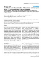
Figure 1. Schematic representation of major genetic flowering
pathways and floral pathway integrators. Four major flowering
pathways, photoperiod, autonomous, GA, and vernalization, are shown. Floral
pathway integrators, SOC1, FT, and LFY integrate flowering signals from
several genetic pathways.
10
called the circadian clock (Thomas and Vince-Prue, 1997). The rhythms of a
circadian clock are generated by a central oscillator, which is coupled to
regulate physiological activities and adjust its pace according to the light and
temperature cycles by multiple pathways (Dunlap, 1999). In Arabidopsis,
CONSTANS (CO), a transcription factor with two B-box zinc-finger domains,
couples the circadian oscillator to the activation of the flowering-time gene FT
(Suarez-Lopez et al., 2001). Plants that overexpress CO flower early in both
short days and long days, whereas loss-of-function co mutants are late
flowering in long days but not short days (Onouchi et al., 2000). The
expression of both CO and its target FT is altered by mutations that influence
circadian rhythms and flowering time (Suarez-Lopez et al., 2001). Under long
days, the coincidence between CO mRNA expression and CO protein stability
allows CO protein accumulation that promotes flowering by inducing
expression of three floral integrators, LFY, FT and SOC1 (Kardailsky et al.,
1999; Kobayashi et al., 1999; Nilsson et al., 1998; Suarez-Lopez et al., 2001).
This coincidence is lacking under short day conditions, which explains why co
mutants flower as wild-type plants in short days (Parcy, 2005).
Vernalization refers to the process that promotes flowering by an extended
exposure to cold temperature. Its requirement is adopted by many
winter-annual Arabidopsis accessions in nature as a reproductive strategy to
ensure that they grow vegetatively through the winter and flower until the
11
favorable spring in the following year (Simpson and Dean, 2002). Dominant
alleles at two loci, FLOWERING LOCUS C (FLC) and FRIGIDA (FRI), are
necessary to confer the vernalization requirement in these natural Arabidopsis
winter-annual accessions (Burn et al., 1993; Clarke and Dean, 1994; Lee et al.,
1993). FLC, which encodes a MADS-box transcription factor, is a potent
repressor of flowering (Michaels and Amasino, 1999; Sheldon et al., 1999).
FRI, encoding a novel protein with two coiled-coil domains, represses floral
transition through its promotive action on FLC mRNA abundance (Johanson
et al., 2000; Michaels and Amasino, 1999; Michaels and Amasino, 2001;
Sheldon et al., 1999; Sheldon et al., 2000). High levels of FLC expression
repress FT expression in leaves and FLC protein also antagonizes meristem
response to flowering signals by inhibiting SOC1 and the FT cofactor FD
expression in meristem (Abe et al., 2005; Corbesier et al., 2007; Searle et al.,
2006; Wigge et al., 2005). The vernalization pathway promotes flowering by
repressing FLC expression and maintaining a repressed state of its chromatin
through various epigenetic mechanisms (Bastow et al., 2004; He et al., 2003;
Sung and Amasino, 2004).
The autonomous pathway is defined by a group of mutants (fca, fy, fpa, ld, fld,
and fve) that are late-flowering independently of photoperiods and highly
sensitive to vernalization treatment (Koornneef et al., 1991; Martinez-Zapater
and Somerville, 1990; Sanda and Amasino, 1996). Much higher levels of FLC
12
mRNA than wild type have also been shown to be common to this group of
mutants, and responsible for their late flowering phenotype that is suppressed
in loss-of-function mutants of FLC (Michaels and Amasino, 1999; Michaels
and Amasino, 2001; Sheldon et al., 1999). Therefore, the autonomous pathway
in wild-type plants promotes flowering and converges with the vernalization
pathway by negatively regulating the transcription of FLC (Mouradov et al.,
2002). Although whether an endogenous input signal to the autonomous
pathway exists remains unknown, recent studies have shown that flowering
control by ambient temperature is mediated by the autonomous pathway in an
FLC-independent manner (Blazquez et al., 2003).
The gibberellin pathway mediates the effect of GA in promoting flowering.
Bioactive GAs are a class of diterpenoid-acid phytohormones that are involved
in regulation of diverse aspects of plant development, such as stem elongation,
seed germination, and floral induction and development (Yamaguchi, 2008).
Exogenous GA application was initially used to demonstrate the promoting
effect of GA on flowering (Langridge, 1957), which was substantiated by the
study on the GA signaling mutant gai that flowers late under both long days
and short days even in the presence of GA (Peng et al., 1997), and GA
biosynthesis mutant ga1-3 that flowers late under long days and extremely late
or never flowers under short days (Blazquez et al., 1998; Wilson et al., 1992).
The complete rescue of the non-flowering phenotype of ga1-3 under short
13