Study on drug delivery for paclitaxel of the lactobacillus strains
Bạn đang xem bản rút gọn của tài liệu. Xem và tải ngay bản đầy đủ của tài liệu tại đây (4.75 MB, 83 trang )
VIETNAM NATIONAL UNIVERSITY – HOCHIMINH CITY
INTERNATIONAL UNIVERSITY
STUDY ON DRUG DELIVERY FOR PACLITAXEL
OF THE LACTOBACILLUS STRAINS
A Thesis submitted to
the School of Biotechnology, International University
in partial fulfillment of the requirements for the degree of
Master of Science in Biotechnology
Student name: Doan Thi Thanh Vinh – MBT04015
Supervisor:
Dr. Nguyen Hoang Khue Tu
July 2013
Study on Drug Delivery for Paclitaxel of the
Lactobacillus Strains
By
DOAN THI THANH VINH
ABSTRACT
The practice of developing molecularly targeted drugs to achieve a higher degree
of cancer therapy and antibiotic resistance is indispensable. Lactobacillus strains
participated in the anti-cancer effects and performed the high-level specificity for
cancer cell lines. This paper discussed the ability of minicells which were
generated
from
Lactobacillus
acidophilus
VTCC-B-871
and
Lactobacillus
rhamnosus JCM 15113 for drug delivery of paclitaxel. L. acidophilus VTCC-B-871
and L. rhamnosus JCM 15113 formed minicells with highly significant number
(1,070,000 and 787,000 minicells, respectively) in modified MRS broth with the
concentration of fructose 10 g/l and lactose 50 g/l, respectively. Nanoparticle
size of obtained minicells ( 400 nm in diameter) was determined using scanning
electron
microscopy.
The
minicells
packaged
paclitaxel
(10
µg/ml)
and
cephalosporin (10 µg/ml) for different times of incubation (10, 15 and 24 hours)
at 37°C. Our results showed that drugs could be completely absorbed for 10
hours by detecting extracted solution from drug packaged minicells on
antimicrobial activities. The significant influence of different concentrations of
loading paclitaxel (5, 10, 20 µg/ml) on drug packaging was studied. The results
indicated that after extraction of the paclitaxel packaged minicells and analysis
with high performance liquid chromatography for determining the number of
paclitaxel presented in a minicell, there was a huge amount of paclitaxel
encapsulated in a minicell (31 million paclitaxel molecules per minicell with
loading paclitaxel (20 µg/ml)). The present work was the first report on the
generation
of
minicells
from
Lactobacillus
acidophilus
VTCC-B-871
and
Lactobacillus rhamnosus JCM 15113 and the drug delivery for paclitaxel of these
minicells.
Key works: Lactobacillus acidophilus, Lactobacillus rhamnosus, minicells,
paclitaxel,
cephalosporin,
antimicrobial
chromatography.
i
activities,
high
performance
liquid
ACKNOWLEDGEMENTS
First of all, it is undeniable that this thesis has taken me a great deal of time and
effort. Moreover, I have to insist that this thesis cannot be well-completed
without the encouragements, the nice ideas and helps from a lot of people. For
these reason, I would like to thank to many people that contributed one way or
another to the realization of this paper.
Remarkably, I would like to express my most sincere gratitude and appreciation
to Dr. Nguyen Hoang Khue Tu for all her enthusiastic supervision, patience and
encouragement of this work. Her critical comments, constructive suggestions,
professional directions also guided effectively my logical ideation and efficient
communication skills throughout the whole research period.
Furthermore, I would love to send my best wishes to all members of the School
of Biotechnology of International University for their tremendous enthusiasms as
well as for their nice behavior towards me, and also for
give
me
the
good
chance to conduct and have my research study completed.
Definitely, it is certain that I really send my thanks to Dr. Ha Dieu Ly and all the
members at Department of Reference Substances, Institute of Drug Quality
Controls and also all members at Scanning Electron Microscopy Laboratory
Room, Vietnam Academy of Science and Technology for their assistance and
technical support.
Words cannot express my respect, profound gratitude for my parents who are
always on me, encourage, and share their great love for me. I love to send my
sincere thanks to my good friends who are being with me during my bad and
good time.
Last but not least, I greatly thank you who spend precious time to read this
document. This actually has profound significance for me as the fruit of my labor
is also gotten your acceptance.
ii
TABLE OF CONTENTS
ABSTRACT ........................................................................................... i
ACKNOWLEDGEMENTS ..........................................................................ii
LIST OF FIGURES ................................................................................ v
LIST OF TABLES .................................................................................. vi
ACRONYMS AND ABBREVIATIONS ........................................................ vii
CHAPTER 1 INTRODUCTION ............................................................. 1
CHAPTER 2 LITERATURE REVIEW ..................................................... 3
2.1 Cancer and tumor-targeted drug delivery systemsError! Bookmark
not defined.
2.1.1 Worldwide cancer burden ............................................................. 3
2.1.2 Cancer treatment and recent advances in cancer therapy ................ 4
2.1.3 Obstacles in cancer therapy and targeted drug delivery systems ...... 5
2.1.4 Approaches to improve the therapeutic index of anti-cancer drugs .... 8
2.2 Bacterially-derived minicells .................................................... 10
2.2.1 Minicells ...................................................................................10
2.2.2 Bacterially-derived mincells as controlled drug delivery for cancer
therapy .............................................................................................11
2.2.4 Current research of minicells in delivering anti-cancer drugs ...........12
2.3 An overview of Lactobacillus species ....................................... 14
2.3.1 The Genus Lactobacillus .............................................................14
2.3.2 Lactobacillus acidophilus .............................................................15
2.3.3 Lactobacillus rhamnosus .............................................................15
2.4 Paclitaxel ................................................................................. 16
2.5 Orientation
of
implementing
to
delivery
paclitaxel
using
minicells derived from the lactobacillus strains .............................. 17
2.6 Process of project .................................................................... 18
CHAPTER 3 MATERIAL AND METHODS ............................................ 19
3.1 Materials .................................................................................. 19
3.1.1 Bacterial strains.........................................................................19
3.1.2 Tools and equipments ................................................................19
3.1.3 Media and chemicals ..................................................................19
3.2 Research methodology ............................................................. 21
3.2.2 Design condition for minicells production from Lactobacillus strains..21
3.2.2 Minicell isolation ........................................................................21
3.2.3 Microscopic studies for characterization of minicell morphology .......21
3.2.4 Packaging of drug into minicells ...................................................22
3.2.5 Drug extraction for measurement ................................................23
iii
3.2.6 Antimicrobial activity tests ..........................................................23
3.2.7 Paclitaxel assay using HPLC-UV/Vis Spectroscopy ..........................23
3.2.8 Calculation of the number of drug molecules .................................24
3.2.9 Data analysis ............................................................................25
CHAPTER 4 RESULTS AND DISCUSSION ......................................... 26
4.1 Screening the carbon sources for study on minicell generation
from Lactobacillus strains .............................................................. 26
4.1.1 Minicells generation in Lactobacillus acidophilus VTCC-B-871 ..........30
4.1.2 Minicells generation from Lactobacillus rhamnosus JCM 15113 ........34
4.2 Microscopic studies: characterization of minicell morphology .. 37
4.3 Package of drug into minicells generated from Lactobacillus
strains ............................................................................................ 39
4.3.1 Antimicrobial activity tests ..........................................................39
4.3.1.1 Drug packaging of minicells produced from L. acidophilus 39
4.3.1.2 Drug packaging of minicells produced from L. rhamnosus ..........41
4.3.2 Paclitaxel quantitation using HPLC-UV/Vis Spectroscopy .................43
4.3.2.1 The minicell drug packaging in response to incubation times ......44
4.3.2.2 The minicell drug packaging in response to concentrations .........45
CHAPTER 5 CONCLUSION AND RECOMMENDATION ........................ 49
REFERENCES ................................................................................... 50
iv
LIST OF FIGURES
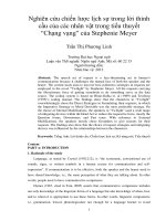
Figure 2.1 Nanoparticle platforms for drug delivery. ..................................... 8
Figure 2.2 Schematic showing minicell formation and bispecific antibodytargeted, drug/siRNA-packaged minicells ..................................11
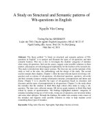
Figure 2.3 Chemical structure of paclitaxel . ...............................................16
Figure 3.1 Process of isolation of bacterially-derived minicells from the parent
cells culture ...........................................................................21
Figure 4.1 The API 50CHL biochemical testing of Lactobacillus acidophilus......26
Figure 4.2 The API 50CHL biochemical testing of Lactobacillus rhamnosus. .....28
Figure 4.3 The number of minicells (×104) produced from L. acidophilus ........32
Figure 4.4 Photomicrographs of Lactobacillus acidophilus VTCC-B-871 and its
minicells (100X)l. ....................................................................33
Figure 4.5 The number of minicells (×104) produced from L. rhamnosus ........35
Figure 4.6 Photomicrographs of Lactobacillus rhamnosus JCM 15113 and its
minicells (100X). .....................................................................36
Figure 4.7 Photomicrograph showing the morphology of minicells from
Lactobacillus acidophilus VTCC-B-871 under the light microscope
after isolation procedure (100 X) ...............................................37
Figure 4.8 Representative SEM images showing the formation of minicells. ....38
Figure 4.9 Representative SEM images of minicells from L. acidophilus VTCC-B871with minicell size after isolation procedure ............................39
Figure 4.10 Drug quantitation in minicells when minicells incubated in the
presence of different drug loading concentrations. .......................46
v
LIST OF TABLES
Table 2.1 Estimated (2008) and projected
numbers (millions) of cancer
cases and deaths, all cancers, both sexes, by development status
or WHO region ............................................................................. 3
Table 2.2 The summary statistics of estimated incidences, mortality in men,
women, and both sexes in Vietnam in 2008. ................................... 4
Table 2.3 Advantages of using nanoparticles as a drug delivery system ........... 9
Table 3.1 Lactobacilli MRS broth ...............................................................20
Table 4.1 The API 50CHL biochemical testing of Lactobacillus acidophilus .......27
Table 4.2 The API 50CHL biochemical testing of Lactobacillus rhamnosus .......29
Table 4.3 The number of minicells produced from L. acidophilus VTCC-B-871 in
modified MRS medium .................................................................32
Table 4.4 The number of minicells produced from L. rhamnosus JCM 15113 in
modified MRS medium .................................................................35
Table 4.5 Antimicrobial spectrum of extracted drugs from drug-packaged
minicells of L. acidophilus VTCC-B-871 and L. rhamnosus JCM 15113
against Gram-positive and Gram-negative bacteria .........................41
Table 4.6 Antibacterial activity of extracted paclitaxel and cephalosporin from
drug-packaged minicells of L. acidophilus against bacterial species ...41
Table 4.7 Antibacterial activity of extracted paclitaxel and cephalosporin from
drug-packaged minicells of L. rhamnosus against bacterial species ....42
Table 4.8 HPLC analysis of paclitaxel in the loading solution in response to
varying times of incubation ..........................................................44
Table 4.9 HPLC analysis of paclitaxel in minicells when incubated in the
presence of different drug concentrations.......................................46
Table 4.10 Paclitaxel quantification in minicells when incubated in the presence
of different drug concentrations ....................................................47
vi
ACRONYMS AND ABBREVIATIONS
ATCC
American Type Culture Collection
BsAbs
Bispecific Antibodies
BSG
Buffer Saline Gelatin
CSCs
Cancer Stem Cells
DDSs
Drug Delivery Systems
EGFR
Epidermal Growth Factor Receptor
EPS
Exoploysaccharide
FDA
Food And Drug Administration
GRAS
Generally Recognized As Safe
HPLC
High Performance Liquid Chromatography
IARC
International Agency For Research On Cancer
IFP
Interstitial Fluid Pressure
JCM
Japanese Collection Microorganism
LAB
Lactic Acid Bacteria
NCI
National Cancer Institute
PBS
Phosphate Buffer Saline
SEM
Scanning Electron Microscope
shRNA
Short Hairpin RNA
siRNA
Small interfering RNA
VTCC
Vietnamese Type Culture Collection
vii
CHAPTER 1
INTRODUCTION
Cancer is the general name for a group of more than 100 diseases which is being
one of the major health problems on over the world. Despite the availability of
several treatment modalities, mortality rates due to cancer are high. Therefore,
effective cancer therapy continues to be a daunting challenge due mainly to
considerable tumor cell heterogeneity, drug resistance of cancer cells, doselimiting toxicity of chemotherapeutics, and difficulties of targeted delivery to
tumors (MacDiarmid et al., 2011).
A key obstacle in the use of current chemotherapeutic anticancer drugs is their
lack of specificity for cancer cells, resulting in severe toxicity when they are
administered systemically (Flemming, 2007). Consequently over the past decade
a significant global effort has focused on the discovery and development of
molecularly targeted drug delivery systems (DDSs) (MacDiarmid et al., 2007a).
DDSs are being developed to achieve a higher degree of tumor cell specificity
and reduce toxic side effects, as well as overcome several challenges to the
treatment of cancer (Chidambaram et al., 2011), including drug resistance and
metastatic disease (Alexis et al., 2010). However, current strategies that use
nanoparticles (Brannon et al., 2004), liposomes (Zheng et al., 2001); or polymer
therapeutics (Duncan, 2003) are hampered by shortcomings such as drug
leakage in vivo, lack of versatility in terms of packaging a diverse range of
different drugs, thereby reducing drug potency, and difficulties in production
scale-up (Ferrari et al., 2005), particularly for nanoparticles.
Recently, a new promising technology for targeted and intracellular delivery of
chemotherapeutic drugs relies on using bacterially derived nano-sized particles
(termed as minicells). Minicells are originated from the normal cell deleting the
min gene (de Boer et al., 1989), but they can package a range of different
chemotherapeutic drugs and specifically targeting the minicells to tumor cell
surface receptors via bispecific antibodies coating the minicells (Flemming,
2007). This technique has been experimented successfully for both Grampositive (Listeria monocytogenes) and Gram-negative bacteria (Salmonella
typhimurium, Escherichia coli, Shigella flexneri, and Pseudomonas aeruginosa);
drug-packaged nano-sized particles effected apoptosis of tumor cells both in
vitro and in vivo; and they also targeted to cancer cells in vivo with high
specificity and, thus, delivered in high concentration in vivo without toxicity
1
(MacDiarmid et al., 2007a). Although minicells currently generated from both
Gram-positive and Gram-negative bacteria and also tested for encapsulating
chemotherapeutic drugs and functioning as nanovectors for drug delivery in
cancer therapy (MacDiarmid et al., 2007b). This mutation may effect on the
growth of bacteria under their control so far (de Boer et al., 1989). Moreover, up
to now, there has not any research on generating bacterially-minicells from
Lactobacillus strains using other method without min gene deletion. Especially,
Lactobacillus strains are GRAS (Generally Recognized as Safe) microorganisms
used as the probiotics that are very important in foods, pharmaceuticals, and
animal husband (Macfarlane and Cummings, 2002). The benefits of probiotics
were found protection against gastrointestinal pathogens, enhancement of the
immune system, reduction of lactose intolerance, reduction of serum cholesterol
level and blood pressure, anti-carcinogenic activity, improved utilization of
nutrients and improved nutritional value of food. Members of the genus
Lactobacillus have also been reported on the significant anti-oxidative, anticarcinogenic, and anti-bacterial activity, as well as the inhibitory effects on
cancer cell growth besides being effect on human immune system (Choi et al.,
2006; Kim et al., 2002). Among the tested Lactobacillus species, Lactobacillus
acidophilus (Choi et al., 2006) and Lactobacillus rhamnosus (Cenci et al., 2002)
strains participated in the anti-cancer effects, anti-carcinogenic ability and
performed the high-level specificity for cancer cell lines.
From those points, this paper presented the generation of bacterially derived
minicells from Lactobacillus strains for drug delivery and investigated if minicells
derived from these strains were able to package with chemotherapeutic drug
(Paclitaxel); in order to detectable a robust and versatile system for in vitro drug
delivery
using
minicell,
a
bacterially-derived
lactic
acid
bacteria
carrier.
Moreover, this study also confirmed the ability of encapsulation of minicells with
other drug as cephalosporin. This study was the primary research on the drug
delivery system which was able to carry many drugs and necessary products for
food and pharmaceutical fields.
Therefore, the main aim of this study was to develop a new drug nano-sized
carrier derived from probiotic Lactobacillus for delivery of chemotherapeutic drug
with fewer side effects, and with further modification, to produce a systemic
complex of molecular targeted drug delivery including probiotics properties.
From this aim, those specific objectives including, performing drug-packaged
bacterially-derived minicells and determining the number of drug molecules
presented in minicell.
2
CHAPTER 2
LITERATURE REVIEW
2.1 CANCER AND TUMOR-TARGETED DRUG DELIVERY SYSTEMS
2.1.1 Worldwide cancer burden
Cancer is a group of diseases characterized by the uncontrolled growth of
abnormal cells that disrupt body tissue, metabolism, etc. and tend to spread
locally and to distant parts of the body. Life-threatening cancer develops
gradually as a result of a complex mix of factors such as complex interactions of
viruses, a person‟s genetic make-up, their immune response and their exposure
to other risk factors which may favor the cancer (Win, 2006). Based on the
GLOBOCAN 2008 estimates (Ferlay et al., 2010a), the standard set of worldwide
estimates of cancer incidence and mortality produced by the International
Agency for Research on Cancer (IARC) for 2008, there were an estimated 12.4
million cases of cancer diagnosed and 7.6 million deaths from cancer and
28 million persons alive with cancer around the world in 2008 (Table 2.1); of
these, 56% of the cases and 64% of the deaths occurred in the less developed
regions of the world,
many
of
which lack the medical resources and health
systems to support the disease burden. By 2030, it could be expected that there
could be 27 million incident cases of cancer, 17 million cancer deaths annually
and 75 million persons alive with cancer within five years of diagnosis (Table
2.1).
Table 2.1 Estimated (2008) and projected numbers (millions) of cancer cases
and deaths, all cancers, both sexes, by development status or WHO region (Boyle
and Levin, 2008)
20301
2008
20302
Region
Cases
Deaths
Cases
Deaths
Cases
Deaths
World
12.4
7. 6
20.0
12.9
26.4
17.0
Africa (AFRO)
0.7
0.5
1.2
0.9
1.6
1.3
Europe (ERO)
3.4
1.8
4.1
2.6
5.5
3.4
East Mediterranean (EMRO)
0.5
0.3
0.9
0.6
1.2
0.9
Pan-America (PAHO)
2.6
1.3
4.8
2.3
6.4
3.1
South-East Asia (SEARO)
1.6
1.1
2.8
1.9
3.7
2.6
Western Pacific (WPRO)
3.7
2.6
6.1
4.4
8.1
5.9
¹ no change in current rates
² with 1% annual increase in rates
3
Overall in 2008, based on the most recently available international data
(GLOBOCAN 2008 estimates) produced by IARC, about 111,600 incident cases of
cancer and 82,000 cancer deaths were estimated to have occurred in Vietnam
(Table 2.2). The most commonly diagnosed cancers were liver (23,251 cases,
20.8% of the total new cancer cases), lung (20,659 cases, 18.5%) and stomach
(15,068 cases, 9.7%) (Figure 2.1). Liver cancer is also the leading cause of
cancer death for both sexes, accounting for 21,748 deaths, comprising 26.5% of
the total cancer deaths (Ferlay et al., 2010a).
Table 2.2 The summary statistics of estimated incidences, mortality in men,
women, and both sexes in Vietnam in 2008; Source : GLOBOCAN 2008 (Ferlay et
al., 2010a).
Vietnam
Male
Female
Both sexes
Population (thousands)
42973
44122
87095
Number of new cancer cases (thousands)
55.0
56.5
111.6
15.9
12.8
14.1
43.7
38.3
82.0
12.7
8.8
10.5
Risk of getting cancer before age 75 (%)
Number of cancer deaths (thousands)
Risk of dying from cancer before age 75 (%)
5 most frequent cancers (ranking defined by total number of cases)
Liver
Liver
Liver
Lung
Lung
Lung
Stomach
Breast
Stomach
Incidence and mortality data for all ages
Proportions per 100,000
2.1.2 Cancer treatment and recent advances in cancer therapy
Although great effort has been made in cancer research, no substantial progress
can be observed in the past fifty years in the USA or almost twenty years in
Vietnam in fighting against cancer. The death rate in the USA was 193.9 per
100,000 in 1950 and remained as high as 194.0 per 100,000 in 2001 (Jemal et
al., 2004). Based on the data from the Hanoi cancer registry, IARC estimated
that in 1990 the mortality in Vietnam was about 82.2 per 100,000 (Pham and
Nguyen, 2002) and actually stayed lower than that 94.1 per 100,000 in 2008
(Ferlay et al., 2010a). It is clear that the progress in cancer treatment has been
slow and inefficient (Win, 2006). It is a multidisciplinary challenge needing more
and closer collaboration between clinicians, medical and biomedical scientists
and biomedical engineers to eventually find a satisfactory solution (Win, 2006).
4
The choice of treatment depends on the type and location of the cancer, whether
the disease has spread, the patient's age and general health, and other factors.
Clinical treatment for cancer therapy is a multidisciplinary therapy consisting of
surgery, radiation therapy, chemotherapy, biological therapy and other methods
(e.g., biological therapy, targeted therapy, or gene therapy for cancer (ACS,
2013). Chemotherapy is most effective against cancers that divide rapidly and
have a good blood supply (Win, 2006). Aims of chemotherapy treatments are to
cure; to maintain long term remission (free of disease); to increase the
effectiveness of surgery or radiotherapy; to help control pain or other symptoms.
However, research still continues in finding ways to make chemotherapy less
toxic and also to minimize the side effects. Molecularly targeted therapy – a new
generation of cancer treatments has emerged as one approach to overcome the
lack of tumor specificity of conventional cancer therapies (Gerber, 2008).
Currently, the pharmaceutical industry has been successful in discovering many
new cytotoxic drugs that can potentially be used for the treatment of cancer, this
life-threatening disease still causes over 7 million deaths every year worldwide
and the number is growing (Ferlay et al., 2010a). Thus, the ongoing obligation to
the design and discovery of new cancer therapy is urgent.
2.1.3 Obstacles in cancer therapy and tumor-targeted drug delivery
systems
There are several serious obstacles in cancer therapy including drug resistance,
high tumor interstitial fluid pressure (IFP), and cancer stem cells (CSCs).
2.1.3.1 Obstacles in cancer therapy
A vast array of resistance mechanisms, involving overexpression of drug
transporters (which are the plasma membrane P-glycoprotein (P-gp) product of
the multidrug resistance (MDR) gene as well
as
other
associated
proteins)
(Gottesman, 2002), mutations or amplification of the target enzyme, decreased
drug activation, increased drug degradation due to altered expression of drug
metabolizing enzymes, diminished drug-target interaction, enhanced DNA repair,
or failure to apoptosis, can defeat single agents, no matter how well designed
and targeted (Chorawala et al., 2012).
High tumor interstitial fluid pressure (IFP) is another barrier for efficient drug
delivery (Heldin et al., 2004). Increased IFP contributes to a decreased
transcapillary transport in tumors leads to a decreased uptake of drugs or
5
therapeutic antibodies. Cancer cells are therefore exposed to a lower effective
concentration of therapeutic agent than normal cells, lowering the therapeutic
efficiency and increasing toxicity. It is now well established that the IFP of most
solid tumors is increased. This increase makes the uptake of therapeutic agents
less efficient in solid tumors (Wu et al., 2006). There are a number of factors
that contribute to increase IFP in the tumour, such as vessel leakiness, lymph
vessel abnormalities, fibrosis and contraction of the interstitial matrix.
The discovery of cancer stem cells (CSCs) in solid tumors has changed our view
of carcinogenesis and chemotherapy. Cancer stem cells are defined as those cells
within a tumour that can self-renew and drive tumorigenesis (Dean et al., 2005).
The CSCs, which are also accurately called „tumor-initiating cells‟, represent a
small population of cancer cells, sharing common properties with normal stem
cells (SCs), that can initiate new tumors following injection into animal models,
while the majority of other cancer cells cannot (Vinogradov and Wei, 2012).
Natural properties of the small group of cancer stem cells involved in drug
resistance to standard chemotherapy agents, metastasis and relapse of cancers
can significantly affect tumor (Dean et al., 2005).
In addition to the obstacles in cancer therapy, current chemotherapeutic drugs
are constrained by severe systemic toxicity due to indiscriminate drug
distribution and narrow therapeutic indices. A key obstacle in the use of
chemotherapeutic anticancer drugs is their lack of specificity for cancer cells,
resulting in severe toxicity when they are administered systemically (Sarosy and
Reed, 1993). This is exacerbated by the fact that systemically delivered cancer
chemotherapy drugs often must be delivered at very high dosages to overcome
poor bioavailability of the drugs and the large volume of distribution within a
patient.
In general, cancer chemotherapy is usually accompanied by severe side
effects and acquired drug resistance.
Therefore, we anxiously await the
development of molecularly targeted therapy that will allow greater tumor
specificity and less toxicity. Over these years, cancer targeting treatment has
been greatly improved by new tools and approaches based on the development
of nanotechnology. Nanotechnology is the creation and utilization of materials,
devices, and systems through the control of matter on the nanometer scale
(Jain, 2005). Nanocarrier systems can be designed to interact with target cells
and tissues or respond to stimuli in well-controlled ways to induce desired
6
physiological responses. They represent new directions for more effective
diagnosis and therapy of cancer (Alexis et al., 2010).
2.1.3.2 Tumor-targeted drug delivery nanoparticles
In recent years, the rapid advent of nanotechnology has stimulated the
development of many novel drug delivery strategies (Wang et al., 2007).
Nanoparticles applied as nanoscale drug delivery vehicles have shown the ability
to encapsulate a variety of therapeutic agents such as small molecules
(hydrophilic and/or hydrophobic), peptides, protein-based drugs, and nucleic
acids, and protect them against enzymatic and hydrolytic degradation (Mohanraj
and Chen, 2006). By encapsulating these molecules inside a nanocarrier, the
known shortcomings of many anticancer drugs can be potentially overcome,
such as low aqueous solubility, stability, high nonspecific toxicity or lack of
selectivity of anticancer drugs (Chidambaram et al., 2011), while at the same
time increasing the circulation time and bioavailability of encapsulated drugs
(Langer, 1998).
Through encapsulation of drugs in a macromolecular carrier, such as a liposome,
the volume of distribution is significantly reduced and the concentration of drug
in a tumor is increased. This causes a decrease in the amounts and types of
nonspecific toxicities, and an increase in the amounts of drug that can be
effectively delivery to a tumor (Moghimi, 2006). The surface of the nanocarrier
can be engineered to increase the blood circulation half-life and influence the
biodistribution, while attachment of targeting ligands to the surface can result in
enhanced uptake by target tissues (Gref et al., 1994; Moghimi et al., 2001). The
small size allows nanocarriers to overcome biological barriers and achieve
cellular uptake (Brigger et al., 2002). The net result of these properties is to
lower the systemic toxicity of the therapeutic agent while increasing the
concentration of the agent in the area of interest, resulting in a higher
therapeutic index for the therapeutic agent (Gilstrap et al., 2011) against the
most difficult cancer challenges (Chidambaram et al., 2011), including drug
resistance and metastatic disease (Alexis et al., 2010).
7
Polymeric
Nanoparticle
Dendrimer
Liposome
Polymeric
Micelle
Inorganic
Nanoparticle
Polymerosome
Protein
Carrier
Biological
Nanoparticle
Polymer-drug
Conjugate
Hybrid
Nanoparticle
Hydrophobic Polymer
Therapeutic load
Hydrophilic Polymer
Targeting Ligand
Lipid
Figure 2.1 Nanoparticle platforms for drug delivery. Nanoparticle platforms are
characterized by their physicochemical structures, including polymer-drug
conjugates, lipid-based nanoparticles, polymeric nanoparticles, protein-based
nanoparticles, biological nanoparticles, and hybrid nanoparticles (Alexis et al.,
2010).
Nanoparticles applied as drug delivery systems are submicronsized particles (10
to 1000 nm) (Shim and Turos, 2007), devices, or systems that can be made
using a variety of materials including polymers (polymeric nanoparticles,
micelles, or dendrimers), lipids (liposomes), magnetic, even inorganic/metallic
compounds (iron, silica) and bacteria (bacterial nanoparticles or “minicells”)
(MacDiarmid and Brahmbhatt, 2011; MacDiarmid et al., 2007b, 2009) (Figure
2.2).
2.1.4 Principle approaches to improve the therapeutic index of anticancer drugs
Nanoparticle drug delivery systems are being studied to overcome limitation of
conventional
therapeutic
areas
particularly
in
cancer
chemotherapy.
As
mentioned in subheading 2.1.3.2, nanomedicine performed a strong potential to
accelerate the development of effective approaches to the treatment of drug8
resistant and recurrent cancers. However, despite the significant progress made
in the development of drug delivery system (DDS) and other nanoplatformbased approaches, serious limitations have also been identified in applications of
these therapies in vivo (Vinogradov and Wei, 2012). Several important
limitations of nanoparticles are highlighted (mostly liposomes (Table 2.4)), such
as ineffective uptake and distribution in tumor tissue, retention in bypassing
organs and by macrophages of the reticuloendothelial system after systemic
administration, and limited oral availability (Yun et al., 2012). Other nanovector
systems such as synthetic biodegradable nanoparticles, polymer micelles, and
several others, are also hampered by drug leakage in vivo, lack of versatility in
terms packaging a diverse range of different drugs, thereby reducing drug
potency, and difficulties in production scale-up (Ferrari, 2005). Despite the
enhanced efficacy demonstrated by many targeted nanoparticles, they also face
three major limitations: immunogenicity or non-specificity of the targeting ligand
leading to accelerated blood clearance; further impaired tumor penetration
compared to the nontargeted nanoparticles; and receptor-mediated endocytosis
and subsequent lysosomal digestion resulting in a major dose loss by the
lysosomal digestion (Chen et al., 2012).
Table 2.3 Advantages and disadvantages of liposome (Anwekar et al., 2011)
Advantages of liposome
Disadvantages of liposome
1. Liposomes increased efficacy and therapeutic
Low solubility
index of drug (actinomycin-D)
2. Liposome increased stability via
Short half-life
encapsulation
3. Liposomes are non-toxic, flexible,
Sometimes phospholipid
biocompatible, completely biodegradable,
undergoes oxidation and
and non-immunogenic for systemic and non-
hydrolysis-like reaction
systemic administrations
4. Liposomes reduce the toxicity of the
Leakage and fusion of
encapsulated agent (amphotericin B, Taxol)
5. Liposomes help reduce the exposure of
encapsulated drug/molecules
Production cost is high
sensitive tissues to toxic drugs
6. Site avoidance effect
Fewer stables
7. Flexibility to couple with site-specific ligands
to achieve active targeting
9
Because problems continue to hamper significantly the success of cancer
therapeutics, an urgent need exists for new targeted drug delivery strategies
that will either selectively deliver drugs to tumor cells and target organs, protect
normal tissues from administered antineoplastic agents, or prevent existing
problems in cancer therapies. The present invention relates to ongoing efforts to
achieve a targeted drug delivery by means of intact bacterial minicells, which are
able to delivery drugs intracellular, within desired target cells in vivo and in vitro
(MacDiarmid et al., 2007a, 2007b, 2009). Minicells containing chemical or
biochemical drugs constitute novel delivery vehicles, capable of being targeted to
specific cells. Because of the benefits of delivering chemotherapeutics drugs for
cancer treatment, the practice of synthesizing and packagage of cytotoxic drug
into minicells is the key point of this study. The present study builds on these
recent discoveries relating to minicells, and addresses the continuing needs for
improved
drug
delivery
strategies,
especially
in
the
context
of
cancer
chemotherapy.
2.2 BACTERIALLY-DERIVED MINICELLS
2.2.1 Minicells
Minicells were first observed and described by Howard Adler and colleagues in
1967, who also coined the term “minicell” with the first description of a mutation
that led to the minicell phenotype in Escherichia coli (Adler et al., 1967), and to
more accurately describe the particle, people propose the new term “nanocell‟
instead of “minicell” since the scale size of the vector is nanometer and is not in
the mini- or micro-range (MacDiarmid et al., 2007a). They are anucleate, nonliving nano-sized cells (100 – 400 nm in diameter) and are produced as a result
of mutations in genes that control normal bacterial cell division (de Boer et al.,
1989; Lutkenhaus, 2007; Ma et al., 2004) there-by depressing polar sties of cell
fission (Figure 2.3). The resultant minicells do not contain any of the original
DNA and the chromosome present in its larger sister, but may contain all of the
molecular components of the parent cell. The minicells are capable of protein
synthesis and normal metabolic functions but are incapable of undergoing further
rounds of cell division. Minicell formation has since been described in a number
of other Gram-positive and Gram-negative species. First discovered over 70
years ago, minicells are becoming of interest to researchers in their potential as
anti-tumor agents.
10
Figure 2.22 Schematic showing minicell formation and bispecific antibodytargeted, drug/siRNA-packaged minicells. Minicells can be loaded with siRNAs
(purple), shRNA (green) or chemotherapeutics (black). Loaded minicells are then
functionalized via bispecific antibody conjugates, with one arm specific for
minicell-surface O-polysaccharide (red) and the other specific for the tumor
cell-surface receptor (blue) (Karagiannis and Anderson, 2009).
2.2.2 Bacterially-derived mincells as controlled drug delivery for cancer
therapy
Minicells are small, semi-spherical, bacterial nano-size particles that contain all
of the components of the parental bacteria, except chromosomes. Without
chromosomes, they cannot divide and are non-infectious, making them highly
suitable for development as in vivo delivery products. So far, clever attempts at
delivering potent drugs straight to the cancer cells using techniques such as
conjugating them to antibodies specific to those cells, have been inconclusive at
best.
The use of molecularly targeted minicell nanovectors affords multiple potential
advantages over conventional cancer therapy (MacDiarmid et al., 2007a). Firstly,
minicells possess the ability to easily package therapeutically significant
concentrations of different cytotoxic or molecularly targeted drugs into the
minicell, ability to encapsulate both
hydrophilic and
hydrophobic drugs.
Secondly, minicells have the ability to readily attach different bispecific
antibodies (BsAbs) on the minicell surface in order to target a receptor found on
the surface of a tumor cell. Thirdly, minicells are able to deliver the drug
intracellularly within a tumor cell and without leakage of drug/siRNA/shRNA from
the vector during systemic circulation. In addition, minicells also provide a
dramatic increase in the therapeutic index with minimal to no toxic side effects.
This also enables the use of potent cytotoxics that have failed toxicity trails but
have the potential to be highly potent anti-cancer drugs. Moreover, minicells are
11
easily purified to homogeneity and the long standing pharmaceutical industry
experience in bacterial fermentation and production of bacterial vaccines shows
that such processes are relatively cheap. The minicells nanovector has the
potential to significantly reduce cost of goods particularly since a minicell-based
anti-cancer therapeutic would carry tiny fractions of the drug and the targeting
antibody compared to free drug or free antibody therapy. Finally, obstacles in
anticancer therapy such as multi-drug resistance of tumor can also be overcome
via receptor-mediated endocytosis which triggered by the adhesion of the
minicells to tumor-surface receptors, or via sequential minicell-mediated delivery
of siRNA followed by drugs (MacDiarmid et al., 2009).
2.2.4 Current research situation of minicells in delivering anti-cancer
drugs
Minicell has been observed and described in the studies on the bacteria cell
division from long time ago. However, it is actually noticeable in recent years as
Himanshu Brahmbhatt, Jennifer MacDiarmid and colleagues firstly showed that
report promising results using bacterial minicells as the drug delivery system in
2007 (MacDiarmid et al., 2007a). The unusual drug delivery vehicle was
generated by inactivating genes that control normal cell division in bacteria.
This led to the formation of anucleate particles that have a uniform diameter of
400 nm, and high yields are readily produced from Gram-positive (Listeria
monocytogenes (L. monocytogenes)) and Gram-negative bacteria (Salmonella
enterica serovar Typhimurium (S. typhimurium), Escherichia coli (E. coli),
Shigella flexneri (S. flesneri), and Pseudomonas aeruginosa (P. aeruginosa)).
These minicells were
loaded
with
a
range of therapeutically significant
concentrations of chemotherapeutics (such as doxorubicin, paclitaxel, irinotecan,
5-flourouracil, cisplatin, carboplatin, and vinblastine) with differing charge,
hydrophobicity and solubility by simple co-incubation in few hours. Minicell has
also been demonstrated that a minicell can be efficiently loaded with si/shRNA
(MacDiarmid et al., 2009). The ability of minicells encapsulated a large number
of drug molecules (1- 10 million per minicell), and they loaded with doublestranded siRNA to an estimated density of nearly 12,000 molecules per minicell.
These minicells selectively targeted to cancer cells via BsAbs.
Cancer-cell
targeting was achieved by coupling minicells to bispecific antibodies, in which
one arm recognizes surface lipopolysaccharide (LPS), and the other a surface
receptor on the targeted cell.
12
In vivo experiments with targeted doxorubicin-loaded minicells led to the
dramatic inhibition and regression of tumour growth in mice that had human
breast, ovarian, leukaemia or lung cancer xenografts. Furthermore, the drug was
undetectable in the plasma of minicell-treated animals, and none of the animals
developed any signs of toxicity. The anticancer efficacy of the minicells was
further evaluated in dogs with advanced T-cell non Hodgkin‟s lymphoma;
treatment led to marked tumour regression and tumour lysis. Importantly, the
treatment was tolerated without adverse side effects despite repeat dosing, and
there was no increase in pro-inflammatory cytokines. Further experiments in
pigs confirmed no adverse reactions in terms of haematological indices, serum
chemistries, food intake or growth, and surprisingly anti-LPS titers remained at
background levels. The team tested this using a form of siRNA designed to
prevent the production of a protein that causes multi-drug resistance in cancer
cells.
Recently, an early phase clinical trial using the platform of minicell nanoparticle
for drug delivery has been tested for the first time on patients with advanced
cancer and found to be safe, well-tolerated and even induced stable disease in
patients with advanced, incurable cancers with no treatment options remaining.
This clinical trial phase I was implemented by Associate Professor Benjamin
Solomon at the Peter MaCallum Cancer Centre in Melbourne, Australia with
colleagues. In a Phase I trial, minicells were loaded with with a cytotoxic
chemotherapy drug called paclitaxel and coated with cetuximab, antibodies that
target the epidermal growth factor receptor (EGFR) which is often overexpressed
in a number of cancers, as a „homing‟ device to the tumor cells. The treatment,
code-named EGFRminicellsPac, was well tolerated, and of the 28 people treated,
10 had stable disease at 6 weeks, and one patient safely received 45 doses over
15 months (ECCO, 2012) . This important study shows for the first time that
these bacterially-derived minicells can be given safely to patients with cancer. It
thereby allows further clinical exploration of a completely new paradigm of
targeted drug delivery using this platform coupled with different concentration of
cell-killing drugs or other treatments such as RNA interference, and with
different targeting antibodies.
After all, the minicell technology is actually a platform for the targeted delivery
of many different molecules, including drugs and molecules for silencing rogue
genes which cause drug resistance in late stage cancer. The technology can also
be viewed as a powerful antibody drug conjugate where up to a million
13
molecules of drug can be attached to targeting antibodies and delivered to the
body in a safe way. In the future this will enable a truly personalized medicine
approach to cancer treatment, since the minicell payload can be adjusted
depending on the genetic profile of the patient. Approaches resulting in selective
delivery of anti-cancer drugs to tumour cells are highly interesting as it may lead
to a reduction in adverse side-effects and improved anti-tumour activity. In this
respect, the use of minicells is a novel and promising technique (ECCO, 2012).
2.3 AN OVERVIEW OF Lactobacillus SPECIES
2.3.1 The Genus Lactobacillus
2.3.1.1 General description of the genus
Taxonomically, the genus Lactobacillus belongs to the phylum Firmicutes, class
Bacilli, order II Lactobacillales, and family Lactobacillaceae and its closest
relatives,
being
grouped
within
the
same
family,
are
the
genera
Paralactobacillus and Pediococcus (Felis et al., 2009). This genus includes a high
number of GRAS species (Generally Recognized as Safe).
Species of genus Lactobacillus are some of the most important taxa involved in
food microbiology and human nutrition: several Lactobacillus species are
remarkably essential in fermented food production and are used as starter
cultures or food preservatives. Moreover, certain strains of human origins are
being exploited as probiotics or vaccine carriers (Goh and Klaenhammer, 2009).
The lactobacilli
strains
can
be
used
as
potential
candidates
for
cancer
prevention (Choi et al., 2006; Liu and Pan, 2010). A few particular strains of L.
acidophilus, L. casei, L. paracasei, L. johnsonii, L. reuterui, L. salivarius and L.
rhamnosus have been extensively studied as candidates of probiotics and their
functional properties and safety have been well-documented.
2.3.1.2 The important role of Lactobacillus species in human life
Many Lactobacillus species are associated with food production, because of
preservative action due to acidification, and/or enhancement of flavour, texture
and nutrition (Jay, 1996; Stiles, 1996). Members of the genus Lactobacillus are
commonly present as members of probiotics (termed as living microorganisms
that are associated with the beneficial effects for humans and animals).
Lactobacilli strains have been used to study the anti-oxidative activity,
antibacterial and anti-cancer discovery besides being effect on human immune
system. This genus evaluated the inhibitory effects of on various human cancer
14
cell lines and attempted to demonstrate whether such effects were cancer cell
selective (Choi et al., 2006). The inhibition of cancer cell growth by Lactobacillus
has also been reported in other studies (Kim et al., 2002). Antioxidant activities
and antiproliferative activities against breast and colon cancer cell lines in vitro
gave a strong evidence of possessing significant anti-cancer activities of several
local lactobacilli strains (Liu and Pan, 2010). Short chain fatty acids produced by
L. acidophilus, L. rhamnosus are reported to inhibit the generation of
carcinogenic products by reducing enzyme activities (Cenci et al., 2002). These
results suggest that Lactobacillus can be used as adjuncts in fermentation of
food and are potential candidates for cancer prevention.
2.3.2 Lactobacillus acidophilus
Among many Lactobacillus species, L. acidophilus is likely the most common
probiotics for dietary use (Parvez, 2006). In addition to its gram positive rod
shape with rounded ends, the typical size of L. acidophilus is 1.5 - 6.0 µm in
length. L. acidophilus is an obligately homofermentative LAB that growth in
anaerobic condition. Because they utilize sugars (e.g. glucose, aesculin,
cellobiose, galactose, lactose, maltose, salicin, and sucrose) as their substrates
for fermentation, they inhabit environments with high sugar abundance, such as
the GI tract in humans and animals (Vijayakumar et al., 2008).
Health benefits of L. acidophilus include providing immune support for infections
or cancer, providing a healthy replacement of good bacteria in the intestinal tract
following antibiotic therapy, reducing occurrence of diarrhea in humans, aiding in
lowering cholesterol and improving the symptoms of lactose intolerance.
Moreover, some of L. acidophilus cell components have potential use in many
different areas of biotechnology such as vaccine development (Jafarei and
Ebrahimi, 2011). It is demonstrated that exoploysaccharide (EPS) from bacteria
specially Lactobacillus sp. may contribute to human health, either as nondigestible
food
fraction
or
because
of
their
immunomodulating or cholesterol lowering activity
anti-tumoral,
anti-ulcer,
(Ganesh, 2006; Vuyst and
Degeest, 1999). EPS has anti-carcinogenic ability mediated by the stimulation of
the mitogenic activity of B lymphocytes (Ruas-Madiedo et al., 2006; Xu et al.,
2010).
2.3.3 Lactobacillus rhamnosus
Lactobacillus rhamnosus GG is a clinically documented bacterial strain which is
used in many countries as a probiotic culture in different dairy products or in
15
pharmaceutical diet supplements (Korpela et al. 1997). L. rhamnosus GG is a rod
shaped, Gram-positive, with 2.0-4.0 µm in length, and often with square ends,
and occur singly or in chains. This bacterium is considered safe (GRAS)
microorganism. L. rhamnosus GG is a homofermentative LAB (Berry et al. 1997).
Lactobacillus rhamnosus has shown antimicrobial activity against Escherichia
coli, Enterobacter aerogenes, Salmonella typhi, Shigella sp., Proteus vulgaris,
Pseudomonas aeruginosa, Serratia marcescens, Staphylococcus aureus, Bacillus
subtilis, Bacillus megaterium, Bacillus cereus, Helicobacter pylori, Campylobacter
jejuni, and Listeria monocytogenes (Ambalam et al., 2009). L. rhamnosus are
reported to inhibit the generation of carcinogenic products by reducing enzyme
activities (Cenci et al., 2002).
2.4 PACLITAXEL
Among the available drugs for chemotherapy, paclitaxel (Taxol®) is one of the
best anti-cancer drugs and also reported to possess radio-sensitizer properties.
Paclitaxel is a white to off-white crystalline powder with empirical formula of
C47H51NO14 and a molecular weight of 853.91. It is highly lipophilic, insoluble in
water, and melts at around 216-217°C. It is a complex, oxygen-rich diterpenoid
(Rowinsky and Donehower, 1995; Rowinsky et al., 1992) and its chemical
structure has been elucidated by chemists as in Figure 2.5. It consists of some
benzene rings and other hydrophobic structures, which lead to its high water
insolubility of paclitaxel.
Figure 2.43 Chemical structure of paclitaxel
16