Identification of genomic alterations in castration resistant prostate cancer using next generation sequencing
Bạn đang xem bản rút gọn của tài liệu. Xem và tải ngay bản đầy đủ của tài liệu tại đây (6.19 MB, 130 trang )
Identification of Genomic Alterations in
Castration Resistant Prostate Cancer using Next
Generation Sequencing
Thesis
Submitted for a Doctoral Degree in Natural Sciences
(Dr. rer. nat)
Faculty of Mathematics and Natural Sciences
Rheinische Friedrich-Wilhelms-
Submitted by
Roopika Menon
from Chandigarh, India
Bonn 2013
Prepared with the consent of the Faculty of Mathematics and Natural Sciences at
the Rheinische Friedrich-Wilhelms-
1. Reviewer: Prof. Dr. Sven Perner
2. Reviewer: Prof. Dr. Hubert Schorle
Date of examination: 19 November 2013
Year of Publication: 2014
Declaration
I solemnly declare that the work submitted here is the result of my own investigation,
except where otherwise stated. This work has not been submitted to any other
University or Institute towards the partial fulfillment of any degree.
____________________________________________________________________
Roopika Menon; Author
Acknowledgements
This thesis would not have been possible without the help and support of many
people. I would like to dedicate this thesis to all the people who have helped make
this dream a reality.
This thesis would have not been possible without the patience, support and guidance
of my supervisor, Prof. Dr. Sven Perner. It has truly been an honor to be his first PhD
student. He has both consciously and unconsciously made me into the researcher that
I am today. My PhD experience has truly been the ‘best’ because of his time, ideas,
funding and most importantly his incredible sense of humor. He encouraged and gave
me the opportunity to travel around the world to develop as a scientist. I cannot thank
him enough for this immense opportunity, which stands as a stepping-stone to my
career in science. I would also like to thank Prof. Roman Thomas and Prof. Hubert
Schorle for their advice on my thesis and their support. I would also like to thank Dr.
Christine Schuberth who played an integral part in guiding me through the PhD
process.
I would then like to thank my family members: my mother, father, and brother who
have been pillars of support at every step of the way and have stood by me throughout
this wonderful journey. I would like to thank them for your advice, guidance, love,
and for being a major source of inspiration throughout my life. Words cannot describe
all that you have done for me, and what you mean to me. I truly believe that my
grandparents’ blessings and good wishes have made me who I am and brought me to
this stage in my life. I would like to thank Dinker Uncle, Pranati Aunty, Brahma
Uncle, Hardi Aunty and Sudhaka for believing in me and for their kinds words of
encouragement.
My fiancé, Vinay, has been a tremendous support during all my times of frustration.
His patience and understanding helped me tackle every hurdle with courage and
strength. My achievements were always his pride. His family has been an incredible
source of encouragement, showering me with words of appreciation and instilling in
me enough faith to carry this journey forward, till the end.
More importantly, I could not have completed my thesis without my ‘German family’.
I would like to specifically thank Diana and Alina for being my ‘besties’. They were
the first people I would run to for sharing all my PhD and non-PhD related happiness
and sorrows. They truly share a very special place in my life. Wenzel’s presence in
the lab brought a smile to my face on each and every day. My students, Kerstin and
Fried, made science a fun and exciting experience. Mario’s bioinformatic analysis
was the heart to my thesis, in him I found a great friend and a wonderful colleague. I
would like to thank Silke, Karen, Anne, Angela, and Michael for their
encouragement. I must mention Zaki Shaikhibrahim, who has been a mentor and a
true friend through every step of the way. His advice and support on every topic has
been invaluable. I must also thank Barny for making my time in the lab memorable
and amusing. Thanks to my ‘German family’, my time in Germany has been a
wonderful experience.
I would like to specifically thank Lynnette Fernandez Cuesta who was a constant
source of optimism through this whole process. She not only guided me
professionally, but has also been a close confidant through the past three years. Her
advice and compassionate attitude have been priceless, and instrumental in my
success. I shall treasure our interactions forever.
I would like to thank all my collaborators, both national and international, all
members of the Institute of Pathology at Tuebingen and Bonn.
I would also like to thank all my friends in Tuebingen and Bonn for the wonderful
time spent in these beautiful cities.
Table of Contents
1.
Summary………………………………………………………………………………………………….....1
2.
Introduction………………………………………………………………………………………………..3
2.1
The
Prostate…………………………………………………………………..……………….4
2.2
Cancer
Stages
and
Cancer
Types……………………………………………………...5
2.3
Genomic
Events
Leading
to
PCa
Initiation
and
Progression.......................7
2.4
Processes
Promoting
PCa
Progression
……………………………..……………11
2.5
Androgen
Receptor
and
PCa……………………………..........................................13
2.6
Available
Treatments
Options
for
PCa……………………………………………14
2.7
Pathology
Archiving
of
PCa
Samples…………………………...………………….16
2.8
Next
Generation
Sequencing
…………………………….………………………..….17
2.9
Next
Generation
Sequencing
Approaches……………………………………….19
2.10
Next
Generation
Sequencing
and
PCa…………………………………………...20
3.
Aims
of
the
Study…………………………………………………………………………...………….23
4.
List
of
Abbreviations………………………………………………………………………………….25
5.
Materials
and
Methods……………………………………………………………………………....27
5.1
Reagents..……………………………………………………………………………………..27
5.2
Apparatus…………………………………………………………………………………….28
5.3
Consumables………………………………………………………………………………...29
5.4
Kits…………………………………………………………………………………………...….30
5.5
Cell
Culture
Reagents………...…………………………………………………………..30
5.6
Cell
Lines……………………………………………………………………………………...31
5.7
Antibodies…………………………………………………………………………………….31
5.8
Primers………………………………………………………………………………………...31
5.9
siRNA…………………………………………………………………………………………...32
5.10
Buffers
and
Solutions……………………………………………………………….….32
5.11
BAC
Clones………………………………………………………………………………….33
6.
Methods…………………………………………………………………………………………………….34
6.1
Next
Generation
Sequencing
(SOLiD4)…………………………………………...37
6.2
Fixation
Protocols
………………………………………………………………………...37
6.3
Fluorescent
In-‐Situ
Hybridization…………………………………………….....…38
6.4
Cell
Culture………………………………………………………………………………..….40
6.5
Protein
Analysis…………………………………………………………………………....42
6.6
Functional
Assays……………………………………………………………………….…44
7.
Results……………………………………………………………………………………………………...49
7.1
Objective
I…………………………………………………………………………………….49
7.2
Objective
II……………………………………………………………………………...……58
7.3
Objective
III
………………………………………………………….………………...……66
8.
Discussion…………………………………………………………………………………………………86
9.
Conclusion………………………………………………………………………………………………...94
10.
References……………………………………………………………………………………………..95
11.
Appendix
I……………………………………………………………………………………………..104
12.
Appendix
II
…………………………………………………………………………………………...111
13.
List
of
Publications
………………………………………………………………………………..115
14.
Curriculum
Vitae
………………………………………………………………………...………...116
1. Summary
Castration resistant prostate cancer (CRPC) is the most aggressive form of prostate
cancer (PCa). For the development of novel therapeutic targets for CRPC, it is key to
decipher the molecular alterations underlying this lethal disease. Next generation
sequencing (NGS) technologies have revolutionized cancer research by detecting
genomic alterations, nucleotide substitutions, insertions, deletions and copy number
alterations. This project was focused on identifying novel genes involved in CRPC by
assessing somatic copy number alterations (SCNA) using whole exome sequencing on
five CRPC and paired normal formalin fixed paraffin embedded (FFPE) samples by
the SOLiD4 next generation sequencing platform.
The central aim of this study was the identification of therapeutic targets for CRPC.
Due to the unavailability and scarcity of fresh frozen CRPC material for research
purposes, the primary aim of this study was to compare the DNA, RNA and protein
integrity in fixed tissues obtained from pathology archives. Secondly, validity of
formalin fixed paraffin embedded (FFPE) and fresh frozen PCa tissue, from the same
patient, was determined by whole exome sequencing. A large data overlap between
both fixed tissue types was observed. This eventually led to the main objective of the
study involving the identification of therapeutic targets for CRPC.
FFPE and HOPE fixed specimen were comparable in DNA quality for downstream
research purposes. Furthermore, FFPE tumor and fresh frozen tumor exome
sequencing data, from the same patient, showed an overlap in the SNV analysis. This
led to the central aim which included the analysis of somatic copy number alterations
(SCNA) using whole exome sequencing on five CRPC and paired normal FFPE
samples by the SOLiD4 next generation sequencing platform. The sequencing data
identified two genes, YWHAZ and PTK2. Both genes, located on chromosome 8, were
1
amplified on all five sequenced patients. Furthermore, the amplification frequency of
both genes increased depending on the stage of PCa: prostate confined or localized
PCa, lymph node metastasized PCa and CRPC. YWHAZ knockdown in the PC-3 cell
line impaired proliferation and migration. Similarly, PTK2 inhibition, using a
pharmacological inhibitor, TAE226 inhibitor, significantly affected both cell
migration and proliferation at a concentration of 10 µM. Overall, these findings
suggest that inhibiting both YWHAZ and PTK2 could potentially delay cancer
progression in patients harboring the amplification of the latter genes. Furthermore,
FFPE tissue could be used as a promising alternative to fresh frozen tissue for NGS
technologies.
2
2. Introduction
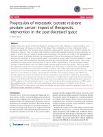
Prostate cancer (PCa) is the second largest cause of cancer related death in men of the
western world, accounting for more than 250,000 deaths a year (Figure 1) (1). It has
been reported that 1 out of every 6 men will be diagnosed with PCa and 1 out of every
3 diagnosed men will die of this disease (2). Various genetic alterations such as
amplifications, deletions, mutations, substitutions, and rearrangements, have been
studied to trigger the onset of disease. Unfortunately, due to its poorly understood
molecular mechanisms, in addition to its highly heterogeneous and complex nature,
treatment options for this disease remain a challenge (3).
Figure 1: Prostate cancer diagnosis world wide.
The most commonly diagnosed cancer among men worldwide in 2008. Prostate cancer (purple) is the
second largest cause of death in men of the western world. (adapted from Ferlay et al. 2010). (4)
3
2.1 The Prostate
The prostate is a walnut sized exocrine gland of the male reproductive organ. It
surrounds the urethra and located at the base of the urinary bladder. The function of
the prostate is the secretion of semen, with spermatozoa and seminal vesicle fluid.
The secretions from the seminal make semen alkaline in nature, which aids in
prolonging the lifespan of sperm (5). The prostate is architecturally defined as having
four zones: the central, periurethral transition, peripheral, and fibromuscular stroma
(6). These prostate zones consist of parenchymal cells, namely luminal epithelial
cells, basal epithelial cells and fibromuscular stromal cells. The luminal epithelial
cells express high levels of androgen receptor (AR). The basal epithelial cells express
AR at low undetectable levels. A rare subset of cells known as the neuroendocrine
cells expressing endocrine markers are also present (7).
In brief, the prostate is regulated by androgens, namely testosterone and 5-alpha-di
hyrdotestosterone, produced by the male testicles. These hormones belong to the
group of steroid hormones. The conversion of cholesterol to testosterone involves
various steps (Figure 2) and studies have shown that 5% of testosterone is converted
to dihydrotestosterone (DHT) through the 5-alpha-reductase catalyzed conversion in
the cells of the external male genitalia, the prostate and the bulbourethral glands. DHT
also exhibits a higher affinity to AR then testosterone. Furthermore, this highly active
metabolite binds to several DNA sequences to initiate transcription. This results in the
development and differentiation of the prostate (8).
4
Cholesterol
StAR
P450c17
17-OH Pregnenolone
P450ssc
3β-HSD
Pregnenolone
4,6
Δ -isomerase
Progesterone
3β-HSD
2,3
Δ -isomerase
17,20 desmolase
Dehydroepiandrosterone (DHEA)
P450c17
17-OH Progesterone
3β-HSD
2,3
Δ -isomerase
17-HSD
17,20 desmolase
Androstenediol
Androstenedione
17-HSD
3β-HSD
4,6
Δ -isomerase
aromatase
Testosterone
Estrone
aromatase
5α -reducatse
Dihydrotestosterone
17-HSD
Estradiol
Figure 2: Conversion of cholesterol to testosterone.
DHT plays an active role in prostate cancer development and progression (adapted from Chen et al,
2006) . (9)
2.2 Cancer Stages and Types
PCa can be broadly categorized into three stages: localized PCa, lymph node
metastasized PCa and distant metastasized PCa (Figure 3). Localized PCa is cancer
that is confined to the prostate gland and is curable in majority of the cases. Lymph
node metastasized PCa includes the spreading of the cancer through the lymph system
consisting of lymph nodes and lymph vessels. The most lethal form of this disease is
distant metastasized PCa, also known as castration resistant prostate cancer (CRPC),
where the cancer has spread to distant tissues through the blood stream (10). The bone
is the most frequent site of metastasis for PCa.
5
Figure 3: Prostate cancer types.
(A) Human schematic representing localized PCa, lymph node metastasized PCa and distant metastasis
to the brain. (B) Metastasis of tumor cells from the site of origin to different locations in the body via
the bloodstream (adapted from Braun et al. 2010) (11)
Additionally, PCa is commonly seen to occur in two types, namely adenocarcinoma
and small cell carcinoma. Adenocarcinoma is the most common form of PCa, which
arises from the cells of the glands. As most of the cells in the prostate are glandular
cells, adenocarcinoma has a tendency to develop metastatic potential. The rare but
highly aggressive form of PCa is small cell carcinoma. Small cell carcinomas are
6
aggressive because they are AR negative and patients do not benefit from hormone
treatments (12).
2.3 Genomic Events Leading to PCa Initiation and Progression
In-depth analysis using comparative genomic hybridization (CGH) studies has
identified various somatic alterations in PCa. These alterations include chromosomal
losses at 3p, 8p,10q, 13q, and 17p. The 8q region is seen to be extensively amplified
in PCa patients (13). Many genes lie in the regions of gains and losses known to be
key players in PCa initiation and progression such as NKX3.1 deletion, MYC
amplification, PTEN (phosphatase and tensin homolog) deletion, EZH2 up-regulation,
and TMRPSS2-ERG gene fusions.
a) NKX3.1 deletion: The NKX3.1 homeobox gene located on 8p is frequently seen to
be down regulated in advanced PCa. Through the progression of PCa, several studies
indicate the complete loss of the gene or a reduction in protein expression. In mice,
NKX3.1 regulates prostate epithelial differentiation and stem cell function. In humans,
the gene protects against DNA damage and regulates inflammation (14). Therefore,
the absence or decrease of NKX3.1 expression in PCa progression suggests a role as a
potential tumor suppressor in PCa.
b) MYC amplification: MYC is an oncogene situated in the 8q24 chromosomal region
and is frequently amplified and over-expressed in advanced PCa. This gene is a
transcription factor regulating metabolism, development, apoptosis, cell proliferation
and differentiation (15). MYC amplifications often occur in combination with other
genetic alterations leading to a cumulative negative effect (16). Studies have also
shown that forced expression of MYC produces immortalized nontumorigenic human
prostate epithelial cells (17).
7
c) PTEN (phosphatase and tensin homolog) deletion: PTEN is a potential tumor
suppressor gene situated on 10q23. This gene is reported to be frequently deleted in
various cancers. Previous publications have reported that the loss of PTEN activates
the AKT and JNK signaling pathways therefore leading to the development of CRPC
(18, 19). In PCa, conflicting data regarding the single allelic deletion of PTEN,
mutation of the second allele, and the reduced expression of PTEN have been
published (20). Interestingly, through the progression of PCa, PTEN copy number loss
correlates to the aggressive nature of the disease. PTEN, MYC and NKX3.1 genetic
alterations often occur together exhibiting a cumulative negative effect on patient
health. With reference to cancer cell lines, human cell lines exhibiting decreased
PTEN expression and PTEN deleted mouse cell PCa lines develop a castration
resistant phenotype (21). The molecular mechanisms behind PTEN alterations in PCa
remain to be investigated.
d) EZH2 up-regulation: The up-regulation of EZH2 through amplification is a
common event in advanced PCa (22). The gene is part of the Polycomb family and
codes for histone lysine methyltransferase. EZH2 targets, which also include NKX3.1,
play an active role in metastasis by the activation of the Ras and NF-ΚB pathways
(23). The polycomb family of genes function as inhibitors of gene expression by
forming multimeric complexes of proteins. These structures alter chromatin structure.
This family of genes regulates expression of cell cycle genes and HOX genes. These
latter genes regulate proliferation (24).
e) TMRPSS2-ERG gene fusions: The most commonly occurring gene fusions in PCa
involve the ETS transcription factors. The ETS transcription factors regulate DNA
binding, both positively and negatively, to transcriptional regulatory properties on the
same domain. Many ETS domain proteins are linked to cancer via various
8
mechanisms, which include their function in chromosomal translocations or
overexpression/down-regulation in cancers. Similarly, the family of transcription
factors also regulate proto-oncogenes or tumor suppressor proteins by their ability to
transduce signals from oncogenically activated signaling cascades (25). The
TMPRSS2-ERG gene fusion is the most common gene fusion occurring in 15-80% of
PCa patients. The large variation is due to the ethnicity, age, family history etc. The
TMPRSS2 gene codes for a serine protease that is expressed in the epithelium of the
prostate (26). The fusion occurs between the 5’exon of TMPRSS2 and the coding
sequence of ERG. The exact location of the fusion is on 21q22.2-22.3 resulting in two
products, an insertion through deletion or a translocation (Figure 4).
(A)
3'
ERG
TMPRSS2
chr. 21
5'
(B)
(C)
ERG
TMPRSS2
chr. 21
3'
3'
3'
ERG
TMPRSS2
chr. 21
chr. ??
5'
5'
5'
Figure 4: ERG rearrangement.
Dual colored fluorescent in situ hybridization (FISH) assay to detect ERG break-apart. (A) A wild type
ERG rearranged nucleus. (B) ERG rearrangement through deletion. (C) ERG rearrangement through
insertion (adapted from Braun et al., 2011). (27)
9
Studies show that in patients harboring the fusion, the TMPRSS2 promoter contains
the responsive promoter elements thus leading to androgen dependent ETS gene
overexpression (28). This fusion may be a result of radiation and other genotoxic
stress leading to double stranded breaks within the DNA. TMPRSS2 is also seen to
fuse with ETV1, ETV4 and ETV5. Another gene fusion seen in very low frequencies is
SLC45A3 gene fusions with ERG, ETV1 and ETV5(29).
The most commonly seen TMPRSS2-ERG gene results in the TMPRSS2 exon 1 fusing
with ERG exon 4, referred to as Type III isoform. This is seen in 80-90% of the gene
fusions. The second most common isoform results in the fusion between TMPRSS2
exon 2 and ERG exon 4, referred to as Type IV (Figure 5). The Type IV fusion result
in enhanced proliferation and invasion of PCa epithelial cells (30).
Figure 5: Role of TMPRSS2-ERG gene fusions.
The most commonly seen isoforms of the gene fusion include the Type III and Type IV. These
fusions lead to the up and down-regulations of various pathways involved in PCa progression (adapted
from Tindall et al, 2011) . (31)
10
Extensive studies on the ERG gene have shown its role in acting as an oncogene and
promoting tumor formation. It interacts with the c-JUN and AP-1 pathways to
promote growth, therefore indication the role of the gene fusion in the same (30). The
VCaP cell line, a PCa cell line derived from a vertebral metastatic lesion, is the only
cell line harboring the Type III gene rearrangement. Furthermore, studies have shown
that the Type III isoform induces increased invasion, proliferation and motility and
decreased differentiation (30).
e) Altered Pathways in PCa: One significant pathway that is altered in PCa is the upregulation of the PI3K/AKT/mTOR signaling cascade. The tumor suppressor gene
PTEN negatively regulates this pathway and loss of PTEN results in increase of cell
growth, proliferation, and survival (32). In addition, the ERK/MAPK signaling
pathway is also frequently activated in advanced PCa. Similarly, the RAS and RAF
pathways are also up-regulated, possibly via mutations, in PCa (32). Inhibition of the
above mentioned pathways has decreased tumor cell development and enhanced
apoptosis. These characteristics have enhanced the role of these pathways as potential
combinational therapeutic targets for treated PCa (33, 34).
2.4 Processes Promoting Prostate Cancer Progression
Various factors lead to PCa progression in men. Amongst these factors, age is
considered to be the most significant risk factor leading to the development of PCa.
Along with age, several environmental and physiological processes contribute to the
progression of the disease. Several studies have identified inflammation, oxidative
stress, telomere shortening and cell senescence also to play a key role in promoting
PCa (7, 35, 36) .
11
a) Inflammation: Chronic inflammation in the presence of the expression of various
chemokines has been reported to be linked to PCa progression (37). In older men,
regions of frequent inflammation have been referred to as “proliferative inflammatory
atrophy (PIA). These regions exhibit increased epithelial proliferation (38).
b) Oxidative Stress: An imbalance in the detoxifying enzymes and the reactive
oxygen species (ROS) leads to lipid, DNA and protein damage, which is a major
cause of oxidative stress. It is speculated that the prostate gland is highly vulnerable to
oxidative stress due to the processes such as inflammation, hormonal dysregulation,
diet, and epigenetic modifications. The epigenetic silencing of the gene, GSTP1,
belonging to the glutathione S-transferase family is a classic example. The function of
this gene is to initiate the detoxification of the ROS. In PCa, the gene is inactivated
due to promoter hypermethylation thereby subjecting DNA to further genome
damaging stress that may lead to malignant cancer progression. Another example is
APE/Ref 1, a multifunctional enzyme controlling other enzymes involved in
detoxification and base excision repair. This enzyme is up-regulated in PCa, but, PCa
patients harboring a polymorphism in the APE gene are susceptible to developing
cancer due to the loss of function of the gene (39).
c) Telomere shortening: Telomeres are repetitive sequencing situated on the ends of
chromosomes to denote chromosomal stability. Studies have correlated the effects of
telomere shortening during prostate carcinogenesis, but the exact mechanism of action
still remains unclear (40).
d) Senescence: In PCa, oncogene driven senescence plays a key role in tumor
suppression. Oncogenes, such as FOXm1, p53, Rb, c-Myc, drive senescence through
replicative stress or the formation of ROS. Several senescence markers such as SA-βGal, p14arf, p16ink4a are used to identify indolent phenotype from the aggressive (41).
12
2.5 Androgen Receptor and PCa
AR plays a key role in prostate cancer. The AR gene is part of the steroid hormone
receptor superfamily of genes. It is a nuclear transcription factor located on the X
chromosome. Structurally, it consists of 8 exons and is divided into three distinct
domains: the N terminal domain (NTD), the deoxyribonucleic acid (DNA) binding
domain, and the ligand binding domain (LBD). The “hinge domain” links the LBD to
the DBD (42) (Figure 6).
Figure 6: Schematic representation of AR on Chromosome X. (43)
Upon the binding of testosterone or DHT to the AR on the target cell, several heat
shock proteins are dissociated to the cytoplasm. This is followed by a conformational
change in the structure of the receptor resulting in its translocation to the nucleus. In
the nucleus the receptor binds to specific DNA sequences and dimerizes resulting in
the recruitment of coactivators such as ARA70, ARA55, ARA54, ARA267-α, Smad3,and AIB1 (44). These steps lead to the activation of transcription of target genes
such Prostate Specific Antigen (PSA) in the prostate, cyclin-dependent kinase
13
inhibitor 1A, Ezrin, Matrix metalloproteinase and SREBF chaperone. AR also recruits
co-repressors such as Cyclin D1, RAD9 homologs, Nuclear receptor co-repressor 1
and others (45-52). Therefore, AR is essential in maintaining homeostasis in both the
epithelial and stromal tissues of the normal prostate.
Apart from the ligand binding activity of AR, several post translational modifications
also play a major role in AR function. AR phosphorylation initiates the activation of
growth factors, which are important for prostate epithelial cell growth and function.
Acetylation of the AR is required for PCa proliferation and survival. This modification
allows for the recruitment of co-regulators of the target genes and inhibits apoptosis
mediated by MEKK1 and JNK (53). Sumoylation of AR is necessary for the
localization, degradation and activation of AR. Furthermore, AR ubiquitination plays
a major role in enhancing the transcriptional of AR in the LNCaP cells (54).
More generalized function of the AR include the regulation of the cell cycle. AR upregulates genes such as Skp2, Cyclin D1, CDK1, and mTOR; and down-regulates
genes such as p21, and p27, thereby facilitating cellular replication and G1/S cell
cycle transition. AR also regulates Cyclin A and CDK2 activity by triggering the S and
G2 phases of the cell cycle (55). Another important function of AR is the inhibition of
apoptosis. It up-regulates Fas/FasL associated death domain protein like inhibitory
protein (FLIP) that in turn inhibits the death induced signaling complex (DISC).
Furthermore, the Wnt pathway downstream modulator, Beta-catenin, also interacts
with AR and enhances gene transcription.
2.6 Available Treatment Options for PCa
PCa can be detected by a digital rectal exam, increased PSA levels in blood, MRI, CT
scan, and surgery. PSA is a part of the kallikrein family and is an androgen regulated
14
serine protease that is secreted by both malignant and benign prostate epithelial cells.
PSA values are influenced by several factors such as age, cancer, race and
inflammation (56). Based on tumor growth and spread, the cancer is characterized
based on the TNM system. T(umor) – measures the size of the tumor, N(odes)accounts for spreading of the cancer to the lymph nodes and M(etastasis) – describes
spreading of the cancer to various distant tissues through the blood stream.
Furthermore, PCa stages I-IV describe the aggressiveness of the disease, with I
denoting cancer confined to the prostate and IV denoting a highly aggressive and
lethal form PCa (57).
Early stage treatment options for the PCa include radiation and surgery (radical
prostatectomy). When closely monitored, 50% of patients benefit from this treatment.
On the other hand, the lethal form of the disease, CRPC, has very limited treatment
options and an average survival rate of a few months to a couple of years (58, 59).
Thus, much needs to be done in developing treatment options for CRPC patients, as it
still remains a major challenge.
The current and most commonly used treatment option for CRPC is androgen ablation
therapy. The therapy is aimed at blocking the production of androgen that in turn
regresses the growth and spread of PCa cells. Some FDA approved drugs include
Abiraterone, a CYP17 inhibitor decreasing testosterone levels, and MDV-3100, an
AR antagonist (60-62). Initially, patients respond well to treatment, but they are never
completely cured of the disease, eventually resulting in recurrence and cancer related
death. The major challenge involving the androgen ablation therapy is the ability of
the PCa cell to survive and proliferate in the absence of androgen. To escape the
androgen blockade, approximately one third of CRPC patient tumors develop AR
amplifications (63). Other studies have shown that androgen ablation therapy results
15
in a gain of function mutation in the AR resulting in increased protein stability,
sensitivity to minute amounts of androgens, sensitivity to other steroid hormones and
increased recruitment of other AR coactivator proteins. Alternative splice forms of
active AR variants often develop in CRPC (64). Lastly, another escape mechanism is
the production of endogenously expressed androgen synthetic enzymes resulting in
the de novo androgen synthesis and conversion of weaker androgens to testosterone
and dihydrotestosterone.
2.7 Pathology Archiving of PCa Samples
For research purposes, PCa samples can be obtained in three forms: fresh frozen,
formalin fixed paraffin embedded (FFPE) and hepes-glutamic acid (buffer mediated
organic solvent protection effect (HOPE) fixation.
Fresh frozen samples are known to be the ideal source of material for genomic and
proteomic analysis, with the least amount of degradation. Unfortunately, they are
sparsely available and require labor-intensive protocols for storage and handling.
Thus, storing fresh frozen material is a very tedious and cost intensive process. On the
other hand, FFPE material is abundantly available in pathology archives. They are
easy to handle and store but their research related applications remain limited (65-68).
Recently, several studies have validated the use of FFPE tissue for DNA, RNA and
protein related research (69, 70).
A promising new alternative is HOPE fixation. This fixation protocol may combine
the benefits of both FFPE and fresh frozen material. The HOPE technique consists of
a solution containing organic buffer, meant to serve as a protectant. Acetone acts as
the dehydrating agent on the tissue, in combination with pure paraffin at 52-54°C
melting temperature. The tissue is passed through the buffer to minimize degradation,
16
then dehydrated and fixed for long term use (71). HOPE fixed material has been used
for several DNA and RNA based studies with promising results (71-73).
With regards to CRPC samples, the availability of fresh frozen CRPC material is
limited. This is mainly because patients with advanced PCa do not undergo biopsies
of metastasis as part of routine medical care (69). And the availably CRPC samples
are FFPE fixed and stored in pathology archives. Due to the fixation protocols, there
are major limitations and hurdles faced in performing a broad spectrum of molecular
analysis with such material.
2.8 Next Generation Sequencing
Next generation sequencing (NGS) has revolutionized science in the past few years. It
has made a profound impact on the understanding of genetics and biology. In
research, NGS has enabled the study of the complete human genome, exome,
transcriptiome and epigenomics, unlike earlier methods, which allowed the study of
only select regions of the genome, exome and transcriptome. The new generations of
sequencing platforms have improved sequencing productivity at an exponential
growth rate, enabling fast, cheap and accurate ways to analyze sequences. The well
known sequencing platforms include the Roche 454 Genome Sequencer, the Illumina
Genome Analyzer and the Life Technologies SOLiD System. Each system has
advantages and disadvantages with reference to its wide range of applications.
The Life Technologies SOLiD system functions on the principle of sequencing by
ligation (74). It utilizes an average read length of 50 bp (base pairs). As FFPE samples
are highly degraded, the short sequencing read length of 50 bp is optimal. In brief, a
library of DNA/RNA fragments is prepared from the sample desired to be sequenced.
The average size for SOLiD sequencing is 50-75 bp. These fragments are then
17
hybridized to beads. Each bead contains one fragment consisting of a universal P1
adapter sequence. This is to maintain homogeneity within all the beads containing the
same P1 adapter sequence.
This method uses the beads to generate amplified
products by emulsion polymerase chain reaction (PCR) using the necessary PCR
reagents. After the PCR reaction, the emulsion is broken and the beads containing
amplified products are then immobilized on a substrate, a glass slide (Figure 7).
Figure 7: Emulsion PCR for the SOLiD platform
An adaptor flanked shotgun library is amplified by PCR that includes beads in water in oil emulsion
reaction. The primer goes and attaches to the surface of the bead. PCR amplicons are captured on the
bead surface that can be enriched after breaking the emulsion. (adapted from Shendure et al. 2008).
(75)
The sequencing occurs on the glass slide where primers that hybridize to the P1
adapter are used. To this reaction, dibase probes, probes consisting of two specific
base pair combinations, compete for ligation to the primer sequence. Next,
fluorescently labeled octamers are added to the mixture. Based on the position of the
nucleotide in the octamer, a fluorescent signals is produced, determining the sequence
of the fragment (Figure 8). Five sets of primers compete for each sequence. Through
the sequencing process, to reduce the sequencing error, each base is read by two
different primers in two independent ligation reactions. Therefore, the advantageous
dual base encoding technology using dibase probes inherently corrects errors.
18