The effect of s1p lyase deficiency on the metabolism of the alzheimer’s related amyloid precursor protein
Bạn đang xem bản rút gọn của tài liệu. Xem và tải ngay bản đầy đủ của tài liệu tại đây (4.76 MB, 133 trang )
Mathematisch-Naturwissenschaftliche Fakultät
Wegelerstr. 10
53115 Bonn
The Effect of S1P-lyase Deficiency on the Metabolism of
the Alzheimer’s related Amyloid Precursor Protein.
Dissertation
Zur
Erlangung des Doktorgrades (Dr. rer. nat.)
der
Mathematisch-Naturwissenschaftlichen Fakultät
der
Rheinischen-Friedrich-Wilhelms-Universität Bonn
Vorgelegt von
Ilker Karaca
aus
Viersen
- Bonn, Juli 2014 -
Angefertigt mit Genehmigung der Mathematisch-Naturwissenschaftlichen
Fakultät der Rheinischen Friedrich-Wilhelms-Universität Bonn.
Gutachter
1. Prof. Dr. rer. nat. Jochen Walter
2. Prof. Dr. rer. nat. Jörg Höhfeld
Eingereicht am: 07.07.2014
In der Dissertation eingebunden:
Zusammenfassung/Abstract
Lebenslauf
An Eides statt versichere ich, dass ich die Dissertation “The Effect of S1P-lyase Deficiency
on the Metabolism of the Alzheimer’s related Amyloid Precursor Protein.“ selbst und ohne
jede unerlaubte Hilfe angefertigt habe und dass diese oder eine ähnliche Arbeit noch an keiner
anderen Stelle als Dissertation eingereicht worden ist.
Auszüge aus dieser Arbeit wurden in „The Journal of Biological Chemistry, 2014 289: 16761
– 16772“ unter dem folgendem Titel publiziert:
„Deficiency of Sphingosine-1-phosphate Lyase Impairs Lysosomal Metabolism of the Amyloid
Precursor Protein“
Die vorliegende Arbeit wurde in der Zeit von September 2010 bis März 2014 in der Klinik
und Poliklinik für Neurologie, Molekulare Zellbiologie, Universitätsklinikum Bonn,
Sigmund-Freud-Str. 25, Bonn unter Leitung von Prof. Dr. Jochen Walter durchgeführt.
Promotionsordnung vom 17. Juni 2011
_________________________________
Ilker Karaca
Contents
List of Figures..…………………………………………………………………………………… I
List of Tables... …………………………………………………………………………………… II
Abbreviations...………………………………………………………………………………….... III
Amino Acids… ………………………………………………………………………………….... VI
Summary/Abstract…… …………………………………………………………………………... VII
1. Introduction…….. ………………………………………………………………………....... 1
1.1. Alzheimer’s disease and the neuropathological hallmarks…….…………………...…... 1
1.1.1. Genetics of AD....………...…………………………………………………………. 3
1.1.2. Metabolism of the Amyloid Precursor Protein. ……………...………………………... 4
1.1.3. Physiological relevance of APP. .……………………………….……………………. 12
1.2. Sphingolipids …………………………………………………………………………... 13
1.2.1. Topology and metabolism of sphingolipids. ………………………..………………… 15
1.2.2. S1P and metabolizing enzymes. ……………………………………………………... 17
1.2.3. Pathological effects of altered Sphingolipid metabolism. ……………………..……….. 20
1.2.4. Sphingolipids in Alzheimer’s disease. ………………………………………….……. 21
1.3. Rationale. ………………………………………………………………………………. 25
2. Material and Methods. …………………………………………………………………….. 26
2.1. Cell biological techniques. ……………………………………………………………... 27
2.1.1. Cell culture.………………………………………………..………………………... 27
2.1.2. Pharmacological treatment. ………………………………………………………….. 28
2.1.3. Immunocytochemistry. …………………………………………...…………………. 29
2.1.4. Transient transfection. …………………………………………………………….… 30
2.1.5. Viral transduction. …………………………………………………..…………….... 30
2.1.6. RNAi transfection. …………………………………………………...……………... 31
2.1.7. Calcium measurement. ………………………………………………...…………….. 31
2.2. Protein biochemical techniques. ……………………………………………………...… 32
2.2.1. Protein extraction. ..………………………………………………………………… 32
2.2.2. Extraction of membrane proteins. …….……………………………………………… 32
2.2.3. Cell fractionation. ………...………………………………………………………… 33
2.2.4. Protein extraction from mouse brain. …………..…………………………………….. 34
2.2.5. Immunoprecipitation. ……………………………………………………………….. 35
2.2.6. Protein estimation. ………………………………………………………………..... 36
2.2.7. Sodium dodecyl sulfate polyacrylamide gel electrophoresis (SDS-PAGE). .…………..... 36
2.2.8. Western immunoblotting. ………………...……………………………………….… 38
2.2.9. Measurement of Aβ. .………………………………………………………………. 40
2.3. Molecular biological techniques. ……………………………………………………….. 41
2.3.1. mRNA extraction and reverse transcription polymerase chain reaction (rt-PCR)……….. 41
2.3.2. Quantitative real time PCR (q-PCR)…. ..…………………………………………….. 41
2.4. Secretase activity measurements. ………………………………………………………. 42
2.4.1. β- and γ-secretase assay in living cells. …….…….………………………………...… 43
2.4.2. β- and γ-secretase assay in purified membranes…… …………………………………. 43
2.4.3. In vitro γ-secretase assay. …………………………………………………………… 44
2.5. Lipid analysis. ………………………………………………………………………...… 44
2.5.1. Lipid extraction and thin-layer-chromatography. ………..…………………………… 44
2.5.2. Mass spectrometry analysis. ……..…………………………………………………. 45
2.6. Statistical analysis. ……………………………………………………………………… 46
3. Results ……………………………………………………………………………………..… 47
3.1. Modulation of intracellular S1P concentration affect the metabolism of APP. ………… 47
3.1.1. Accumulation of S1P in S1P-lyase deficient cells. ……………………………………. 47
3.1.2. Genetic deletion of S1P-lyase results in increased levels of APP-FL and APP-CTFs…..... 48
3.1.3. Pharmacological inhibition of sphingosine kinases decreases APP-FL and APP-CTFs...… 49
3.1.4. Overexpression of S1P-lyase increases APP-FL and APP-CTFs. …………………….... 51
3.2. Modulation of S1P-recepor activity has no effect on APP. …………………………….. 52
3.3. S1P-lyase deficiency affects proteolytic processing of APP. …………………………... 54
3.3.1. Lack of S1P-lyase modulates the generation of Aβ in APP695swe overexpressing cells. …. 54
3.3.2. Elevation of S1P concentration decreases the activity of γ- and β-secretase……………… 55
3.3.2.1. Direct modulation of β-secretase BACE1 through S1P…………………………. 55
3.3.2.2.
S1P impairs γ-secretase activity……………………………………………….. 58
3.4. S1P-lyase deficiency impairs lysosomal function. ……………………………………... 62
3.4.1. Accumulation of APP-CTFs in lysosomal compartments. …………………………….. 62
3.4.2. Increased stability in S1P-lyase deficient cells. ……………………………………….. 63
3.4.3. Deletion of the S1P-lyase impairs the maturation of cathepsin D. ………………...…….. 64
3.4.4. Impaired autophagic turnover in S1P-lyase deficient cells. ………………………….… 66
3.5. Distribution of subcellular compartments is altered in S1PL-KO cells. ………………... 68
3.6. Alteration in lipid metabolism in S1P-lysase deficient cells. …………………………... 70
3.7. Immediate elevation of intra.cellular Ca2+ reduces APP-FL and APP-CTF levels. ……. 72
3.8. Alteration in protein kinase C signaling in S1P-lyase deficient cells. ………………….. 73
3.8.1. Lack of S1P-lyase affects the localization of activated PKC. ……………………….….. 73
3.8.2. Inhibition of PKC causes its translocation into membrane fractions and increases APP…..74
3.8.3. Sphingosine causes PKC translocation and increases APP levels………………………...76
4. Discussion.....………………………………………………………………………………… 78
4.1. The role of S1P metabolism in the proteolytic processing of APP. …………………...... 78
4.2. Deficiency of the S1P-lyase impairs the lysosomal turnover. ………………………..… 82
4.3. Potential role of S1P-lyase in vesicular trafficking. ……………………………………. 85
4.4. Role of PKC in the Processing of APP. ………………………………………………… 87
5. Outlook. ……………………………………………………………………………………... 92
6. References. …………………………………………………………………………………... 93
7. Acknowledgment. …………………………………………………………………………… 117
8. Curriculum vitae. ………………………………………………………………………….... 118
I
Index
List of Figures
Fig. 1:
Aβ positive plaques and tau positive NFTs in human AD brains.
Fig. 2:
Proteolytic processing pathways of APP.
Fig. 3:
γ-secretase complex and Aβ producing sequential cleavage lines.
Fig. 4:
Intracellular trafficking of APP and subcellular sites for processing.
Fig. 5
Inter-conversion of the sphingoid bases ceramide, sphingosine and sphingosine-1phosphate.
Fig. 6:
Topological biosynthesis of sphingolipids in the de novo and the recycling pathway. .
Fig. 7:
Similiarities between trafficking and localization of APP and GSLs.
Fig. 8:
Effect of S1P-lyase knock-out on S1P concentration.
Fig. 9:
Genetic deletion of the S1P-lyase gene results in accumulation of APP-FL and APP-CTFs.
Fig. 10:
Pharmacological inhibition of sphingosine-kinases.
Fig. 11:
Reconstitution of S1P-lyase variants elevates the levels of APP-FL and APP-CTF.
Fig. 12:
Inhibition of S1PR1 and S1PR2 using potent antagonists.
Fig. 13:
Decreased secretion of Aβ in S1P-lyase KO cells.
Fig. 14:
S1P reduces BACE1 activity.
Fig. 15:
Determination of the sAPPβ/sAPPα ratio using APP695swe -overexpressing cells.
Fig. 16:
Immunoprecipitation of APP-FL and APP-CTFs.
Fig. 17:
Presence of high S1P concentrations selectively affects the γ-secretase activity in living
cells.
Fig. 18:
In vitro γ-secretase assay revealed a reduced generation of AICD in S1P-lyase deficient
cells.
Fig. 19:
Sphingosine kinase inhibition reduces PS1-CTFs.
Fig. 20:
Accumulation of APP-CTFs in lysosomal compartments.
Fig. 21:
APP-FL is more stable in S1P-lyase deficient cells than in WT cells.
Fig. 22:
S1P-lyase affects the maturation of cathepsin D.
Fig. 23
Accumulation of Lamp2 and Gm2a in S1P-lyase deficient cells.
Fig. 24:
Impaired turnover of radiolabeled proteins during shorter chasing times in S1P-lyase KO
cells.
Fig. 25:
Impaired autophagic turnover in S1P-lyase deficient cells.
Fig. 26:
Co-staining of endoplasmic reticulum reveals increased reactivity for calnexin in S1P-lyase
deficient cells.
Fig. 27:
Co-immunostaining of early and late golgi marker.
Fig. 28:
Co-immuno staining revealed strong differences in EEA1 and cathepsin D between WT
and S1P-lyase deficient cells.
Fig. 29:
S1P-lysase deficient cells show several alterations in lipid homeostasis in comparison to
WT.
II
Index
Fig. 300: Increase of intracellular Ca2+ affects the metabolism of APP-FL and APP-CTFs.
Fig. 311: Selective release of lysosomal Ca2+affects the APP metabolism.
Fig. 32:
Analysis of PKC localization in WT and S1PL-KO cells.
Fig. 33:
Analysis of PKC localization and APP metabolism in WT and S1P-lyase deficient cells
upon PKC inhibition.
Fig. 34:
Time-dependent treatment of WT and S1P-lyase deficient cells with 10 µM sphingosine
causes APP-FL elevation.
Fig. 35:
Hypothetical scheme of the effects induced by S1P-lyase deficiency.
List of Tables
Table 1:
Equipment and Material.
Table 2:
Cell lines.
Table 3:
List of pharmacological compounds.
Table 4:
Dilution scheme for the Optiprep (iodixanol) gradient.
Table 5:
Composition of the SDS gels for protein separation.
Table 6:
List of the primary antibodies used for western immunoblotting, immunocytochemistry and
immunoprecipitation.
Table 7:
List of secondary antibodies used for western immunoblotting and immunocytochemistry.
Table 8:
List of primers used for rt-PCR and q-PCR.
III
Index
Abbreviations
AB
Antibody
ACSF
Artificial Cerebrospinal Fluid
AD
Alzheimer's Disease
ADAM
A Disintegrin And Metallo Proteinase
AICD
Amyloid Intracellular Domain
Aph1
anterior pharynx defective 1
APLP 1/2
APP like Protein 1/2
ApoE
Apolipoprotein E
APP
Amyloid Precursor Protein
APP-FL
Amyloid Precursor Protein - Full Length
APPswe
APP - Swedish Variant
APS
Ammoniumpersulfate
Aβ
Amyloid β
BACE 1/2
β-site APP Cleaving Enzyme 1/2
BIM I
Bisindolylmaleimide I
BSA
Bovine Serum Albumin
Cat.D
Cathepsin D
Cer
Ceramide
CERT
Ceramide Transfer Protein
Chx
Cycloheximid
COPI
Coat Protein Complex I
CTF
C-Terminal Fragments
DAG
Diaglycerol
DEAE
Diethylaminoethylcellulose
DHS
Dihydrosphngosine
DMEM
Dulbecco's Modified Eagle's Medium
DMSO
Dimethylsulfxoide
DNA
Desoxyribonucleic Acid
DTT
Dithiothreitol
EBSS
Earle’s Balanced Salt Solution
ECL
Enhanced Chemiluminescence Solution
EDTA
Ethylendiamintetraacetat
EEA1
Early Endosomal Adaptor Protein 1
EOAD
Early Onset Alzheimer's Disease
IV
Index
ER
Endoplasmic Reticulum
ERK 1/2
Extracellular Signal-Regulated Kinases 1/2
FACS
Fluorescence-Activated Cell Sorting
FCS
Fetal Calf Serum
FTLD
Fronto-Temperal Lobe Dementia
FTY720
Fingolimod
GFP
Green Fluorescent Protein
GGA
Golgi associated, γ-adaptin ear containing, ARF
binding protein
GluCer
Glucosyl-Ceramide
GM2a
GM2 activator Protein
GPN
Gly-Phe β-naphtylamide
GSK3β
Glycogen Synthase Kinase 3 β
GSL
Glycosphingolipids
HEK
Human Embryonic Kidney Cells
Hex A/B
Hexosaminidase A/B
HRP
Horseradish peroxidase
ICC
Immunocytochemistry
IR
Infrared
IRES
Internal Ribosomal Entry Site
KO
Knockout
KPI
Kunitz Protease Inhibitor Domain
LC/MS/MS
Liquid chromatography coupled to triple-quadruple
mass spectrometry
LOAD
Late Onset Alzheimer's Disease
LRP1
Low density lipid protein receptor related protein
LSD
Lysosomal Lipid Storage Disorders
MAM
Mitochondria Associated Membranes
MAPK
Mitogen Activated Protein Kinase
MEF
Mouse Embryonic Fibroblasts
mRNA
messenger Ribonulceic Acid
mTOR
mammalian Target
MTT
4,5-dimethylthiazol-2-yl)-2,5-diphenyltetrazolium
MW
Molecular Weight
N9
Murine Microglial Cells
NCT
Nicastrin
NFT
Neurofibrillary Tangles
V
Index
NP A/B/C
Niemann Pick A/B/C Disease
NPC 1/2
Niemann Pick C Cholesterol Transporter 1/2
NRG-1
Neuregulin 1
NTF
N-Terminal Fragment
PBS
Phosphate Buffered Saline
PCR
Polymerase Chain Reaction
PEN2
Presenilin Enhancer 2
PFA
Paraformaldehyde
PHF
Paired Helical Filaments
PIP2
Poshpatidylinositol 4,5-Bisphosphate
PKA
Protein Kinase A
PKC
Protein Kinase C
PLC
Phospholipase C
PM
Plasma Membrane
PS 1/2
Presenilin 1/2
qPCR
quantitative-real-time-Polymerase Chain Reaction
RACK
receptor for activated c-kinases
RNAi
RNA interference
rt-PCR
reverse-transcriptase-Polymerase Chain Reaction
S1P
Sphingosine-1-Phosphate
S1PL
S1P-lyase
S1PR 1 - 5
S1P Receptor 1 - 5
sAPP
soluble APP
SDS
Sodiumdodecylsulfate
SDS-PAGE
Sodiumdodecylsulfate Polyacrylamide Gel
Electrophoresis
siRNA
small interfering Ribonucleic Acid
SM
Sphingomyelin
Sph
Sphingosine
SphK 1/2
Sphingosine Kinase 1/2
SPP 1/2
S1P-Phosphatase 1/2
TBE
TRIS-Borat-EDTA
TEMED
Tetramethylethylendiamine
TGN
Trans Golgi Network
TM
Transmembrane
TNFα
Tumor Necrosis Factor α
TREM2
Triggering Receptor On Myeloid Cells 2
VI
Index
TRPML 1 -3
TRP-mucolipidosis type IV associated 1 - 3
WB
Westernblot
WT
Wild type
Amino Acids, Abbreviation and Single Letter Code
Aminno Acid
3-Letter code
1-Letter Code
Alanine
Ala
A
Arginine
Arg
R
Asparagine
Asn
N
Aspartic acid
Asp
D
Cysteine
Cys
C
Glutamic acid
Glu
E
Glutamine
Gln
Q
Glycine
Gly
G
Histidine
His
H
Isoleucine
Ile
I
Leucine
Leu
L
Lysine
Lys
K
Methionine
Met
M
Phenylalanine
Phe
F
Proline
Pro
P
Serine
Ser
S
Threonine
Thr
T
Tryptophan
Trp
W
Tyrosine
Tyr
Y
Valine
Val
V
VII
Index
Summary/Abstract
Alzheimer’s disease is neuropathologically characterized by intracellularly accumulated tau protein
and by extracellular plaques, mainly composed of the small hydrophobic peptide amyloid β (Aβ).
Sequential cleavage of the amyloid precursor protein (APP) by the transmembrane enzymes β- and γsecretase generates Aβ. In addition to the proteolytic processing, APP can further undergo metabolic
processing by acidic hydrolases in lysosomal compartments.
Membrane lipid composition is of great importance for the proper function of secretases, as well as for
lysosomal activity. Disturbances in the lipid homeostasis can cause severe accumulations of different
lipids and thereby also impair the metabolism of APP. Several lysosomal lipid storage disorders
(LSDs) show pathological accumulation of lipids and share similarities to the pathological features of
AD.
Here it is shown that accumulation of intracellular sphingosine-1-phosphate (S1P) impairs the
metabolism of APP. Lack of the S1P cleaving enzyme S1P-lyase induces a LSD-like phenotype and
causes an accumulation of full-length APP and its potentially pathogenic C-terminal fragments (CTFs)
which was partially rescued by the inhibition of sphingosine phosphorylation. Genetic deletion of S1Plyase impairs the β- and γ-secretase dependent processing of APP on one hand, but also decreased the
lysosomal degradation of APP and its CTFs on the other hand. The increase of lysosomal marker
proteins like cathepsin D or lamp2 indicated a general impairment of the lysosomal activity.
Accumulation of APP and CTFs was also partially reversed when Ca2+ was selectively mobilized from
endoplasmic reticulum or lysosomes. Additional results further indicate an involvement of protein
kinase C in the altered lysosomal metabolism upon inhibition of S1P lyase.
Taken together, the data demonstrate that S1P-lyase plays a critical role in the regulation of lysosomal
activity and the processing of APP. S1P and other sphingolipid metabolizing enzymes could therefore
be further explored to dissect molecular pathways underlying the pathogenesis of AD and represent
potential targets in disease progression or prevention.
1
Introduction
1. Introduction
1.1 Alzheimer’s disease and the neuropathological hallmarks.
When Alois Alzheimer presented his discoveries on “A peculiar disease of the brain cortex.” in 1906,
the overall interest at the south-west-German conference for psychiatrists was very low (Maurer and
Maurer, 2010). However, nearly 110 years later Alzheimer’s disease (AD) has become the most
common cause of dementia. AD is a progressive neurodegenerative disorder characterized by severe
brain atrophy. Patients suffer from cognitive and functional impairments in their brain activity, and
show a loss of memory and language skills (Arnaiz & Almkvist, 2003; Forstl & Kurz, 1999). Aging is
a major risk factor for developing AF and with the continuous increase in life expectancy, the number
of affected people will rise. Currently more than 24 million people are diagnosed with dementia, from
which about 60% are affected by AD. According to predictions, the numbers will double every 20 year
(Ferri et al, 2005). This makes AD the sixth most leading cause of death in the United States of
America and represents a major liability on medical care (www.alz.org/facts).
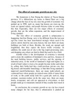
AD is characterized by two distinct neuropathological hallmarks presented as intracellular
neurofibrillary tangles (NFT) that contain hyper phosphorylated tau protein, and extracellular amyloid
plaques mainly composed of the Aβ peptide (Fig. 1) (Masters & Beyreuther, 1991; Selkoe, 2001a).
The relationship of these two independent protein accumulations is poorly understood and under
extensive investigation.
A
B
Figure 1. Plaques and tangles in the AD brain. (a) A representativemicrophotograph
of amyloid plaques in the AD brain. Amyloid plaques were visualized by
using an anti-Aβ42 specific antibody. Scale bar represents 125 mm. (B) Immuno histochemical staining of neurofibrillary
immunostaining with an anti-Ab42 specific antibody. Scale bar: 125 mm. (b) A
tangles using an anti-tau PHF-1 antibody. Scale bar represents 62.5 mm. (LaFerla & Oddo, 2005).
representative microphotograph of neurofibrillary tangles. Tangles were visualized
by immunostaining with an anti-PHF1 specific antibody. Scale bar: 62.5 mm. Note
the prominent tau immunoreactivity in the somatodendritic compartment,
characteristic of tau mislocalization.
Fig. 1: Aβ positive plaques and tau positive NFTs in human AD brains. (A) Immuno histochemical staining of plaques
2
Introduction
Tau is a microtubule associated protein that stabilizes polymerized microtubules by binding to the αand β-tubulin subunits via its 3-repeat or 4-repeat binding domains. Hyper-phosphorylation of tau in
this domains due to increased kinase activity or lowered phosphatase activity, leads to its detachment
from the microtubule and the formation of so-called paired helical filaments (PHFs) with a diameter of
~20 nm (Goedert et al, 2006). Continuous formation of PHFs leads to the accumulation of NFTs
within neurons. The decreased binding of tau results in destabilization of the microtubule network,
causing an impaired retrograde transport in neuronal cells. As a result, tau accumulates and aggregates
in somatodentric compartments (Grundke-Iqbal et al, 1986). The formation of PHFs and NFTs could
induce retrograde degeneration of neurons and cell death. The tau hypothesis assumes that formation
of NFTs initiates and promotes the pathogenesis of AD.
The second distinct neuropathological hallmark of AD brains are Aβ plaques that show a
heterogeneous appearance with a diameter of up to 20 - 50 µm. Plaques are spherical extracellular
multi-protein aggregates that are mainly composed of the peptides Aβ40 or Aβ42 and surrounded by
abnormal neuronal processes. Plaques can be classified into diffuse and neuritic (senile) types. The
involvement of diffuse plaques in early stages of AD is discussed controversially. They lack
association with altered neurites and glial cells when compared to neuritic plaques (Joachim et al,
1989). Diffuse plaques are not necessarily indicative for AD patients, since they are also detected in
cognitively normal individuals as well (Hardy & Selkoe, 2002). They are mainly composed of nonfibrillar Aβ and can be found in most brain regions (Tagliavini et al, 1988; Yamaguchi et al, 1988).
Neuritic or senile plaques, on the other hand occur in a brain region specific sequence and increasing
number during disease progression (Thal et al, 2002). The cortex and the hippocampus are mainly
affected by neuritic plaques. In later stages, neuritic plaques can also be found to a lesser extend in the
brainstem and the molecular layer of the cerebellum (Thal et al, 2002). Neuritic plaques are more
heterogeneous and dense than diffuse plaques, and consist of fibrillar and non-fibrillar Aβ variants as
well as degenerated neurites (Braak & Braak, 1996). Further components of neuritic plaques are
complement factors, glucosaminoglycans, ApoE, cholesterol or cytoskeletal proteins (Liao et al,
2004).
3
Introduction
The amyloid-hypothesis states that the accumulation of Aβ triggers the pathological development of
AD, including neuronal dysfunction and neuroinflammation. Aβ induced damage of neuronal cells and
alterations in phosphatase or kinase activities are suspected to induce hyper-phosphorylation of tau
and the formation of NFTs (Hardy & Selkoe, 2002). Interestingly, mutations that are associated with
early onset AD have been identified in three different genes directly involved in Aβ generation,
strongly supporting the amyloid hypothesis. In contrast no mutations in tau have been identified so far
to cause AD. However, tau mutations are associated with other neurodegenerative diseases, including
frontotemporal dementia (Goedert & Spillantini, 2000) or progressive supranuclear palsy (Stanford et
al, 2000)suggesting an important role of tau in neurodegeneration.
1.1.1
Genetics of AD.
The majority of AD cases occur sporadically at a higher age (>65 years) without a known causative
gene mutation. This form is known as late onset AD (LOAD). The genetic factors underlying the
pathogenesis of LOAD are not fully understood. However, the apolipoprotein allele ε4 (ApoE4) was
discovered as the major genetic risk factor for developing LOAD (Strittmatter et al, 1993). The chance
to develop AD is increased by threefold in the presence of one ApoE4 allele and approximately by 12fold when two alleles are present, as compared to individuals with no ε4 allele (www.alzgene.org).
While the exact molecular mechanism remains elusive, several studies indicated a decreased ability of
ApoE4 for the clearance of Aβ (Bu, 2009) or an impaired endocytosis of Aβ by microglial cells
(Carter, 2005). A recent study has furthermore identified TREM2 (triggering receptor expressed on
myeloid cells 2) as an additional risk factor for the pathogenesis of AD (Guerreiro et al, 2013; Jonsson
et al, 2013). Loss of a single TREM2 copy had no effects on the Aβ deposition, but altered the
morphology of plaque-associated microglial cells, which highlights its role in microglial response
(Ulrich et al, 2014). In genome wide association studies a number of further genes like PICALM or
CLU have been shown to be associated with the AD risk (Harold et al, 2009). However, all of these
risk factors have much lower impact on the AD development compared to ApoE.
A minor percentage of all AD cases are linked to mutations in PS1, PS2 and APP, and cause an early
onset of AD (EOAD). A high number of different mutations in these genes were identified (>25 APP;
4
Introduction
>10 PS2; >150 PS1) and shown to cause either a misbalance in the processing of APP or to promote
the aggregation propensity of ACai et al, 1993; Citron et al, 1992; Goate et al, 1991). Commonly,
these mutations lead to elevated generation of total Aβ or a change in the relative ratio of Aβ40 to
Aβ42, and thus, could promote plaque formation (Tanzi & Bertram, 2001). The so-called Swedish
mutations of APP at position 670 and 671 (KMNL) favors processing by the β-secretase and
increases generation of Aβ40 and Aβ42 (Citron et al, 1992). Further mutations such as the Austrian
(T714A), German (V715A) or Florida (I716V), lie within the trans-membrane domain and cause an
increased production of the longer Aβ42 species that are more hydrophobic and more neurotoxic
(Suzuki et al, 1994). All APP mutations identified so far, are mainly located close or within the Aβ
domain.
Mutations in the presenilin genes on the other hand occur in different regions of the encoded proteins,
but are enriched in hydrophobic trans-membrane or membrane associated domains (Tanzi & Bertram,
2005). Most mutations in the PS1 and PS2 genes are missense mutations and affect their endoproteolytic cleavage (see 1.1.2), the generation of different splice variants and also the overall
enzymatic activity. A missense mutation in the splice acceptor site of exon 9 causes a deletion of this
exon (Δexon9) and results in the loss of the endo proteolytic cleavage site of PS1 (De Strooper, 2007).
A double transgenic mouse model expressing PS1 Δexon9 and the human variant of APPswe, also
known as APP/PS1, is a commonly used mouse model for AD. These mice show severe plaque load
with increasing age, as well as cognitive and behavioral deficits (Dewachter et al, 2001).
Interestingly, even though more than 60 mutations for tau have been identified, none is associated with
the pathogenesis of AD. Most of the tau mutations are mainly associated with fronto-temperal lobe
dementia (FTLD). An interactive diagram with all mutations in APP, PS1, PS2 and tau can be found at
/>
1.1.2 Metabolism of the Amyloid Precursor Protein.
Aβ peptides derive from the sequential cleavage of the amyloid precursor protein (APP). APP is a 100
– 140 kDa, ubiquitously expressed type I transmembrane protein. The molecular weight varies due to
5
Introduction
differences in maturation states, alternative splicing and post-translational modifications. Alternative
splicing of the 19 different exons generates three predominant length variants that show tissue specific
distribution. APP695, also known as the neuronal APP, consists of exons 1 – 6 and 9 – 18, whereas
APP751 (exon 1 – 7, 9 – 13) and APP770 (exons 1 – 18) are predominately expressed in peripheral
tissues (Selkoe, 2001b). Heterogeneity in APP is further caused by co- and posttranslational
modifications (Hung & Selkoe, 1994; Tomita et al, 1998; Walter et al, 1997a; Walter & Haass, 2000).
First, immature full-length APP (APP-FLim) undergoes co-translational N-glycosylation in the
endoplasmic reticulum and is then transported to cis-Golgi compartments. During transport in the
Golgi, APP undergoes further maturation by O-glycosylation, sulfation and phosphorylation. Mature
APP (APP-FLm) is then transported via secretory vesicles to the plasma membrane for proteolytic
cleavage or re-internalization into endocytic vesicles (Weidemann et al, 1989). APP can undergo
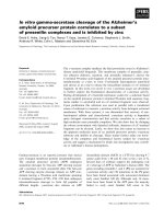
sequential cleavage mediated by three distinct intramembranous proteases, called α-, β- and γsecretases. Processing of APP can take place in two principal cleavage pathways: the nonamyloidogenic and the amyloidogenic pathway (Fig. 2).
amyloidogenic
non-amyloidogenic
extracellular
space/lumen
N
N
sAPPα
N
γ
α
C
αCTF
γ
β
Aβ
α
C
N
N
p3
AICD
sAPPβ
APP
C
β
γ
C
βCTF
C
AICD
cytosol
Fig. 2: Proteolytic processing pathways of APP. Initial cleavage of APP by α-secretase in the non-amyloidogenic pathway
precludes the generation of Aβ, but liberates sAPPα into extracellular fluids. Membrane associated αCTFs are further
processed by the γ-secretase complex producing the amyloid-intracellular domain (AICD) and p3. Alzheimer’s associated Aβ
variants are generated in the amyloidogenic pathway by β-secretase (BACE1) cleavage. Initial β-secretase cleavage leads to
secretion of sAPPβ into the extracellular milieu. βCTFs are then further processed by the γ-secretase complex, resulting in the
generation of Aβ and the AICD. Secreted Aβ can undergo dimerization (dAβ) or moreover oligomerization (oAβ), which
might result in the formation of amyloid plaques.
6
Introduction
α-Secretase
The predominant cleavage of APP is initiated by the α-secretase in the non-amyloidogenic pathway
between Lys16 and Leu17 within the Aβ domain. α-secretase cleavage precludes the generation of Aβ
and results in the secretion of soluble APPα (sAPPα) and the generation of the membrane-tethered Cterminal fragment (αCTF) (Esch et al, 1990; Sisodia et al, 1990; Wang et al, 1991) (Fig. 2). This
cleavage occurs predominantly at the cell surface and suggests a plasma-membrane localization of the
α-secretases (Sisodia, 1992b). Studies identified at least four enzymes to have α-secretase cleavage
properties that belong to the family of “a disintegrin and metallo proteinases”: ADAM9, ADAM10,
ADAM17 and ADAM19 (Allinson et al, 2003). All ADAM-proteins are type I transmembrane
proteins and require zinc as a co-factor for their activity (Sisodia, 1992a). The predominant form in
neuronal cells was recently discovered to be ADAM10 (Kuhn et al, 2010). α-Secretase activity can be
regulated by protein kinase C (PKC). Phorbol esters stimulate PKC activity and increase the αsecretory cleavage of APP resulting in both, elevated secretion of sAPPα (Buxbaum et al, 1990) and
decreased generation of Aβ (Hung et al, 1993). However, ADAM proteins not only cleave APP, but
also several other proteins like Notch receptors, tumor necrosis factor α (TNFα), cadherins and IL-6
(Seals & Courtneidge, 2003). This highlights the physiologic relevance of ADAM-proteases also
documented by the in utero lethality of ADAM10 or ADAM17 knockout mice (Hartmann et al, 2002;
Peschon et al, 1998).
β-Secretase
β-secretase or BACE1 (β-site APP cleaving enzyme) (Sinha et al, 1999; Vassar et al, 1999; Yan et al,
1999) initiates the generation Aβ and is the rate limiting enzyme in the amyloidogenic pathway.
Cleavage of APP by BACE1 leads to secretion of sAPPβ and the generation of βCTF containing the
Aβ domain (Fig. 2). BACE1 is a type I transmembrane aspartyl protease, consisting of a cytosolic cterminus, a transmembrane domain and a luminal/extracellular domain (Hussain et al, 1999; Vassar et
al, 1999). The latter contains the proteolytically active site and shows similarities to other members of
the pepsin family (Hong et al, 2004). Two distinct DTGS and DSGT motifs form the catalytic center
7
Introduction
of BACE-1. Mutations of either motif lead to complete loss of enzymatic activity (Hussain et al, 1999;
Vassar et al, 1999). Two distinct cleavage sites for BACE1, Asp1 and Glu11 have been identified in
APP. In human, the predominant BACE1 cleavage takes place at Glu11 of the Aβ domain and
precludes the generation of Aβ (Liu et al, 2002). A recent study claimed, that Glu11 cleavage by
BACE1 is favored, but shifting its activity towards Asp1, may be the pathologically more relevant
process (Deng et al, 2013). An enzyme with 55% homology to BACE1 was identified and termed
BACE2. However, BACE2 cleaves APP within the Aβ domain between Phe19 and Phe20 and thus,
likely does not contribute to amyloid generation (Farzan et al, 2000; Fluhrer et al, 2002).
BACE1 is ubiquitously expressed, but with the highest expression in pancreatic and neuronal cells
(Ehehalt et al, 2002). The high expression rate of BACE1 and APP in neuronal cells, explains why
neurons mainly contribute to the generation of Aβ. BACE1 as well as BACE2, contain a pro-peptide at
their n-terminal domains, which undergoes furin mediated cleavage in the Golgi compartment. Block
of the forward transport with brefeldin A or monensin reduces the propeptide cleavage (Bennett et al,
2000). N-glycosylation at Asp residues in the ectodomain takes place in the ER, while the
restructuring and trimming of the glycol-moieties occurs in Golgi compartments, from where BACE1
is routed to the plasma membrane (Capell et al, 2000). Trafficking of BACE1 is regulated by its
phosphorylation at Ser468. While phosphorylation facilitates retrograde transport of BACE1 to juxta
nuclear Golgi compartments, non-phosphorylated BACE1 accumulates in peripheral early endosome
antigen 1 (EEA1) positive vesicles (Walter et al, 2001). Phosphorylation of BACE1 regulates the
interaction with adapter proteins of the Golgi associated, γ-adaptin ear containing, ARF binding
protein (GGA) family that mediate sorting between endosomal/lysosomal compartments and the transGolgi Network (TGN) (Tesco et al, 2007; von Arnim et al, 2006; Wahle et al, 2005; Wahle et al,
2006). Most BACE1 protein can be found in these particular compartments. Especially its pH
optimum of 4.5 – 5 indicates a pronounced activity of the enzyme in endosomal and lysosomal
compartments (Vassar & Citron, 2000). BACE1 was also shown to undergo degradation in acidic
organelles (Koh et al, 2005).
Generation of BACE1 knockout mice helped to identify the physiologic role of BACE1. Initial
findings indicated no deficits in viability or fertility (Cai et al, 2001; Luo et al, 2003; Roberds et al,
8
Introduction
2001). However, later studies with BACE1 KO mice showed subtle effects on behavior with impaired
memory function or spontaneous hyperactivity (Dominguez et al, 2005; Harrison et al, 2003).
Moreover, severely reduced myelination of neurons was present as well, probably caused by a
precluded cleavage of neuregulin-1 (NRG1), a mediator for Schwann-cell myelination (Willem et al,
2006). Further substrates of BACE1 are voltage-dependent sodium channels (Kim et al, 2007), the
type II α-2,6-sialyltransferase (Kitazume et al, 2003), the platelet selectin glycoprotein ligand-1,
(Lichtenthaler et al, 2003), LRP1 (von Arnim et al, 2005), APLP1/2 (Li & Sudhof, 2004) and the
interleukin receptor II (Kuhn et al, 2007).
γ-secretase
Intramembranous cleavage of both APP CTF variants, αCTFs and βCTFs, is mediated by the γsecretase. Cleavage of αCTFs by the γ-secretase, results in the generation of the APP intracellular
domain (AICD) and the secretion of the small peptide p3 (Fig. 2) (Haass et al, 1993). However, γsecretase dependent cleavage of the βCTFs induces the generation of the AICD and 37 – 49 amino
acid long Aβ peptide variants (Fig. 2). The predominant variant is Aβ40 and to a lesser extent Aβ42
(Citron et al, 1996; Wiltfang et al, 2002). Aβ42 is more hydrophobic and has increased propensity to
aggregate as compared to A40. The additional γ-secretase product AICD on the other hand is
released into the cytosol and may have a role in nuclear signaling (Cao & Sudhof, 2001; von Rotz et
al, 2004). A series of other proteins like ErbB4 (Lee et al, 2002), Notch (Kimberly et al, 2003), CD43
(Andersson et al, 2005), ephrin B1 (Tomita et al, 2006), LRP1 (Lleo et al, 2005) and TREM2
(Wunderlich et al, 2013) also undergo cleavage by γ-secretase. In general, γ-secretase has little
substrate specificity. Because -secretase requires short ectodomains and single transmembrane CTFs
of the respective protein substrates, the cleavage of the different γ-secretase substrates is mainly
regulated by ectodomain shedding of type I membrane proteins (Hemming et al, 2008)
γ-Secretase is a multimeric multi-transmembrane enzyme-complex composed of presenilin 1 or
presenilin 2 (PS1/PS2), nicastrin (NCT), anterior pharynx defective 1 (Aph1) and the presenilin
enhancer 2 (PEN2) (Francis et al, 2002; Yu et al, 2000). A minimal stoichiometric ratio of 1:1:1:1 of
9
Introduction
the components is necessary for its activity. The essential γ-secretase component NCT is required for
substrate selection and transport of the -secretase complex in the secretory pathway (Dries et al, 2009;
Shah et al, 2005; Yu et al, 2000). PEN2 facilitates the endo proteolytic cleavage of the presenilins and
confers their stability (Hasegawa et al, 2004; Hu & Fortini, 2003; Prokop et al, 2004). The role of
Aph1 is still elusive, but it is suspected to act as a scaffolding protein in the complex (LaVoie et al,
2003). Interestingly, the molecular weight of the whole γ-secretase complex is higher than the additive
and predicted size of the single components, suggesting an involvement of more associated proteins or
protein complexes. Some additional proteins like TMP21 (Chen et al, 2006), CD147 (Zhou et al,
2005) or the γ-secretase activating protein GSAP (He et al, 2010a) were recently identified. However,
it could be demonstrated that these proteins are not essential for the γ-secretase activity (Winkler et al,
2009). The proteolytic activity of the γ-secretase is carried out by PS1 or PS2. The major presenilin
involved in the APP cleavage is PS1, although PS2 has the ability to cleave APP as well (De Strooper
et al, 1998; Wolfe et al, 1999). PS1 and PS2 have 9 transmembrane domains. The 50 kDa full-length
forms of these proteins undergo autocatalytic cleavage to form 30 kDa N-terminal fragment (NTF) and
a 20 kDa CTF (Thinakaran et al, 1996; Walter et al, 1997b). Both CTF and NTF form a heterodimer
with one Asp residue in each fragment (Fig. 3A). These neighboring Asp residues in the sixth and
seventh transmembrane domains form the catalytic center of the γ-secretase complex (Wolfe et al,
1999). Knock out of PS1 in mice, causes embryonic lethality, due to impaired processing of Notch
(Herreman et al, 1999; Shen et al, 1997). The knock-out of PS2 does not lead to overt phenotypes.
However, the double KO of PS1 and PS2 causes a more severe phenotype and earlier embryonic
lethality as compared to the PS1 single KO, indicating a physiological relevance of PS2 (De Strooper
et al, 1998).
10
Introduction
A
B
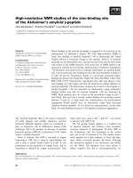
Fig. 3: γ-secretase complex and Aβ producing sequential cleavage lines. (A) The γ-secretase complex with subunits and
membrane topology. Full length presenilin is autocatalytically cleaved into a CTF and NTF. Aspartyl residues are indicated
by D. Further γ-secretase complex subunits nicastrin, Aph-1 and Pen-2 have supporting properties and are necessary for
catalytic activity of the presenilin (De Strooper et al, 2012).
Subcellular trafficking of APP and its metabolizing enzymes.
As mentioned before, maturation of APP involves N- and O-glycosylation, as well as phosphorylation,
during its transport from the ER to the Golgi compartments and forth the plasma membrane in the
secretory pathway. Mature full length APP is either rapidly processed in the secretory pathways or at
the plasma membrane by the α-secretases or internalized into endocytic vesicles. Following the
endocytosis, APP is either transported back to the plasma membrane, or delivered into endosomal or
lysosomal compartments for degradation (Fig. 4). The initial internalization from the plasma
membrane was shown to be dependent on a YENPTY motif at the c-terminus (Lai et al, 1995;
Marquez-Sterling et al, 1997). Mutations in this motif selectively inhibited the internalization and
prevented the binding of adaptor proteins like Fe65 (Borg et al, 1996; Perez et al, 1999). Fe65 binding
also facilitates BACE1 and γ-secretase mediated processing of APP. The phosphorylation at Thr688
residue of APP introduces a conformational change and precludes interaction with Fe65 (Ando et al,
2001; Chang et al, 2006).
In neurons, APP undergoes polarized trafficking. Various proteins and lipids are involved in this
regulation. After leaving the ER, APP is first transported to Golgi compartments. Interestingly, in a
model for polarized cells (Madin-Darby canine kidney cells: MDCK), a substantial pool of APP can
undergo cleavage already in these compartments as it was shown (Haass et al, 1995). However, non-